ABSTRACT. Lukas J. Farbiak. Director: Darryn S. Willoughby, Ph.D.
|
|
|
- Rudolf Russell
- 6 years ago
- Views:
Transcription
1 ABSTRACT Effects of Lower- and Higher-Volume Resistance Exercise on Serum Total and Free Testosterone, Skeletal Muscle Testosterone and Dihydrotestosterone Content, and Skeletal Muscle Androgen Receptor mrna Expression and Protein Content Lukas J. Farbiak Director: Darryn S. Willoughby, Ph.D. Testosterone is the primary sex steroid hormone within males. Its effects are ubiquitous, and can be categorized as either anabolic or androgenic. Testosterone exerts its effects on a specific nuclear androgen receptor (AR). Upon binding testosterone, the AR translocates to the nucleus of the cell. Once in the nucleus of the cell, the active AR complex binds to the androgen response element on DNA resulting in an up-regulation of gene expression. Androgen receptors are found in skeletal muscle which is responsive to testosterone. The binding of testosterone to the AR results in DNA binding, and subsequently promotes protein synthesis (anabolism) and a decrease in the breakdown of muscle tissue (catabolism). Both AR mrna and protein expression and testosterone levels affect muscle protein balance. It is known that high intensity resistance exercise increase endogenous serum testosterone levels. Therefore, the purpose of this study was to examine the ability of a resistance exercise-induced elevation in serum and free testosterone to increase skeletal muscle testosterone, 5α-dihydrotestosterone (DHT), AR mrna expression and protein content. In a randomized cross-over design, venous blood was obtained in male participants immediately before, after, and 30min, 1 hr, 2 hr, 3 hr, and 24 hr after a single bout of exercise. Muscle samples were also obtained immediately before, after, and 3 hr, 24 hr after exercise. Exercise bouts were either lower volume (LV) and consisted of a lower body resistance exercise program (knee extensions) or higher volume (HV) consisting of an upper body/lower body resistance exercise program (bench press, seated rows, shoulder press, knee extensions). Exercise bouts were separated by one week. From each blood sample, the levels of serum and total testosterone were determined. From each muscle sample, the concentration of testosterone, and dihydrotestosterone (DHT) was determined, along with the mrna expression and protein content of the androgen receptor. Statistical analysis was performed by utilizing separate 2x4 and 2x7 (Session x Test) factorial analyses of variance (ANOVA) with repeated measures for muscle and blood analyses, respectively. Further analysis of the main effects was performed by separate one-way ANOVAs. Significant between-group differences were then determined involving the Tukey s Post Hoc Test.
2 APPROVED BY DIRECTOR OF HONORS THESIS: Dr. Darryn Willoughby, Department of Health and Human Performance APPROVED BY THE HONORS PROGRAM: Dr. Andrew Wisely, Director DATE:
3 EFFECTS OF LOWER- AND HIGHER-VOLUME RESISTANCE EXERCISE ON SERUM TOTAL AND FREE TESTOSTERONE, SKELETAL MUSCLE TESTOSTERONE AND DIHYDROTESTOSTERONE CONTENT, AND SKELETAL MUSCLE ANDROGEN RECEPTOR MRNA EXPRESSION AND PROTEIN CONTENT A Thesis Submitted to the Faculty of Baylor University In Partial Fulfillment of the Requirements for the Honors Program By Lukas J. Farbiak Waco, Texas May 2013
4 TABLE OF CONTENTS Table of Contents... ii List of Figures... iii List of Tables... iv Acknowlegments... v Dedication... vi Chapter One Introduction... 1 Chapter Two Literature Review... 7 Chapter Three Methods Chapter Four Results Chapter Five Discussion Appendix References ii
5 LIST OF FIGURES Figure Study Outline iii
6 LIST OF TABLES Table Study Visit Outline Table Subject Baseline Demographics Table Subject Body Water and Hydration Status Table Subject Blood Pressre and Heart Rate Table Serum Total Testosterone Table Serum Free Testosterone Table Skeletal Muscle Testosterone Table Skeletal Muscle DHT Table Skeletal Muscle AR Protein Content Table Skeletal Muscle AR mrna Expression iv
7 ACKNOWLEDGMENTS I would like to thank Lord and Savior Jesus Christ for empowering me with the strength to complete this thesis. Without Him, none of this would be possible. -2 Corinthians 12:9-10 I am truly indebted and thankful to Dr. Darryn Willoughby, Ph.D., for his relentless patience and guidance in helping me complete this project. Without his direction and dedication completion of this thesis would be impossible. I owe sincere and earnest thankfulness to Mr. Mike Spillane, M.S.Ed., Ph.D.(c), for his countless hours of help, constant patience, genuine support and encouragement. v
8 DEDICATION To my parents, Scott and Karen Farbiak, And my friends, Without whom none of my success would be possible vi
9 CHAPTER ONE Introduction Testosterone is the primary androgenic sex steroid hormone in males. Testosterone production is tightly regulated by the hypothalamic pituitary gonadal axis, wherein the Leydig cells of the testis account for 95% of the testosterone produced within adult males (Rommerts, E. Nieschlag et al., 2004; Kicman, 2010). Testosterone effects are ubiquitous and categorized as either anabolic (i.e. growth) or androgenic (i.e. male sex characteristics). Testosterone s primary anabolic effects occur via adaptation of skeletal muscle increase in protein synthesis. This increase in protein synthesis is accomplished via numerous biochemical and molecular pathways. Specifically, testosterone binds to a nuclear androgen receptor (AR) which then translocates to the nucleus. Following translocation, the AR complex binds to the androgen response element on DNA which then causes an up-regulation of gene expression (Kicman, 2010). The rate of protein synthesis is regulated by the binding ability of testosterone to the AR. Also, it has been shown that AR expression will fluctuate in response to varying serum testosterone levels (Bamman, Shipp et al., 2001; Lee, McClung et al., 2003; Willoughby and Taylor, 2004). Both short and long term resistance exercise is known to produce an increase in muscle protein synthesis leading to a promotion of skeletal muscle hypertrophy (Abernethy, Jurimae et al., 1994; Fitts and Widrick, 1996). Depending on the protocol, resistance exercise has shown to increase testosterone levels, with hormonal alterations 1
10 occurring both locally and systemically (Kraemer and Ratamess, 2005; Spiering, Kraemer et al., 2008; Vingren, Kraemer et al., 2010). Also, the AR protein expression within skeletal muscle has shown to be responsive to resistance exercise (Willoughby and Taylor, 2004; Vingren, Kraemer et al., 2009). Resistance exercise mediates androgen signaling through several mechanisms. A transient increase in endogenous testosterone levels in response to resistance exercise increased the probability of testosterone AR interactions, and muscle contraction/overload has shown to cause an up-regulation of skeletal muscle AR content by increasing the transcription of AR mrna (Bamman et al., 2001; Lee, Thompson et al., 2003; Willoughby and Taylor, 2004). Also, it is known that the administration of androgens via muscular injection in the absence of muscular contraction/overload increases AR mrna translation which results in an up-regulation of AR content, and also increases AR half-life (Syms et al., 1985). Muscle contraction combined with elevated endogenous testosterone levels constitutes the bases of enhanced AR content in skeletal muscle (Spiering, Kraemer et al., 2008). In order to maximize the effects of these androgens it is necessary to perform resistance exercise bouts high in intensity and volume; comprised of the number of sets, exercises, rest periods, and weight used. In accordance with this reasoning, it has been shown that protocols high in volume, moderate to high in intensity, using short rest intervals and stressing a large muscle mass, tend to produce the greatest acute hormonal elevations, suggesting that elevations in testosterone appear to dependent on volume/intensity (Ratamess and Kraemer et al., 2005; Kraemer and Ratamess, 2005). 2
11 Testosterone/AR interactions are largely responsible for changes in skeletal muscle adaptation, which ultimately affects muscle hypertrophy. It is known that resistance exercise causes a transient increase in endogenous circulating serum testosterone levels. The impact of this elevation of serum testosterone seen during resistance training to increase testosterone levels within the skeletal muscle in addition to the interaction with the AR still needs to be determined. Problem Statement How does an endogenous elevation in testosterone resulting from a single resistance exercise bout involving either lower-volume (LV) or higher-volume (HV) alter androgen levels within the blood and muscle tissue, and androgen receptor mrna expression and protein content. Purpose of Study The serum testosterone increase in response to HV resistance exercise, which involves upper-body resistance exercise performed immediately prior to lower-body exercise, is typically greater than that of the increase in serum testosterone resulting from LV exercise, which involves only lower-body resistance exercise. Therefore, the purpose of this study was to examine the effect of possible elevations in endogenous testosterone levels immediately before, after, and 30 min, 1 hr, 2 hr, 3 hr, and 24 hr after a single bout of either LV or HV resistance exercise. Heavy resistance exercise of the upper-body performed immediately prior to lower-body resistance exercise should result in a greater elevation in serum testosterone level compared to resistance exercise involving only the lower-body. Specifically, the purpose of this study was to determine if elevated blood 3
12 testosterone would result in an increase in muscle testosterone, dihydrotestosterone (DHT), and androgen receptor mrna expression and protein content. Hypotheses H 1 : Following the HV exercise bout involving both upper- and lower-body resistance exercise, a significant increase in serum testosterone will occur compared to the LV exercise bout only involving lower-body resistance exercise. H 2 : Following the HV exercise bout involving both upper- and lower-body resistance exercise, a significant increase in muscle testosterone and DHT content will occur compared to the LV exercise bout only involving lower-body resistance exercise. H 3 : Following the HV exercise bout involving both upper- and lower-body resistance exercise, a significant increase in AR mrna expression and protein content will occur compared to the LV exercise bout only involving lower-body resistance exercise. Delimitations Ten apparently healthy males between the ages of who had consistent resistance training (at least thrice weekly) for at least one year prior to the study. Participants were recruited from Baylor University and within the surrounding Waco, Tx area by flyers and online advertisements. Participants were excluded from the study if prior ingestion (within 6 months) of any dietary supplement or pharmaceutical aid used as a potential ergogenic aids. All participants were considered low risk for cardiovascular disease, with no contraindication to exercise as outlined by the American College of Sports Medicine (ACSM). Participants were in a euhydrated state prior to participation in both exercise bouts. All participants were tested at the Baylor Laboratory for Exercise Science and Technology (BLEST) and Exercise Nutritional Biochemical Laboratory (EBNL) in accordance with Helsinki Code after signed university approved informed consent documents. 4
13 Limitations The study utilized a convenience sampling of those individuals who were within the city of Waco, as well as a small sample size (n=10), external validity to the greater population of resistance trained males (18-30 y) may be reduced; although, it is unlikely to be a significant concern Participants were expected to maximally exert themselves during both the upper body/lower body and lower body only resistance exercise bouts Each participant can have inherent circadian rhythms that will alter hormonal levels throughout the day; this variation was minimized by testing during the morning (am) hours for each participant The biopsy procedure can cause trauma (inflammation) to the site of extraction; to minimize any possible stress response, additional samples were taken from incision 0.5cm medial or lateral to the original biopsy site Assumptions All laboratory equipment would be functioning properly with validity and reliability measurements being established. Proper calibration and the use of trained research staff would minimize any potential for errors. All participants would follow the guidelines provided and performed the exercises at maximal effort during the testing sessions All participants would be truthful in training status: consistent resistance training (3 times/week) for at least one year prior to the study. All participants would arrive at each testing session in a fasted state ( >8 hours) All participants would arrive with adequate sleep (7-8 hours) before each of the testing sessions Definitions AR Androgen Receptor, type of nuclear receptor that is activated by binding of androgenic hormones testosterone or 5α-dihydrotestosterone ARE Androgen Response Element, specific genomic site to which androgen receptor binds and modulates transcription of nearby genes 5
14 BIA Bioelectrical Impedance Analysis, method to measure body composition specifically body water through the use of a low level electrical current by measuring the resistance to the current DEXA- Dual Energy X-ray Absorptiometry, imaging technique that uses two lowdose x-ray beams to measure the density of specific tissues (bone mineral, lean tissue, adipose tissue) DHT - 5α-dihydrotestosterone, derivative of testosterone having androgenic and anabolic activities. Responsible for male primary sex characteristics DNA Deoxyribonucleic Acid, is a nucleic acid containing the genetic instructions used in the development and functioning of all known living organisms Free Testosterone Testosterone not bound to binding proteins IGF-1 Insulin Like Growth Factor -1, polypeptides hormone similar to insulin which stimulates protein synthesis MGF Mechano Growth Factor also known as IGF-1Ec, splice variant of insulin like growth factor which is produced in response to mechanical stress within skeletal muscle mrna- Messenger Ribonucleic Acid, A coding molecule that provides genetic information for the cell to produce new proteins Myofibrillar Protein - a muscle fibril, one of the slender threads of a muscle fiber, composed of numerous myofilaments Myostatin Member of the transforming growth factor-β (TGF-β) superfamily, functions as negative regulator of skeletal muscle SHBG Sex hormone binding globulin, glycoprotein that binds to testosterone Testosterone androgenic (Steroid) hormone, primarily produced within the Leydig's cells of the testis in men 1-RM 1 repetition maximum, the maximum amount of weight one can lift in a single repetition for a given exercise 6
15 CHAPTER TWO Literature Review Testosterone Production The major circulating androgen within males is testosterone (17β-hydroxy-4- androstenen-3-one), which is a kd C 19 steroid hormone produced from cholesterol (Rommerts, E. Nieschlag et al., 2004; Vingren, Kraemer et al., 2010). Several enzymatic reactions within the Leydig cells of the testes utilize cholesterol in the formation of testosterone (Mendelson, Dufau et al., 1975; Cigorraga, Dufau et al., 1978). The testes account for greater than 95% of the testosterone production within an adult male, which translates to roughly 3-7 mg per day (Rommerts, E. Nieschlag et al., 2004; Kicman, 2010). However, testosterone has a secondary site of production in the zona reticularis of the adrenal cortex in combination with brain and nervous system cells where it is produced in small quantities (Baulieu, 1997; King, Manna et al., 2002; Marouliss and Triantafillidis, 2006). The biosynthesis of testosterone from cholesterol within the gonads and adrenal gland requires two major classes of enzymes (cytochrome P450 and hydroxysteriod dehydrogenase). Specifically, a cholesterol side chain is catalyzed by enzyme CYP11A to yield the C 21 steroid pregnenolon, which is the rate limiting step in testosterone biosynthesis. An enzymatic reaction of 3βHSD can catalyze pregenolone into progesterone. Pregnenolone and/or progesterone will further yield either 17αhydroxypregnenolone or 17α-hydroxyprogesterone via enzyme CYP17. Additional cleavage will take place yielding the C 19 steroids dehydroepiandrosterone (DHEA) or 7
16 androstenedione. Within the gonads androstenedione is catalyzed into active steroid hormone testosterone via 17HSD3 (Payne and Hales, 2004; Miller and Auchus, 2011). Testosterone Regulation HPG axis The hypothalamic- pituitary- gonadal axis (HPG axis) is responsible for the regulation of testosterone production. The hypothalamus is innervated by the central nervous system where the release of three neuropeptides (kisspeptin, neurokinin B, dynorphin) controls the production and secretion of gonadotropin-releasing hormone (GnRH) (Lehman, Coolen et al., 2010). GnRH is released from the hypothalamus into the hypophyseal portal circulation system where GnRH subsequently binds to receptors on gonadotropes located in the pituitary (Kaiser, Sabbagh et al., 1995; Veldhuis, Keenan et al., 2009). Upon binding to gonadotropes, gonadotropin stimulation results in the synthesis and release of luteinizing hormone (LH) and follicle-stimulating hormone (FSH) from the anterior pituitary (Kaiser, Sabbagh et al., 1995; Keenan and Veldhuis, 1998; Veldhuis, Keenan et al., 2009). GnRH is released in pulsatile bursts from the hypothalamus (Keenan and Veldhuis, 1998; Veldhuis, Keenan et al., 2009). The pulsatile burst release of GnRH results in diurnal fluctuation in testosterone production; thus, testosterone levels have shown to be 30-35% higher during the morning hours ( hours) compared to mid to late afternoon (Brambilla, Matsumoto et al., 2009). Once released from the anterior pituitary, LH and FSH enter systemic circulation and have their site of action in the gonads. LH binds to G-protein coupled LH receptors within the Leydig cells of the testes resulting in activation of cyclic AMP. Receptor activation via LH binding results in the stimulation of testosterone production (Mendelson, Dufau et al., 1975; Cigorraga, Dufau et al. 1978; Miller and Auchus, 2011). 8
17 FSH does not directly result in testosterone production in males, but does stimulate the production of sex hormone binding globulin (SHBG) (Rosner, Hryb et al., 2010). Synthesized testosterone is not stored, and instead is rapidly released. A negative feedback loop tightly regulates central nervous stimulation of the pituitary and the HPG axis. CNS stimulation of the hypothalamus results in GnRH release and subsequently FSH and LH release from the anterior pituitary, which leads to testosterone production within the gonads. Elevation in testosterone levels will result in a negative feedback action upon the hypothalamus, reducing the release of GnRH which then decreases the release of LH and FSH in the anterior pituitary, inhibiting testosterone production within the gonads (Cigorraga, Dufau et al., 1978; Veldhuis, Keenan et al., 2009; Sharma, Nett et al., 2012). Testosterone Transport Testosterone is a lipid-based hydrophobic steroid hormone and thus must be transported by specific hydrophilic transporter proteins within the blood (Burton and Westphal, 1972). Total blood testosterone levels consist of both transport protein bound testosterone as well as a small amount of free unbound testosterone. Testosterone in the blood can be bound to SHBG which accounts for 44% to 60% of total serum levels (Heinlein and Chang, 2002; Rommerts, E. Nieschlag et al., 2004; Kicman, 2010; Vingren, Kraemer et al., 2010). The remaining serum testosterone is either weakly bound to albumin or unbound which accounts for roughly 2% of total serum levels (Hammond, Nisker et al., 1980; Rommerts, E. Nieschlag et al., 2004; Vingren, Kraemer et al., 2010). Testosterone that is bound to SHBG may not be transported into target tissues for androgen receptor binding, rendering this testosterone inactive (Pardridge, 1986). 9
18 Contrary to SHBG bound testosterone, bioavailability of albumin bound testosterone has shown to be high, with approximately 55% of albumin-bound testosterone being able to enter the target tissues (Manni, Pardridge et al., 1985). Free testosterone has the highest binding potential and as such is the most biologically active form of testosterone found in the bloodstream (Rommerts, E. Nieschlag et al., 2004; Vingren, Kraemer et al., 2010). Testosterone Metabolism There are several naturally occurring endogenous androgens, the most active of which are testosterone and 5α-dihydrotestosterone (DHT). Other androgens [5αandrostanediol, Androstenedione, Epitestosterone, Dehydroepiandrosteone (DHEA), Androsterone, Etiocholanolone] may be produced by the oxidation of 17β-hydroxyl group and/or reduction of 3-oxo group of testosterone and/or DHT. The androgens formed by this oxidative process exhibit either a reduced level of androgenic activity, or a complete loss of androgenic activity altogether (Kicman, 2010). Androgen levels within the body are determined by the rate of synthesis and degradation, with serum concentration of testosterone in eugonadal men ranging from 3-10 ng ml -1 (Kicman, 2010). The metabolism (aromatization) of testosterone is accomplished via the enzyme cytochrome P450 and may take place in various tissue (i.e. placenta, ovary, testis, adipose tissue, liver, hair follicles, brain, blood vessels, bone, and cartilage) within the body (Rommerts, E. Nieschlag et al., 2004; Czajka-Oraniec and Simpson, 2010). Despite such diverse distribution of aromatization, the major site of androgen metabolism is in the liver (Kicman, 2010). The aromatization of circulating androgens account for 85% of 17 β- estradiol and 95% of the estrone produced in males (Simpson, Clyne et al., 2002; Simpson 2003; Czajka-Oraniec and Simpson, 2010). Most aromatization of testosterone 10
19 into estrogen takes place within adipose tissue; however, some production of estrogen occurs in skeletal muscle tissue (Larionov, Vasyliev et al., 2003). Testosterone is also aromatized into 5α-dihydrostestosterone (DHT) by 5α-reductase which exists as either type 1 and 2 isoforms (Thigpen, Silver et al., 1993; Simpson, 2003). The enzymes involved in aromatization are NADPH-dependent and are located in the microsomes of the cell. These enzymes act by reducing the bonds of C 19 and C 21 steroids (Imperato- McGinley and Zhu, 2002). DHT and testosterone bind to the same intracellular receptor, but DHT has a higher affinity for the androgen receptor (Imperato-McGinley and Zhu, 2002; Kicman, 2010). Biological effects of Testosterone Due to the hydrophobic lipid soluble nature of free testosterone, it was traditionally hypothesized that testosterone free from binding proteins passively diffused across the plasma membrane of the cell to reach intracellular targets (Adams, 2005; Hammes, Andreassen et al., 2005; Kicman, 2010). However, research in cultured cells conducted by Hammes et al demonstrated that SHBG can bind to megalin (lowdensity lipoprotein receptor-related protein). The binding of SHBG to megalin internalizes SHBG into the cytoplasm where it can then be degraded by lysosomes. This process of internalization results in the release of steroids within the cellular environment. This proposed mechanism of steroid hormone release requires further investigation, and the traditionally proposed hypothesis allows for rapid entry of steroids into the intracellular environment. Testosterone, now translocated into the intracellular environment may undergo two different paths. It can bind to a specific nuclear receptor without disruption or can first be converted into 5α-dihydrotestosterone (DHT) by 5α- 11
20 reductase and then, post conversion may bind to the same nuclear receptor (Kicman, 2010). The activity of 5α-reductase in skeletal muscle is comparably much less than that in other tissues such as the skin and prostate (Thigpen, Silver et al., 1993; Hsiao, Thin et al., 2000; Zouboulis, Chen et al., 2007). Despite the higher affinity of DHT for the nuclear androgen receptor, these results indicate that due to the lower DHT levels within skeletal muscle, testosterone is the primary androgen binding to the nuclear receptor. Endogenous androgen alters androgen receptor expression, thereby mediating physiological effects on skeletal muscles, intracellular metabolism, and genomic and nongenomic pathways (Kicman, 2010). Androgen Receptor The androgen receptor is a member of ligand activated nuclear hormone receptor super family (Li and Al-Azzawi, 2009; Kicman, 2010). The androgen receptor consists of an N-terminal regulatory domain (NTD), DNA binding domain (DBD), variable hinge region (H), a ligand binding domain (LBD), and two transcriptional activation domains AF-1 and AF-2 (Li and Al-Azzawi, 2009; Kicman, 2010; Askew, Minges et al., 2012). The androgen receptor is expressed in two different isoforms; androgen receptor A and androgen receptor B. Androgen receptor A is a 87kDa isoform with a reduced NTD region while Androgen receptor B isoform NTD region is full length 110 kda (Wilson and McPhaul, 1994; Li and Al-Azzawi, 2009). The predominate isoform is androgen receptor B and is expressed in a variety of both fetal and adult human tissues (Wilson and McPhaul, 1996). While androgen receptor A may be expressed in the same tissues, it is not able to mediate all the effects when androgens bind, while this is not the case with androgen receptor B (Wilson and McPhaul, 1994). Elevation in testosterone levels have 12
21 shown to have an effect on the expression of androgen receptors. Ferrando et al. (2002) have shown that during pharmaceutical treatment with intramuscular testosterone injection in elderly men, the expression of androgen receptors is up-regulated after 1 month of treatment. However, after continued intramuscular injection of testosterone for a period of 6 months, the expression of androgen receptors returned to baseline levels. Additional studies have shown similar results in men with acute increases in testosterone leading to an up-regulation of the AR and a return to baseline AR expression after extended exposure (Kadi, Eriksson et al., 1999; Ferrando, Sheffield-Moore et al., 2001; Carson, Lee et al., 2002; Lee, Thompson et al., 2003). Shinha-Hikim et al. (2004) demonstrated that cycling on and off 600 mg of testosterone enanthate for 20 wk can lead to a long term increase in AR protein in older men (Sinha-Hikim, Taylor et al., 2004). Testosterone AR binding DNA binding Once testosterone translocates into the cell, the steroid hormone will bind to the AR. The AR is sequestered by specific chaperone proteins known as heat shock proteins (HSPs). Specifically HSP90, HSP70, and HSP56 are bound to the AR. When the androgens, testosterone or DHT, bind to the AR a transformation of the receptor occurs in which HSPs are dissociated resulting in the activation of the testosterone-ar bound complex (Veldscholte, Berrevoets et al., 1992; Gelmann, 2002). The bound active complex will translocate from the cytosol into the nuclei where it will bind to the androgenic response element (ARE) on DNA (Bennett, Gardiner et al., 2010). The DNA binding domain on the AR contains two zinc finger-like motifs that allow for insertion in a groove within the ARE (Claessens, Verrijdt et al., 2001; Helsen, Kerkhofs et al., 2012). The expression of targeted genes is generated by the activation of the androgen receptor 13
22 by testosterone or DHT which subsequently binds to the ARE on DNA within the nucleus (Maurer, Trajanoski et al., 2001). Attachment of the testosterone/ar complex to the ARE on DNA triggers formation of transcription complexes which activates gene sequences that alter the transcription and/or translation of that gene (Kicman, 2010). This mechanism is the classic explanation of testosterone function. Contrary to this process, however, testosterone may also play a role in more rapid, non-genomic effects. It is possible that androgens may bind to specific sites on specific molecules in the absence of the androgen receptor, and additionally androgens may alter membrane fluidity via binding transmembrane G-protein coupled receptors. Mediation of such changes occurs through increases in intracellular calcium or activation of signaling cascades (MAPK, PI- 3K) (Michels and Hoppe, 2008). Exercise Hormonal/Molecular Responses Testosterone Resistance exercise has shown to elicit a testosterone response in numerous studies (Kraemer, Marchitelli et al., 1990; Kraemer, Gordon et al., 1991; Kraemer, Hakkinen et al., 1999; Spiering, Kraemer et al., 2008; Roberts, Dalbo et al., 2009). The testosterone response seen within resistance exercise depends on numerous factors including age of exercise participant and type of exercise performed. Specifically, younger age men (20-30 yr) will have a greater testosterone response when compared to adolescent (14-18 yr), middle age (38-53 yr), older ( 59 yr) men and women (Vingren, Kraemer et al., 2010). To alter testosterone response, exercise bouts will need to have high intensity (load) (85%-95%) of one repetition max and meet a minimum threshold, 14
23 and moderate to high volume (set x number of reps x intensity) is also required. Volume alterations can be achieved by changing the number of sets or number of exercises, with exercises that utilize large muscle groups (i.e. power clean, squats, and dead lifts) eliciting the greatest response. Performing exercise in order with large muscle groups first, along with utilization of short rest periods (30-60 sec), will also result in the largest testosterone response (Kraemer, Marchitelli et al., 1990; Spiering, Kraemer et al., 2008; Vingren, Kraemer et al., 2010). Androgen Receptor Alteration of AR mrna and protein content has shown to be a consequence of resistance exercise. Following a single bout of resistance exercise, immediate down regulation of the AR has been observed (Vingren, Kraemer et al., 2010). Ratamess et al. (2005) showed a 46% down regulation of the AR immediately following 6 sets of 10 repetition squat exercises (Ratamess, Kraemer et al., 2005). Using similar exercise protocol, Vingren et al. (2009) produced comparable results, showing a down regulation 70 min post exercise (Vingren, Kraemer et al., 2009). Despite initial decline in AR mrna post exercise, when measured 48 hr after an acute exercise bout a significant increase in mrna has been shown (Hulmi, Ahtiainen et al., 2008). Spiering et al. (2009) showed a similar results with a significant increase in AR content occurring only 3 hr post-exercise (Spiering, Kraemer et al., 2009). Willoughby and Taylor (2004) have also shown a significant increase in AR mrna and protein content 48 hr post exercise after sequential exercise bouts (Willoughby and Taylor, 2004). These findings indicate that immediately post exercise AR may be down regulated, but as recovery time increases AR are up-regulated. However, not all studies have shown similar results. No significant 15
24 change was shown by Kvorning et al. (2007) in AR mrna expression 4 and 24 hr after a strength training session in young men. Similar results have been shown in young (25-30 yr) and older (60-65 yr) men with no significant difference in AR mrna expression or protein concentration 1 and 48 hr after heavy resistance exercise bout although it was noted that significant individual differences were observed within the groups. (Ahtiainen, Hulmi et al., 2011). In some but not all studies, the AR mrna and protein expression appears to show a phasic response. It has been shown that circulating testosterone and/or resistance exercise influences AR expression, however, it is clear that additional mechanisms responsible for regulating AR expression must be further investigated. Conclusion The primary androgenic hormone, testosterone, has ubiquitous effects within the male body. The primary site of testosterone synthesis is within the testis of adult males. Production is tightly regulated by the hypothalamic-pituitary-gonadal axis. Due to the lipid based nature of testosterone, a transport protein is required (sex hormone binding globulin and albumin) for transportation within the blood. However, a small percentage (~2-3%) of total testosterone is not bound to transport proteins and is considered free testosterone; the most biologically active form (Hammond, Nisker et al., 1980; Rommerts, E. Nieschlag et al., 2004). Free testosterone exudes its effects by binding to a ligand activated nuclear hormone (androgen) receptor, which upon binding translocates into the nucleus of the cell. Within the nucleus of the cell, the now active testosterone/ar complex will bind to a specific region of the DNA known as the androgen response element in the promoter region of the gene. Binding to the ARE 16
25 allows control over regulation of transcription and/or translation for specific genes (Kicman, 2010). Thus, testosterone has the ability to directly control the rate of skeletal muscle adaptation by regulating the rate of transcription and/or translation required for protein synthesis. Both acute and chronic bouts of resistance exercise have shown to promote skeletal muscle adaptation via an increase in muscle protein synthesis (Abernethy, Jurimae et al., 1994; Fitts and Widrick, 1996). Specific exercise protocols which emphasize moderate to high metabolic demand (intensity) have shown to increase testosterone levels systemically (Kraemer, Marchitelli et al., 1990). In addition to the hormonal response with respect to resistance exercise, a fluctuation in AR expression has been observed. The fluctuation is phasic; consisting of an initial down regulation immediately post exercise, followed by an up-regulation as recovery time increases (Hulmi, Ahtiainen et al., 2008; Vingren, Kraemer et al., 2010). Due to a lack of congruence in results from a number of studies, the extent to which an endogenous serum testosterone is preferentially affected by either lower-volume or higher-volume resistance exercise, and the extent in which it may influence increases in skeletal muscle testosterone, DHT, AR mrna expression and protein content still requires further research. 17
26 CHAPTER THREE Methods Participants Ten apparently healthy resistance trained [regular, consistent resistance training (i.e. thrice weekly) for at least 1 year prior to the onset of the study], men between the ages of volunteered to serve as participants in this study. Enrollment was open to men of all ethnicities. Only participants considered as low risk for cardiovascular disease and with no contraindications to exercise as outlined by the American College of Sports Medicine (ACSM), and who have not consumed any nutritional supplements (excluding multi-vitamins) three months prior to the study were allowed to participate. All eligible subjects signed university-approved informed consent documents and approval was granted by the Institutional Review Board for Human Subjects. Additionally, all experimental procedures involved in the study conformed to the ethical consideration of the Helsinki Code.. Study Site Within the Department of Health, Human Performance, and Recreation at Baylor University, all familiarization and testing sessions were performed in the Baylor Laboratories for Exercise Science & Technology (BLEST). All sample analyses were completed in the Exercise and Biochemical Nutrition Laboratory (EBNL) at Baylor University. 18
27 Study Design Table 1 provides an outline of the study. In a randomized, cross-over design, participants visited the laboratory on 5 separate occasions in the following manner: visit 1 = entry/familiarization session, visit 2 = testing/resistance exercise session 1, visit 3 = 24 hour follow-up for session 1, visit 4 = testing/resistance exercise session 2, visit 5 = 24 hour follow-up for session 2. Relative to the testing sessions (visits 2 & 4), participants performed a resistance exercise session involving the knee extension exercise on two occasions separated by one week. One session constituted the control session and was preceded by rest and the other was preceded by the experimental session and preceded by a bout of high-volume, moderate-intensity upper-body resistance exercise using short rest periods. 19
28 Table 1. Overview of Research Design Visit 1 (Familiarization and Entry) Testing Session 1 (Visit 2) 24 Hour Follow-Up (Visit 3) Testing Session 2 (Visit 4) 24 Hour Follow-Up (Visit 5) Explanation of Study Procedures Urine Specific Gravity Heart Rate and Blood Pressure Urine Specific Gravity Heart Rate and Blood Pressure Demographic and Health History Form Activity Form General Exam to Determine Qualifications to Participate in Study Heart Rate and Blood Pressure Blood Collection Muscle Biopsy Diet Log Analysis Blood Collection Muscle Biopsy Heart Rate and Blood Pressure Blood Collection Muscle Biopsy Diet Log Analysis Blood Collection Muscle Biopsy Informed Consent Form Determination of Height and Body Weight LV or HV Resistance Exercise Session LV or HV Resistance Exercise Session Determination of Resting Heart Rate and Blood Pressure Body Composition Assessment Muscle Strength Assessments 20
29 Independent and Dependent Variables The independent variable was the resistance exercise protocol (control vs. experimental). Dependent variables in serum included free and total testosterone. In skeletal muscle, the variables included testosterone, DHT and AR receptor mrna expression and protein expression. Entry and Familiarization Session Participants expressing interest in participating in this study were interviewed on the phone to determine whether they appeared to qualify to participate in the study. Participants believed to meet eligibility criteria were then invited to attend an entry/familiarization session. Once reporting to the lab, participants completed a medical history questionnaire and underwent a general physical examination to determine whether they met eligibility criteria. Participants meeting entry criteria were well familiarized with the study protocol via a verbal and written explanation outlining the study design and then underwent assessments for body composition and muscle strength assessments. At the conclusion of the familiarization session, participants were given an appointment in which to attend their first testing session. In addition, each participant was instructed to refrain from exercise for 48 hours, fast for 8-hours, and record their dietary intake for 4 days prior to each of the two testing sessions involved in the study. Anthropometric and Body Composition Testing Total body mass (kg) was determined by using a calibrated electronic scale with a precision of ± 0.02 kg (Detecto, Webb City, MO). Total body water (total, intracellular, and extracellular) was determined through use of bioelectrical impedance analysis (BIA) 21
30 (Xitron 4200, San Diego, CA). The subjects were instructed to lie in a supine position on a table and then were swabbed with an alcohol pad on their right hand and foot. Four electrodes were placed on the body to allow a low energy high frequency of 500 micro 50 khz to flow through the body which measured resistance to the current within the body. The positive electrodes were placed on the hand. One electrode was placed on the posterior surface of the right wrist, between the radial and ulna styloid processes. The other was placed on the posterior surface of the right hand and the distal base of the second metacarpal. The negative electrodes were placed on the foot. One was placed on the anterior surface of the right foot with the other placed at the distal end of the first metatarsal. Once connected, the subject s age, gender, weight, and height were entered into the unit and the analysis was then started. Percent body fat, fat mass, and fat-free mass was determined using dual-energy x-ray absorptiometer [(DEXA) Hologic Discovery, Bedford, MA]. The subjects were asked to lie in a supine position in only shorts and t-shirt. The subjects were asked to lie motionless for approximately six minutes while the scan is being performed. The subjects were exposed to a low dosage of radiation at each scan. Approximately 1.5 mr of radiation was emitted during the scan. The maximal amount of x-ray radiation exposure per year for non-occupation exposure is 500 mr; thus the radiation exposure was not significantly more than the background radiation in the local Waco area. Once the scan was completed it was then analyzed following completion of the entry session. The DEXA scans were segmented into regions (right & left arm, right & left leg, and trunk). Each of these segments was analyzed for fat mass, lean mass, and bone mineral content. 22
31 Muscle Strength Assessments In order to determine muscular strength, participants performed one-repetition maximum (1-RM) tests on the bench press, overhead shoulder press (Nebula, Versailles, OH), seated row, and knee extension (Cybex, Medway, MA) exercises while attending the familiarization session. Participants warmed up by completing 5 to 10 repetitions at approximately 50% of the estimated 1-RM. The participant then rested for 1 minute, and subsequently completed 3 to 5 repetitions at approximately 70% of the estimated 1-RM. The weight was then increased conservatively, and the participant attempted to lift the weight for one repetition. If the lift was successful, the participant rested for 2 minutes before attempting the next weight increment. This procedure was continued until the participant failed to complete the lift. The 1-RM was recorded as the maximum weight that the participant was able to lift for one repetition. Test-retest reliability of performing these strength assessments on subjects within our laboratory has demonstrated low mean coefficients of variation and high reliability for the bench press (1.9%, intraclass r = 0.94). Heart Rate and Blood Pressure At visits 1-5, heart rate and blood pressure were assessed. At the entry and familiarization session, these variables were obtained as part of the health history assessment. At visits 2 and 4, heart rate and blood pressure were obtained at each of the 7 time points where blood samples were obtained. Heart rate and blood pressure were also obtained at visits 3 and 5. Heart rate was determined by palpation of the radial artery using standard procedures. Blood pressure was assessed in the supine position after resting for 5-min using a mercurial sphygmomanometer using standard procedures. 23
32 Resistance Exercise Protocol During the LV resistance exercise session, participants performed 5 sets of 5-RM (90%-95% 1-RM) of the bilateral knee extension exercise with 3 minutes of rest between sets. However, during the HV resistance exercise session, participants performed in the following order, an upper-body resistance exercise protocol of 4 sets of 10-RM each of the bench press, seated row, and overhead shoulder press exercises immediately prior to the knee extension protocol. For the upper-body protocol, the initial load was set at 80% 1-RM for each participant. If muscle fatigue/failure occurred during a set, a spotter provided assistance until the participant completed the remaining repetitions and resistance was reduced for subsequent sets. In all cases, 2 minutes of rest separated sets and exercises. Within 2 minutes, participants began the knee extension exercise protocol identically as performed during the control trial. All training sessions were conducted in the Baylor Laboratories for Exercise Science & Technology (BLEST) and supervised by study personnel. Hydration Status During both LV and HV resistance exercise sessions, hydration status was assessed through a urine sample provided immediately prior to each testing sessions. The urine sample was measured for urine specific gravity determined by Clinitek Status+ Analyzer (Siemens, Tarrytown, NY). Urine specific gravity is the relative density of urine vs. water which is measured via urine refractometry. This method has been an established as an accurate measurement of hydration status in both athletic and normal populations (Armstrong, Maresh et al. 1994; Oppliger, Magnes et al. 2005). Previous research has demonstrated individuals who are hypohydrated will have an attenuated 24
33 testosterone response with resistance exercise (Judelson, Maresh et al. 2008). Adequate hydration was established if urine specific gravity was (<1.02). If participants were classified as dehydrated (urine specific gravity >1.02) participants ingested water until hydration status was met or the testing session was rescheduled. Blood Sampling Venous blood samples were obtained into 10 ml vacutainer tubes from a 20 gauge intravenous catheter inserted into the antecubital vein. Blood samples were allowed to stand at room temperature for 10 min and then were centrifuged for fifteen minutes. The serum was removed and frozen at -80 C for later analysis. Eight blood samples were obtained at each of the two resistance exercise sessions, with a total of 16 blood samples being obtained during the course of the study. At each testing session, blood samples were obtained: immediately prior to the commencing the testing session, immediately prior to lower-body exercise, immediately after lower-body exercise, 0.5 hour after exercise, 1 hour after exercise, 2 hours after exercise, and 3 hours after exercise. However, 24 hours after the exercise session blood venous blood samples were obtained from the antecubital vein into a 10 ml collection tube using a standard Vacutainer apparatus. Muscle Biopsies Percutaneous muscle biopsies (50-70 mg) were obtained from the middle portion of the vastus lateralis muscle of the leg (4 from each leg), at the midpoint between the patella and the greater trochanter of the femur at a depth between one and two cm. The skin was topically anesthesized (1.5 ml 1% Lidocaine) prior to the incision. After the 25
34 initial biopsy, for the remaining biopsies attempts were made to extract tissue from approximately the same location as the initial biopsy by using the pre-biopsy scar, depth markings on the needle, and a successive incision that was made approximately 0.5 cm to the former from medial to lateral. After removal, adipose tissue was trimmed from the muscle specimens and the specimens were then immediately frozen in liquid nitrogen and stored at -80 C for later analysis. Four muscle samples were obtained at each of the two resistance exercise sessions, with a total of eight muscle samples being obtained during the course of the study. At each testing session, muscle samples were obtained: immediately prior to commencing the testing session, immediately after lower-body exercise, 3 hours after exercise, and 24 hours after exercise. Blood Analyses Serum Free and Total Testosterone From the 16 total blood samples obtained from the 2 resistance exercise sessions, total and free testosterone levels were determined using commercially available enzymelinked immunoabsorbent assays (ELISA) kits (Alpha Diagnostics, San Antonio, TX) with a microplate reader (xmark Microplate Absorbance Spectrophotometer, Bio-Rad, Hercules, CA). The sensitivity for these particular ELISA assays was reported by the manufacturer to be 0.17 pg/ml and ng/ml for free and total testosterone, respectively. To begin the assay for free testosterone, 25 μl of standards, control, and serum samples were pipetted in duplicate into designated wells on a microplate which contained an immobilized monoclonal anti-insulin antibody. 100 μl of diluted enzyme conjugate (free testosterone-horseradish peroxidase) was then pipette into each well and 26
35 gently mixed. The plate was subsequently covered and allowed to incubate in the microplate shaker for 60 minutes at 37ºC with gentle shaking. Following the incubation period, the microplate was aspirated and washed three times with approximately 300 μl of wash buffer (phosphate buffered saline and Tween-20). After the plate was washed, 150 μl of horseradish peroxidase substrate mix (HRP, H2O2 and TMB) was added to each well and gently mixed. The microplate was then incubated a second time for 15 minutes on the microplate shaker at 37ºC. Next, 50 μl of a stop solution (0.2 M sulfuric acid) was supplied to each individual well at the same timed intervals as that of the addition of 150 μl of horseradish peroxidase substrate mix (HRP, H2O2 and TMB) and gently mixed. A set of five testosterone standards which ranged from 0 to 100 pg/ml were utilized to construct standard curve by plotting the net absorbance values of the standards against their respective protein concentrations. All samples were run in duplicate and the assays were performed at 450 nm wavelength, each against a known standard curve. Data analysis was performed using Microplate Manager 6 Software (Bio-Rad, Hercules, CA). To begin the assay for total testosterone, 50 μl of standards, control, and serum samples were pipetted in duplicate into designated wells on a microplate which contained an immobilized monoclonal anti-insulin antibody. 100 μl of diluted enzyme conjugate (horseradish peroxidase) was then pipetted into each well and gently mixed. The plate was subsequently covered and allowed to incubate in the microplate shaker for 60 minutes at 25ºC (room temperature) and approximately 200 rpm. Following the incubation period, the microplate was washed three times with approximately 300 μl of wash buffer (phosphate buffered saline and Tween-20). After the plate was washed,
36 μl of horseradish peroxidase substrate mix (HRP, H2O2 and TMB) was added to each well and gently mixed for 5-10 seconds. The microplate was then covered and incubated a second time for 15 minutes on the microplate shaker at 25ºC (room temperature) and approximately 200 rpm. Next, 50 μl of a stop solution (0.2 M sulfuric acid) was supplied to each individual well and gently mixed. A set of six free testosterone standards which ranged from 0 to 20 ng/ml were utilized to construct standard curve by plotting the net absorbance values of the standards against their respective protein concentrations. All samples were run in duplicate and the assays were performed at 450 nm wavelength, each against a known standard curve. Data analysis was performed using Microplate Manager 6 Software (Bio-Rad, Hercules, CA). Skeletal Muscle Analyses Total RNA Isolation Approximately mg of muscle tissue was used for biochemical analysis. Total cellular RNA was extracted from homogenate of biopsy samples with a monophasic solution of phenol and guanidine isothiocyanate contained within the TRIreagent (Sigma Chemical Co., St. Louis, MO). The total RNA isolation methods were designed for smaller muscle samples to yield approximately μg/mg muscle tissue. 1 ml per mg of tissue or ~500 μl of TRI-Reagent was added to each tube, and then muscle samples were homogenized using a pestle. 0.2 ml per ml of TRI Reagent used or ~100 μl of chloroform was added to each tube and shaken, then allowed to sit for 15 minutes. The samples were separated into three distinct phases, a lower (pink) organic phase which contains the protein, a middle (gray) interphase containing the DNA, and an 28
37 upper (clear) aqueous phase containing the RNA. Using a sterile transfer pipette, the clear aqueous phase was transferred into a new microfuge tube. The remaining interphase and organic phase were stored in an ultra-low freezer at -80 o C. Subsequently, 0.5 ml per ml of TRI Reagent used or ~250 μl of 100% isopropanol was added to each tube and allowed to sit at room temperature for 5-10 minutes. Samples were then centrifuged at 12,000 x g at 2-8 o C for 10 minutes, allowing for the formation of a RNA pellet. The supernatant was discarded, then 1 ml per 1 ml of TRI Reagent used in sample preparation or ~500 μl of 75% ethanol was added then vortexed to wash the pellet. The samples were centrifuged at 7500 x g at 2-8 o C for five minutes then the supernatant was discarded. The washing proceeded was repeated twice. The pellet was allowed to air dry for 5-10 minutes, then 50 μl of nuclease free water was added. The total RNA concentration was determined spectrophotometerically (SmartSpec Plus, Bio-Rad, Hercules, CA, USA) by optical density (OD) at 260 nm using an OD 260 equivalent to 40 g/ l and the final concentration expressed relative to muscle wet-weight. Test-retest reliability of performing this procedure of total RNA expression on samples in this laboratory has demonstrated low mean coefficients of variation and high reliability (1.8%, intraclass r = 0.96). Aliquots of total RNA (5μl) were separated with 1% agarose gel electrophoresis, ethidium bromide stained, and monitored under an ultraviolet light (Chemi-Doc XRS, Bio-Rad, Hercules, CA) to verify RNA integrity and absence of RNA degradation, indicated by prominent 28s and 18s ribosomal RNA bands, as well as an OD 260 /OD 280 ratio of approximately 2.0. The RNA samples were stored at -80 C until later analysis. 29
38 Reverse Transcription and cdna Synthesis Two g of total skeletal muscle RNA were reverse-transcribed to synthesize cdna using the iscript cdna Synthesis Kit (Bio-Rad, Hercules, CA, USA). Each reverse transcription reaction mixture was incubated at 25 C for 5 min, 42 C for 30 min, heated to 85 C for 10 min, and then quick-chilled on ice. The cdna concentration was determined by using an OD 260 equivalent to 50 g/ l and starting cdna template concentration was standardized by adjusting all samples to 200 ng prior to amplification. Oligonucleotide Primers for PCR The mrna sequences of human skeletal muscle -actin (NM_001101) and AR (NM_000044) published in the NCBI Entrez Nucleotide database ( were used to construct PCR primers using Beacon Designer software (Bio-Rad, Hercules, CA, USA), and then commercially synthesized (Integrated DNA Technologies, Coralville, IA). These primers amplified fragments of 145 bp for AR. Due to its consideration as a constitutively expressed "housekeeping gene," and the fact that it has been shown to be an appropriate external reference standard in human skeletal muscle using real-time PCR, β-actin was used for detecting the relative change in the quantity of mrna in response to resistance exercise. For -actin, these primers amplified a PCR fragment of 135 bp. Real-Time PCR Amplification and Quantitation Two hundred ng of cdna template were added to iq SYBR Green Supermix (Bio-Rad, Hercules, CA, USA) and each PCR reaction was amplified using real-time quantitative PCR (icycler IQ Real-Time PCR Detection System, Bio-Rad, Hercules, CA, 30
39 USA). The amplification profile was run for 40 cycles employing a denaturation step at 95 C for 30 s, primer annealing at 58 C for 30 s, and extension at 72 C for 30 s. Fluorescence was measured after each cycle resulting from the incorporation of SYBR green dye into each amplicon. The expression of mrna was determined from the ratio of the C T values relative to β-actin. The specificity of the PCR was demonstrated with an absolute negative control reaction containing no cdna template, and a single gene product was confirmed using DNA melt curve analysis. Positive amplification of the amplicons will be assessed with agarose gel electrophoresis illuminated with UV transillumination (Chemi-Doc XRS, Bio-Rad, Hercules, CA, USA). Skeletal Muscle Androgen Receptor mrna Expression From the 8 muscle tissue samples obtained at the 2 resistance exercise sessions, the mrna expression of the androgen receptor gene was performed using real-time PCR based on our previously established guidelines. Oligonucleotide primers were designed using Primer Express from known human mrna sequences available online through the NCBI database. The quantity of mrna was determined relative to the expression of β- actin, and ΔC T values will be used to compare gene expression. The specificity of the PCR was demonstrated with an absolute negative control reaction containing no cdna template, and single gene products confirmed using DNA melt curve analysis. Total Muscle Protein Isolation The remaining organic phase from the RNA isolation was isolated for total protein content. Specifically, 1.5ml per 1ml of TRI Reagent used in sample preparation or ~ 750 μl of isopropanol was added and allowed to sit at room temperature for 10 31
40 minutes. The soluation was centrifuged at 12,000 x g for 10 minutes at 2-8 o C. The resulting supernatant was discarded, and the outstanding pellet was washed in 2 ml per 1ml TRI Reagent used in sample preparation or ~ 1 ml of 0.3 M guanidine / 95% ethanol, then was allowed to stand for 20 minutes at room temperature and centrifuged for five minutes at 7,500 x g at 2-8 o C (the process was repeated three times). The supernatant was again discarded, followed by the addition of 1 ml 100% ethanol, vortexed, and allowed to stand at room temperature for 20 minutes. The mixture was then centrifuged for 5-minutes at 7,500 x g at 2-8 o C. The supernatant was removed and the pellet was air-dried for 10 minutes. 1 ml of 1% SDS was then added to dissolve the pellet aided by grinding with a plastic pestle. The supernatant was subjected to centrifugation for 10 minutes at 10,000 x g at 2-8 o C. The supernatant was transferred to new a microfuge tube and used immediately or stored at -20 o C (Sigma-Aldrich; Willoughby et al., 2007). Skeletal Muscle Testosterone and DHT From the 8 muscle samples obtained at the 2 resistance exercise sessions, free testosterone and DHT levels were determined using commercially available enzymelinked immunoabsorbent assays (ELISA) kits (Alpha Diagnostics, San Antonio, TX) with a microplate reader (xmark Microplate Absorbance Spectrophotometer, Bio-Rad, Hercules, CA). The sensitivity for these particular ELISA assays was reported to be ng/ml and 6 pg/ml, for skeletal muscle testosterone and skeletal muscle dihydrotestosterone (DHT), respectively. To begin the assay for skeletal muscle testosterone, 50 μl of standards, control, and serum samples were pipetted in duplicate into designated wells on a microplate which contained an immobilized monoclonal anti- 32
41 insulin antibody. 100 μl of diluted enzyme conjugate (horseradish peroxidase) was then pipetted into each well and gently mixed. The plate was subsequently covered and allowed to incubate in the microplate shaker for 60 minutes at 25ºC (room temperature) and approximately 200 rpm. Following the incubation period, the microplate was washed three times with approximately 300 μl of wash buffer (phosphate buffered saline and Tween-20). After the plate was washed, 150 μl of horseradish peroxidase substrate mix (HRP, H2O2 and TMB) was added to each well and gently mixed for 5-10 seconds. The microplate was then covered and incubated a second time for 15 minutes on the microplate shaker at 25ºC (room temperature) and approximately 200 rpm. Next, 50 μl of a stop solution (0.2 M sulfuric acid) was supplied to each individual well and gently mixed. A set of six testosterone standards which ranged from 0 to 20 ng/ml were utilized to construct standard curve by plotting the net absorbance values of the standards against their respective protein concentrations. All samples were run in duplicate and the assays were performed at 450 nm wavelength, each against a known standard curve. Data analysis was performed using Microplate Manager 6 Software (Bio-Rad, Hercules, CA). To begin the assay for skeletal muscle DHT, 50 μl of standards, control, and serum samples were pipetted in duplicate into designated wells on a microplate which contained an immobilized monoclonal anti-insulin antibody. 100 μl of diluted enzyme conjugate (DHT-horseradish peroxidase) was then pipetted into each well and gently mixed. The plate was subsequently covered and allowed to incubate in the microplate shaker for 60 minutes at 25ºC (room temperature) and approximately 200 rpm. Following the incubation period, the microplate was aspirated and washed three times with approximately 300 μl of wash buffer (phosphate buffered saline and Tween-20). 33
42 After the plate was washed, 150 μl of horseradish peroxidase substrate mix (HRP, H2O2 and TMB) was added to each well and gently mixed. The microplate was then covered and incubated a second time for 15 minutes on the microplate shaker at 25ºC (room temperature) until a blue color developed in standard A. This reaction may be stopped sooner or prolonged until desired color is obtained. Next, 50 μl of a stop solution (0.2 M sulfuric acid) was supplied to each and every individual well and gently mixed until the blue color turned yellow. A set of six DHT standards which ranged from 0 to 2500 pg/ml were utilized to construct standard curve by plotting the net absorbance values of the standards against their respective protein concentrations. All samples were run in duplicate and the assays were performed at 450 nm wavelength, each against a known standard curve. Data analysis was performed using Microplate Manager 6 Software (Bio-Rad, Hercules, CA). Skeletal Muscle Androgen Receptor Protein Expression From the eight muscle tissue samples obtained at the two resistance exercise sessions, activated androgen receptor protein was determined by a transcription factor ELISA (Active Motif, Carlsbad, CA) which utilizes a consensus sequence of the androgen response element located within the promoter of the androgen receptor gene and a specific polyclonal antibody for the androgen receptor (Santa Cruz Biotech, Santa Cruz, CA) and using ELISA. All samples were run in duplicate and the assays were performed at 450 nm wavelength. Data analysis was performed using Microplate Manager 6 Software (Bio-Rad, Hercules, CA), and the final concentration expressed relative to muscle wet-weight. 34
43 The sensitivity for this particular ELISA assay was reported to be 0.6 μg nuclear extract/well. To begin the assay, nuclear extract was first prepared. All cells were washed with 10 ml of ice-cold PBS/PIB. 10 ml of ice-cold PBS/PIB was then added and the cells were scraped off the dish with a cell lifter and transferred into a pre-chilled 15 ml tube and spun at 300 x g for 5 minutes at 4ºC. The Pellet was re-suspended in 1 ml of ice-cold HB buffer via gentle pipetting and the cells were then transferred into a prechilled 1.5 ml tube. The cells were allowed to the swell on ice for 15 minutes. 50 μl of 10% Nonidet P-40 (0.5% final) was gently pipetted into the mix. The homogenate was then centrifuged for 30 seconds at 4ºC in a microcentrifuge. The supernatant was discarded carefully as to not disturb the pellet. The nuclear pellet was then re-suspended in 50 μl Complete Lysis Buffer and rocked gently on ice for 30 minutes on a shaking platform. Afterwards, the nuclear pellet was centrifuged for 10 minutes at 14,000 x g at 4ºC. The supernatant (nuclear extract) was saved and stored at -80ºC. The protein concentration of the extract was determined by using a Bradfordbased assay. The assay was divided into four steps, A, binding of AR to the capture antibody, B, binding of the detecting antibody, C, binding of the secondary antibody, and D, colorimetric detection. To begin step A, 50 μl of sample diluted in Diluent Buffer was added to each sample well. 5 μg of LNCaP nuclear extract diluted in 50 μl of Diluent buffer was then added to the control wells so that there was 1 μl of extract in 49 μl of Diluent Buffer per well. 50 μl Diluent Buffer was added to the remaining blank wells. The provided adhesive cover was then used to seal the plate, and the plate was incubated for 1 hour at 25ºC (room temperature) with mild agitation (100 rpm on rocking platform). 35
44 To begin step B, 50 μl of diluted AR antibody (1:2000 dilution in Diluent Buffer) was added to all used wells. The plate was then covered and allowed to incubate for 1 hour at room temperature with gentle rocking. After incubation, the wells were washed 3 times with 200 μl 1X Wash Buffer. Step C began by adding 50 μl diluted horseradish peroxidase-conjugated antibody (1:1000 dilution in Diluent Buffer) to all used wells. The plate was again covered and incubated for 1 hour at 25ºC (room temperature) with gentle rocking. During this incubation, the Developing Solution was placed at 25ºC (room temperature). After the incubation, the wells were washed 4 times with 200 μl 1X Washing Buffer. Finally, step D began by transferring an aliquot of Developing Solution into a secondary container. 100 μl of Developing Solution was then added to all wells used. The plate was then allowed to incubate for 10 Minutes 25ºC (room temperature) while being protected from direct light, until the positive control wells turned medium to dark blue. At this point, 100 μl of stop solution was then added to the wells and the blue control wells turned yellow. All samples were run in duplicate and the absorbance was read at 450 nm wavelength. 36
45 Statistical Analyses Statistical analyses were performed by utilizing separate 2x4 and 2x7 (Session x Test) factorial analyses of variance (ANOVA) with repeated measures for muscle and blood analyses, respectively. Further analysis of the main effects was performed by separate one-way ANOVAs. Any significant between-group differences were then determined involving the Tukey s Post Hoc Test. All statistical procedures were performed using SPSS 19.0 software and a probability level of < 0.05 was adopted throughout. 37
46 CHAPTER FOUR Results Subject Demographics Nine participants who were initially recruited for the study completed consent forms and participated in an initial familiarization session. All nine of the participants completed the study. Table 2 shows the sample size, along with the baseline means (±SD) for height, weight, age, and average years of resistance training experience for the participants. Table 2 Subject Baseline Demographics N Size Height (cm) (±5.08) Weight (kg) (±14.73) Age (years) Resistance Training (years) (±4.58) 8.38 (±4.75) Body Composition of Subjects Body composition, involving percent body fat, fat mass, and lean mass, was measured during baseline testing, and revealed a body fat of (±8.14) %, a fat mass of (±9.81) kg, and a lean mass of (±5.05) kg. 38
47 Hydration Status Hydration status was also assessed prior to performing both the LV and HV resistance exercise sessions. Measurements of body water (total, intracellular, and extracellular) were assessed using bioelectrical impedance analysis (BIA). Table 3 shows the means (±SD) for body water (total, intracellular, and extracellular) with respect to the LV and HV resistance exercise sessions. All participants met urine specific gravity (<1.02), indicative of a euhydrated state. Table 3 Body Water and Hydration Status Total Body Water (L) Intracellular (L) Extracellular (L) LV (±4.70) (±2.50) (±2.70) HV (±4.18) (±1.77) (±2.68) Blood Pressure and Heart Rate Systolic and diastolic blood pressure and heart rate were recorded at 7 time points (pre, post, 30 minutes post, 1 hour post, 2 hours post, 3 hours post, and 24 hours post) during both the LV and HV exercise sessions. Table 4 shows the means (±SD) for systolic and diastolic blood pressure and heart rate at each of the seven time points for both the LV and HV exercise sessions. 39
48 Table 4 LV and HV Blood Pressure and Heart Rate Systolic PRE POST 30 MIN 1 HR 2 HR 3 HR 24 HR LV 128 (±15) 127 (±17) 121 (±7) 122 (±7) 120 (±9) 125 (±10) 125 (±11) HV 128 (±12) 124 (±10) 121 (±11) 129 (±24) 126 (±11) 124 (±10) 124 (±10) Diastolic PRE POST 30 MIN 1 HR 2 HR 3 HR 24 HR LV 80 (±8) 78 (±8) 76 (±9) 78 (±9) 78 (±9) 78 (±10) 75 (±10) HV 81 (±7) 80 (±8) 76 (±8) 79 (±7) 80 (±8) 79 (±10) 78 (±7) Heart Rate PRE POST 30 MIN 1 HR 2 HR 3 HR 24 HR LV 5 (±9) 63 (±8) 60 (±14) 59 (±10) 57 (±10) 55 (±8) 60 (±10) HV 59 (±11) 85 (±9) 72 (±7) 64 (±8) 63 (±7) 61 (±5) 61 (±8) 40
49 Total and Free Serum Testosterone levels The means (±SD) for serum total and free testosterone are shown in Tables 5 and 6, respectively. Results showed no significant group main effects for total (p = 0.100) and free TEST (p = 0.886) with respect to LV and HV. However, a moderate trend for an increase in free TEST (p=0.066) within HV was observed. No significant main time effects were observed for either total (p = 0.142) or free TEST (p = 0.987). Table 5 Serum Total Testosterone Levels (ng/dl) Test Time Mean ±SD LV PRE POST MIN HR HR HR HR HV PRE POST 30MIN 1HR 2HR 3HR 24HR
50 Table 6 Serum Free Testosterone Levels (pg/ml) Test Time Mean ±SD LV PRE POST MIN HR HR HR HR HV PRE POST 30MIN 1HR 2HR 3HR 24HR Skeletal Muscle Testosterone and Dihydrotestosterone (DHT) The means (±SD) for skeletal muscle testosterone and DHT are shown in Table 7 and 8, respectively. Results showed no significant group main effect for skeletal muscle TEST (p = 0.507) and DHT (p = 0.335) with respect to LV or HV. Additionally, no significant time main effect was observed for skeletal muscle testosterone (p= 0.057) or skeletal muscle DHT (p = 0.118). 42
51 Table 7 Skeletal Muscle Testosterone Levels (pg/mg) Test Time Mean ±SD LV PRE POST HR HR HV PRE POST 3HR 24HR Table 8 Skeletal Muscle DHT Levels (pg/mg) Test Time Mean ±SD LV PRE POST HR HR HV PRE POST 3HR 24HR
52 Skeletal Muscle Androgen Receptor (AR) Protein and mrna Expression The means (±SD) for skeletal muscle AR protein content are shown in Table 9, and the data for AR mrna expression are shown in Table 10. Results showed no significant group main effect for AR protein content (p = 0.874) or mrna expression (p = 0.536) with respect to LV and HV. Also, no significant time main effect for AR protein content (p = 0.133) or mrna expression (p = 0.507) was observed. Table 9 Skeletal Muscle AR Protein Content (μg/mg) Test Time Mean ±SD LV PRE POST HR HR HV PRE POST 3HR 24HR Table 10 Skeletal Muscle AR mrna Expression Test Time Mean ±SD LV PRE POST HR HR HV PRE POST 3HR 24HR
53 CHAPTER FIVE Discussion Introduction The purpose of this study was to determine if higher volume (HV) resistance exercise (involving upper- and lower-body exercise) produced a differential response in its ability to elevated serum testosterone when compared to lower volume (LV) resistance exercise (involving only lower-body exercises). A secondary purpose was to determine if any preferential effect in serum testosterone occurred, was there any associated effects in elevating skeletal muscle levels of testosterone and DHT they may impact AR mrna expression and protein content. In a cross-over design wherein participants followed both a LV and HV resistance exercise protocol, total and free serum testosterone levels, skeletal muscle testosterone and DHT levels, and skeletal muscle AR mrna expression and protein content were examined in response to both bouts of resistance exercise. Total Serum Testosterone Several studies have shown that acute resistance exercise bouts elicit a testosterone response (Kraemer, Marchitelli et al., 1990; Kraemer, Gordon et al., 1991; Kraemer, Hakkinen et al., 1999; Spiering, Kraemer et al., 2008; Roberts, Dalbo et al., 2009). Such exercise bouts shown to elicit a testosterone response need to consist of a high intensity (load) (85%-95%) of one repetition max and meet a minimum threshold, and moderate to high volume (set x number of reps x intensity). Exercises that utilize large muscle groups (i.e. power clean, squats, and dead lifts) as well as performing 45
54 exercises involving large muscle groups first, with short rest periods (30-60 sec) have shown to elicit the greatest response (Kraemer, Marchitelli et al., 1990; Spiering, Kraemer et al., 2008; Vingren, Kraemer et al., 2010). In a study by Spiering and Kraemer et al., subjects performed a similar exercise protocol to the current study; consisting of one LV trial of knee extensions only, preceded by rest, and another trial separated by one to three weeks in which the leg extensions were preceded by a bout of HV upper-body resistance exercise with short rest periods. Results from this study showed no significant changes in endogenous testosterone levels in response to the LV protocol, and that HV resistance exercise transiently augmented endogenous testosterone levels above resting levels (+16%). However, exclusion criteria for participants in this study included any previous involvement in resistance training protocol within the last 6 months (Spiering, Kraemer et al., 2008). Contrary to these results, in the present study we employed the exact same experimental protocol in an attempt of preferentially elevated serum testosterone with HV resistance exercise. However, we found no significant (p >.05) difference in total serum testosterone after the LV and HV resistance exercise sessions. An integral difference between previous studies and the present study is the participant s previous resistance training experience. Wherein other studies the subjects were untrained, with resistance exercise experience usually amounting to less than one year (Kraemer, Marchitelli et al., 1990; Kraemer, Gordon et al., 1991; Kraemer, Hakkinen et al., 1999; Spiering, Kraemer et al., 2008; Roberts, Dalbo et al., 2009), in the current study participants averaged ~8 years of extensive resistance training involving three or more days of resistance training per week. A study by Tremblay, Copeland, and 46
55 Van Helder (2003) showed a differential hormonal response among resistance trained, endurance trained, and sedentary individuals with respect to performance of resistance exercise or endurance exercise protocol. Specifically, the results indicated that in response to resistance exercise, sedentary, non-resistance trained subjects had significantly greater total testosterone concentrations after completion of resistance exercise when compared with resistance trained subjects (Tremblay, Copeland et al., 2003). These results provide a possible explanation as to why no significant increases in serum testosterone levels were observed. The subjects used in studies depicting an increase in total serum testosterone levels in response to resistance exercise had either none or very little resistance exercise experience; therefore, rendering the untrained individuals resistance exercise experience analogous to that of the sedentary individuals used in the study of Tremblay et al. (2003). It is known that the testosterone response to resistance exercise is highly variable (Kraemer, 1988). Thus, it is possible that over multiple years of resistance training, the initial phasic response of the hypothalmogonadal axis (aka. testosterone axis) response elicited by resistance exercise bouts in untrained individuals has become blunted from habitual resistance exercise. However, it is necessary that further research be conducted to elucidate why this blunted response occurs. Serum Free Testosterone Free testosterone comprises ~2% of total circulating testosterone, has the highest bioavailability, and is able to serve as the ligand for AR binding and activation (Hammond, Nisker et al., 1980; Rommerts, E. Nieschlag et al., 2004; Vingren, Kraemer 47
56 et al., 2010; Willoughby and Taylor, 2004). In a study performed by Willoughby and Taylor (2004), 18 untrained males were assigned to either a control group, in which participants performed no resistance exercise, or a resistance training group in which participants performed three sets of 8-10 repetitions at 75-85% one repetition max (1RM) using squat, leg press and leg extension exercises, respectively. Free androgen index (FAI) was measured in order to estimate free testosterone levels in response to the three sequential bouts of resistance exercise. It was shown that serum testosterone and FAI was significantly increased in response to the exercise protocol; with the increase in FAI indicating significant increases in estimated free testosterone (Willoughby and Taylor, 2004). Other studies have demonstrated that an increase in total serum testosterone, (as observed by Willoughby and Taylor (2004)) should lead to an increase in free serum testosterone (Durand et al., 2003; Kraemer, Marchitelli et al., 1990; Kraemer, Gordon et al., 1991; Kraemer, Hakkinen et al., 1999; Spiering, Kraemer et al., 2008; Roberts, Dalbo et al., 2009). In the HV exercise session, a moderate trend (p= 0.066) was observed with respect to an increase in free testosterone levels, irrespective of time. It has been shown that an increase in total serum testosterone should lead to an increase in free serum testosterone (Durand et al., 2003). No significant increase in total serum testosterone was observed in response to acute resistance exercise bouts, which may serve as a possible explanation as to why there was not a significant (p >.05) increase in free serum testosterone levels among subjects. 48
57 Skeletal Muscle Testosterone At present, very limited research has been conducted regarding skeletal muscle testosterone levels in response to resistance exercise in human subjects. In a study by Vingren and Kraemer et al. (2008), fifteen highly resistance-trained men performed 6 sets of 10 repetitions of Smith machine squats using 80% of their 1RM. Muscle biopsies were obtained from the vastus lateralis before the exercise bout, 10 minutes and 70 minutes after the exercise bout, and the muscle samples were then analyzed for testosterone content. Results showed that in these highly resistance trained subjects, skeletal muscle testosterone content was unaffected by an acute bout of heavy resistance exercise. These results are contrary to previous findings in animal studies, suggesting a species-specific difference in skeletal muscle testosterone content in response to bouts of resistance exercise (Vingren, Kraemer et al., 2008). In concordance with Vingren and Kraemer et al. (2008), we observed no significant change (p >.05) in skeletal muscle testosterone content in response to either LV or HV resistance exercise. As stated previously, more investigation into skeletal muscle testosterone levels in response to resistance exercise is necessary. Skeletal Muscle Dihydrotestosterone As is the case with skeletal muscle testosterone, minimal research currently exists with respect to skeletal muscle DHT levels in response to resistance exercise. Testosterone is aromatized into 5α-dihydrostestosterone (DHT) by 5α-reductase which exists as either type 1 and 2 isoforms (Thigpen, Silver et al., 1993; Simpson, 2003). The enzymes involved in aromatization are NADPH-dependent and are located in the 49
58 microsomes of the cell. Testosterone is necessary for DHT production. It was shown that no significant difference (p >.05) in skeletal muscle testosterone levels occurred in response to either HV or LV resistance exercise. Further research is necessary to understand the correlations between skeletal muscle DHT and resistance exercise. Skeletal Muscle Androgen Receptor mrna Expression Resistance exercise mediates androgen signaling through several mechanisms. A transient increase in endogenous testosterone levels in response to resistance exercise increased the probability of testosterone AR interactions, and muscle contraction/overload has shown to cause an up-regulation of skeletal muscle AR content by increasing the transcription of AR mrna (Mora and Mahesh, 1999; Syms et al., 1985). Also, it is known that the administration of androgens via muscular injection in the absence of muscular contraction/overload increases AR mrna translation which results in an up-regulation of AR content, and also increases AR half-life. Muscle contraction combined with elevated endogenous testosterone levels constitutes the bases of enhanced AR content in skeletal muscle (Spiering, Kraemer et al., 2008). Elevation in testosterone levels have shown to increase the expression of androgen receptors. In multiple studies, a phasic response consisting of acute increases in testosterone cause an initial up-regulation in AR mrna expression, which is then followed by a return to baseline levels of expression after extended exposure (Kadi, Eriksson et al., 1999; Ferrando, Sheffield-Moore et al., 2001; Carson, Lee et al., 2002; Lee, Thompson et al., 2003). It has also been shown that muscle contraction/overload caused an up-regulation of skeletal muscle AR content via increased transcription of AR 50
59 mrna (Bamman et al., 2001; Lee, Thompson et al., 2003; Willoughby and Taylor, 2004). In response to a single bout of resistance exercise, immediate down regulation in AR mrna expression has been observed (Kraemer et al., 2010). However, after the initial decline in AR mrna post exercise, as recovery time increased, a significant increase in AR mrna expression has been shown (Hulmi, Ahtiainen et al., 2008). One specific study by Willoughby and Taylor (2004) noted earlier, showed 35% and 43% increases in AR mrna expression 48 hours after the first and third resistance exercise bouts, with a peak increase of 68% in AR mrna expression occurring 48 hours after the second resistance exercise bout within the resistance exercise group. The increased AR receptor expression was mediated by consistent elevations in serum testosterone levels which subsequently resulted in an increased capacity for ligand (free testosterone) binding (Willoughby and Taylor, 2004). In support of these findings, multiple other studies have shown that acute increases in testosterone levels in men are correlated with an initial up-regulation of the AR mrna expression (Kadi, Eriksson et al., 1999; Ferrando, Sheffield-Moore et al., 2001; 2003, Lee et al., 2002; Lee, Thompson et al., 2003). Also, when treated pharmaceutically with intramuscular testosterone, it has been shown that AR mrna expression was up-regulated after a treatment period of 1 month (Ferrando et al., 2002). Thus, it is well documented that an increase in serum testosterone has downstream effects on the regulation of AR mrna expression; showing a phasic response consisting of an initial increase in AR mrna expression, followed by a decrease to baseline levels of AR mrna expression after extended exposure to increased levels of serum testosterone. 51
60 However, in response to LV and HV exercise, no increase in serum testosterone levels was observed; contrary to what has been shown in other studies (Kadi, Eriksson et al., 1999; Ferrando, Sheffield-Moore et al., 2001; Carson, Lee et al., 2002; Lee, Thompson et al., 2003; Willoughby and Taylor, 2004). As a result, no significant increase in AR mrna expression was observed, indicative of the previous results which show no significant increase in serum testosterone levels. If the serum testosterone levels were increased, this would likely result in an increase in AR mrna expression. In addition, an increase in serum testosterone would also increase the probability of testosterone binding to the AR and subsequently translocating into the nucleus of the cell where it would then bind to the androgenic response element (ARE) in DNA promoter region would occur (Speiring et al., 2008; Bennett, Gardiner et al., 2010). Activation of the AR by testosterone or DHT, which subsequently binds to the ARE on DNA within the nucleus, triggers formation of transcription complexes which activates gene sequences that alter the transcription and/or translation of that gene (Kicman, 2010; Maurer, Trajanoski et al., 2001). By elevating endogenous testosterone through resistance exercise, it is conceivable that the interaction between testosterone and the AR interaction would be increased (Spiering et al., 2008). Specifically, muscle contraction/overload renders an increase in AR mrna transcription via the action of RhoA (a member of the Rho family of small GTPases) as well as serum response factor signaling (Lee et al., 2003). In conjunction with muscle contraction/ overload, testosterone increases skeletal muscle AR via two mechanisms. First, testosterone causes an increase in AR mrna coalition with polyribosomes serving to increase AR mrna translation. Second, the presence of elevated testosterone levels 52
61 have shown to increase the half life of AR from 3.1 to 6.6 hours, resulting in a longer timeframe wherein testosterone may bind to AR (Spiering et al., 2008). Thus, a possible explanation for no significant change in AR mrna expression may be that because no significant changes in serum testosterone levels were observed, there was no increase in probability of testosterone binding to the AR; therefore, no increase in the binding to the ARE on DNA. If ARE binding is not altered, no change in transcription complexes occurs and thus there is no alteration in AR mrna expression. Whether or not the up-regulation of AR mrna expression post resistance exercise occurs is still being debated as some studies have shown no significant change in mrna expression post resistance exercise being observed (Ahtiainen, Hulmi et al., 2011; Kvorning et al., 2007). The mechanisms responsible for regulating AR expression warrant further research. Skeletal Muscle Androgen Receptor Protein When either DHT or testosterone bind to the AR, the bound active complex translocates into the nucleus of the cell where it binds to the androgenic response element on DNA. This attachment of the testosterone/ar complex to DNA elicits the formation of transcription complexes which then serve to activate gene sequences that alter the transcription or translation of that particular gene (Bennett, Gardiner et al., 2010; Maurer, Trajanoski et al., 2001). In the study by Willoughby and Taylor (2004), measurements of AR protein content at different time points (immediately before the first resistance exercise bout and 48 hours after each of the three resistance exercise bouts) were obtained from the nine participants in the exercise group. It was shown that heavy 53
62 resistance exercise was correlated with 40% and 100% increases in AR protein expression in response to analysis performed 48 hours after the first and second resistance exercise bouts, respectively. 48 hours after the third exercise bout, a (202%) peak increase in AR protein expression was observed. As noted earlier, this study showed significant increases in serum testosterone levels and AR expression, which is likely responsible for mediating enhanced muscle protein synthesis (Willoughby and Taylor, 2004). Elevated AR protein content results from an increase in interaction between the active AR-androgen complex and the ARE found within the promoter region of muscle specific genes, which then triggers an increase in translation (Willoughby and Taylor, 2004). Testosterone causes an increase in AR mrna coalition with polyribosomes which is correlated with an increase AR mrna translation, and subsequently increased AR protein content (Spiering et al., 2008). Clinical and animal studies have demonstrated that fluctuations in AR protein occur in response to increased testosterone levels, and resistance exercise in men and women. Specifically, it was shown that AR content decreased 1 day post resistance exercise returned to baseline levels after 3 days and increased 7 days post resistance exercise. AR content remained elevated through 21 days following resistance exercise (Lee, Thompson et al., 2003). However, despite these conclusions by previous studies, we showed no significant alteration (p >.05) in AR protein content in response to either LV or HV bouts of resistance exercise (Saleh et al., 2004; Vingren et al., 2009). 54
63 Conclusions No significant changes in total or serum testosterone levels, skeletal muscle testosterone or DHT levels, and skeletal muscle mrna expression or protein were observed. Other studies have failed to find significant differences in such variables as well (Kraemer et al., 1990). The cause of such observations is multifaceted, and these results may stem from differences in age as well as training status among subjects. Factors such as these have the ability to profoundly influence the release of testosterone and thus may account for variability between studies (Schoenfeld, 2013). As noted earlier, the average training experience among subjects in this study was (~8) years; this may be a factor influencing testosterone response, as other studies showing significant changes in total or serum testosterone levels, skeletal muscle testosterone, and AR mrna expression or protein content largely used untrained (resistance exercise training amounting to less than 1 year) participants (Kraemer, Marchitelli et al., 1990; Kraemer, Gordon et al., 1991; Kraemer, Hakkinen et al., 1999; Spiering, Kraemer et al., 2008; Roberts, Dalbo et al., 2009). The variation in time of day at which the testing procedures commenced should be noted. For the control session, the average start time was 7:25:00 AM, ranging from 5:00:00 AM to 10:00:00 AM; and for the experiment session the average start time was 7:09:23 AM, ranging from 5:00:00 AM to 10:00:00 AM. Such fluctuations in start time may influence testosterone release, as pulsatile burst release of GnRH results in diurnal fluctuation in testosterone production; thus, testosterone levels have shown to be 30-35% higher during the morning hours ( hours) compared to mid to late afternoon (Brambilla, Matsumoto et al., 2009). Although the average start time for each of the 55
64 sessions falls within hours, the range exceeds the 0800 hours limit. Also of note is the age range of participants. The average age of participants was ±4.58 years old. It is known that age has a drastic influence on testosterone response to resistance exercise. Studies have shown that younger age men (20-30 yr) will have a greater testosterone response when compared to adolescent (14-18 yr), middle age (38-53 yr), older ( 59 yr) men and women (Vingren, Kraemer et al., 2010). Therefore, it may be concluded that a culmination of numerous factors such as age, time of day, and training experience influence, and thus may account for discrepancies in testosterone response to acute resistance exercise. 56
65 APPENDIX 57
66 Application to the Baylor IRB For Review of Research/Activity Proposal Part 1: Signature Page 1. Name Darryn S. Willoughby, Ph.D., FACSM, FISSN 2. Address (optional) 3. Complete Mailing Address P.O. Box Position Associate Professor 5. Faculty Advisor (if researcher is Graduate Student) 6. Department/School HHPR/SOE & Biomedical Science Institute 7. Telephone # x3504 FAX # x Are you using participants in research (Y or N) or in teaching exercises (Y or N)? 9. Title of the research project/teaching exercise: Effects of Endogenous Elevations in Testosterone on Resistance Exercise-Induced Skeletal Muscle Androgen Receptor -Mediated Signaling and DNA Binding in Men. 10. Please return this signed form along with all the other parts of the application and other documentation to the University Committee for Protection of Human Subjects in Research; Dr. Michael Sherr, Chairman, School of Social Work, P.O. Box 97320, Waco, Texas 76798, phone number (254) If you have questions, or if you would like to see a copy of the OHRP Report on protection of human subjects in research, contact Dr. Sherr at extension Signature of Principal Investigator 02/02/11 Date Signature of Faculty Advisor (required if researcher is a Graduate Student) Departmental Review: Department Chair or the Chair's Designate 58
67 Part 2: Introduction & Rationale Resistance exercise stimulates skeletal muscle growth which results from protein accretion within existing muscle fibers, thus promoting muscle fiber hypertrophy (1). There are a number of physiological mechanisms known to contribute to this process such as intrinsic factors within skeletal muscle, immune/inflammatory, and endocrine. Of the endocrine factors, a number of studies have shown the ability of resistance exercise to increase the endogenous level of testosterone (1, 2, 3, 4). Testosterone is an androgenic hormone with robust anabolic capabilities in skeletal muscle due to its ability to increase muscle protein synthesis. Being a lipophilic hormone, testosterone will bind with its androgen receptor within skeletal muscle. Upon binding, this hormone-receptor complex migrates into the nucleus where it binds to a specific sequence within various muscle-specific genes, thereby up-regulating gene expression. Resistance exercise affects androgen signaling via several mechanisms. First, resistance exercise transiently elevates endogenous testosterone concentrations (2). Transient elevations in testosterone due to resistance exercise are known to increase muscle strength following long-term training (5). Second, muscle contraction up-regulates muscle androgen receptor content due to up-regulation in androgen receptor gene expression (6, 7, 8). Third, evidence indicates that transient elevations in endogenous testosterone following resistance exercise could feasibly increase muscle androgen receptor content independently of the influence of muscle contraction (4). This process of androgen signaling represents an important target for research aimed at promoting muscle growth and/or attenuating the muscle atrophy characteristic of aging, inactivity/immobilization, and various diseases. Therefore, the purpose of this study is to determine the influence of transiently elevated endogenous circulating testosterone levels on skeletal muscle testosterone, androgen receptor protein, and myofibrillar protein concentration, as well as the mrna expression of the androgen receptor. Methods Experimental Approach Part 3: Methodology In a randomized, cross-over design, participants will visit the laboratory on 5 separate occasions in the following manner: visit 1 = entry/familiarization session, visit 2 = testing/resistance exercise session 1, visit 3 = 24 hour follow-up for session 1, visit 4 = testing/resistance exercise session 2, visit 5 = 24 hour follow-up for session 2. Relative to the testing sessions (visits 2 & 4), participants will perform a resistance exercise session involving the knee extension exercise on two occasions separated by three weeks. One session will constitute the control session and be preceded by rest and the other will be preceded by the experimental session and preceded by a bout of high-volume, moderate-intensity upper-body resistance exercise using short rest periods (Figure 1). This approach is based on the premise that the resistance exercise session immediately preceded by upper-body resistance exercise will elevate 59
68 endogenous testosterone compared to the resistance exercise session preceded by rest (4). This will help to determine if endogenous elevations in testosterone will be involved in regulating androgen receptor gene and protein expression due to facilitating androgen-receptor mediating up-regulations in DNA binding capacity. Participants Ten apparently healthy, recreationally active, but non-resistance trained [no regular, consistent resistance training (i.e. thrice weekly) for at least 1 year prior to the onset of the study], men between the ages of will volunteer to serve as participants in this study. Enrollment will be open to men of all ethnicities. Only participants considered as low risk for cardiovascular disease and with no contraindications to exercise as outlined by the American College of Sports Medicine (ACSM), and who have not consumed any nutritional supplements (excluding multi-vitamins) 3 months prior to the study will be allowed to participate. All eligible subjects will sign university-approved informed consent documents and approval will be granted by the Institutional Review Board for Human Subjects. Additionally, all experimental procedures involved in the study will conform to the ethical consideration of the Helsinki Code. Study Site All supervised testing and supplement assignment will be conducted in the Exercise & Sport Nutrition Laboratory (ESNL) at Baylor University. All sample analyses will be completed in the Exercise and Biochemical Nutrition Laboratory (EBNL) at Baylor University. 60
69 Independent and Dependent Variables The independent variable will be the resistance exercise protocol (control vs. experimental). Dependent variables in serum will include: free and total testosterone. In skeletal muscle, the variables will include: testosterone, dihydrotestosterone, androgen receptor mrna expression, androgen receptor protein expression, androgen receptor/dna binding capacity, and myofibrillar protein content. Entry and Familiarization Session (Visit 1) Participants expressing interest in participating in this study will be interviewed on the phone to determine whether they appear to qualify to participate in this study. Participants believed to meet eligibility criteria will then be invited to attend an entry/familiarization session. Once reporting to the lab, participants will complete a medical history questionnaire and undergo a general physical examination to determine whether they meet eligibility criteria. Participants meeting entry criteria will be familiarized to the study protocol via a verbal and written explanation outlining the study design and will undergo assessments for body composition and muscle strength assessments. At the conclusion of the familiarization session, participants will be given an appointment in which to attend their first testing session. In addition, each participant will be instructed to refrain from exercise for 48 hours, fast for 8-hours, and record their dietary intake for 4 days prior to each of the two testing sessions involved in the study. Urine Sampling Immediately prior to exercise sessions 1 and 2, urine samples will be obtained in midstream into a collection container using a standard collection protocol. Urine samples will be immediately analyzed (Bayer Clinitek Status +) for urine specific gravity to verify adequate hydration status. Urine specific gravity must be 1.02 in order to the participant to be able to proceed with the exercise session since dehydration may affect the testosterone response to exercise (4). Otherwise, they will be re-scheduled testing at a later date. Muscle Biopsies (Exercise Session 1/Visit 2 & Exercise Session 2/Visit 4, Visits 3 & 5) Percutaneous muscle biopsies (50-70 mg) will be obtained from the middle portion of the vastus lateralis muscle of the dominant leg at the midpoint between the patella and the greater trochanter of the femur at a depth between 1 and 2 cm. After the initial biopsy, for the remaining biopsies attempts will be made to extract tissue from approximately the same location as the initial biopsy by using the pre-biopsy scar, depth markings on the needle, and a successive incision that will be made approximately 0.5 cm to the former from medial to lateral. After removal, adipose 61
70 tissue will be trimmed from the muscle specimens and will be immediately frozen in liquid nitrogen and then stored at -80 C for later analysis. Four muscle samples will be obtained at each of the two resistance exercise sessions, with a total of eight muscle samples being obtained during the course of the study. At each testing session, muscle samples will be obtained: immediately prior to commencing the testing session, immediately after lower-body exercise, 3 hours after exercise, and 24 hours after exercise. Blood Sampling (Exercise Session 1/Visit 2 & Exercise Session 2/Visit 4, Visits 3 & 5) Venous blood samples will be obtained into 10 ml vacutainer tubes from a 20 gauge intravenous catheter inserted into the antecubital vein. Blood samples will be allowed to stand at room temperature for 10 min and then centrifuged. The serum will be removed and frozen at -80 C for later analysis. Eight blood samples will be obtained at each of the two resistance exercise sessions, with a total of 16 blood samples being obtained during the course of the study. At each testing session, blood samples will be obtained: immediately prior to the commencing the testing session, immediately prior to lower-body exercise, immediately after lower-body exercise, 0.5 hour after exercise, 1 hour after exercise, 2 hours after exercise, and 3 hours after exercise. However, 24 hours after the exercise session blood venous blood samples will be obtained from the antecubital vein into a 10 ml collection tube using a standard Vacutainer apparatus. Body Composition Testing (Entry and Familiarization Session/Visit 1) At the entry and familiarization session, total body mass (kg) will be determined on a standard dual beam balance scale (Detecto). Total body water (total, intracellular, and extracellular) will be determined with bioelectrical impedance [(BIA) Xitron 4200, San Diego, CA]. Percent body fat, fat mass, and fat-free mass, will be determined using using a calibrated Hologic 4500W dual-energy x-ray absorptiometry (DEXA). The DEXA will segment regions of the body (right arm, left arm, trunk, right leg, and left leg) into three compartments. Dietary Analysis (Exercise Session 1/Visit 2 & Exercise Session 2/Visit 4) Participants will be required to record their dietary intake for 4 days prior to each of the two resistance exercise sessions. The participants diets will not be standardized and participants will be asked not to change their dietary habits during the course of the study. The dietary recalls will be evaluated with the Food Processor dietary assessment software program to determine the average daily macronutrient consumption of fat, carbohydrate, and protein in the diet for the duration of the study. 62
71 Muscle Strength Assessments (Entry and Familiarization Session/Visit 1) In order to determine muscular strength, participants will perform one-repetition maximum (1-RM) tests on the bench press, seated row, overhead shoulder press and knee extension exercises while attending the familiarization session. Participants will warm up by completing 5 to 10 repetitions at approximately 50% of the estimated 1- RM. The participant will rest for 1 minute, and then complete 3 to 5 repetitions at approximately 70% of the estimated 1-RM. The weight will then be increased conservatively, and the participant will attempt to lift the weight for one repetition. If the lift is successful, the participant will rest for 2 minutes before attempting the next weight increment. This procedure will be continued until the participant fails to complete the lift. The 1-RM will be recorded as the maximum weight that the participant is able to lift for one repetition. Resistance Exercise Protocol (Exercise Session 1/Visit 2 & Exercise Session 2/Visit 4) During the control session, participants will perform 5 sets of 5-RM (90%-95% 1- RM) of the bilateral knee extension exercise with 3 minutes of rest between sets. This heavy-load knee extension protocol will involve low total work and long rest periods which has been previously shown to induce minimal increases in testosterone response (2, 4). However, during the experimental session, participants will perform in the following order, an upper-body resistance exercise protocol of 4 sets of 10-RM each of the bench press, seated row, and overhead shoulder press exercises immediately prior to the knee extension protocol (3, 4). For the upper-body protocol, the initial load will be set at 80% 1-RM for each participant. If muscle fatigue/failure occurs during a set, a spotter will provided assistance until the participant completes the remaining repetitions and resistance will be reduced for subsequent sets. In all cases, 2 minutes of rest will separate sets and exercises. Thigh high-volume, short rest period, upper-body protocol has been shown to maximize endogenous testosterone levels in response to resistance exercise (2, 4). Within 2 minutes, participants will begin the knee extension exercise protocol identically as performed during the control trial. All training sessions will be conducted in the ESNL at Baylor University and supervised by study personnel. Assessment of Heart Rate & Blood Pressure (Visits 1, 2, 3, 4, & 5) At visits 1-5, heart rate and blood pressure will be assessed. At the entry and familiarization session, these variables will be obtained as part of the health history assessment. At visits 2 and 4, heart rate and blood pressure will be obtained at each of the 7 time point where blood samples are obtained. Heart rate and blood pressure will also be obtained at visits 3 and 5. Heart rate will be determined by palpation of the radial artery using standard procedures. Blood pressure will be assessed in the supine position after resting for 5-min using a mercurial sphygmomanometer using standard procedures. 63
72 Serum Free and Total Testosterone Assessments From the 16 total blood samples obtained from the 2 resistance exercise sessions, total and free testosterone levels will be determined using commercially available enzyme-linked immunoabsorbent assays (ELISA) kits (Diagnostic Systems Laboratories, Webster, TX) with a microplate reader (Wallac Victor 1420, Perkin Elmer, Boston MA). All samples will be run in duplicate and the assays will be performed at 450 nm wavelength, each against a known standard curve. Data analysis will be performed using MicroWin microplate data-reduction software (Mikrotek Laborsysteme, Germany). Skeletal Muscle Testosterone and DHT Assessments From the 8 muscle samples obtained at the 2 resistance exercise sessions, free testosterone and DHT levels will be determined using commercially available enzyme-linked immunoabsorbent assays (ELISA) kits (Diagnostic Systems Laboratories, Webster, TX) with a microplate reader (Wallac Victor 1420, Perkin Elmer, Boston MA). All samples will be run in duplicate and the assays will be performed at 450 nm wavlength, each against a known standard curve. Data analysis will be performed using MicroWin microplate data-reduction software (Mikrotek Laborsysteme, Germany). Assessment of Skeletal Muscle Androgen Receptor mrna Expression From the 8 muscle tissue samples obtained at the 2 resistance exercise sessions, the mrna expression of the androgen receptor gene will be performed using real-time PCR based on our previously established guidelines (8). Oligonucleotide primers will be designed using Primer Express from known human mrna sequences available online through the NCBI database. The quantity of mrna will be determined relative to the expression of β-actin, and ΔC T values will be used to compare gene expression. The specificity of the PCR will be demonstrated with an absolute negative control reaction containing no cdna template, and single gene products confirmed using DNA melt curve analysis. Assessment of Skeletal Muscle Androgen Receptor Protein Expression From the 8 muscle tissue samples obtained at the 2 resistance exercise sessions, activated androgen receptor protein will be determined by a transcription factor ELISA (Active Motif, Carlsbad, CA) which utilizes a consensus sequence of the androgen response element located within the promoter of the androgen receptor gene and a specific polyclonal antibody for the androgen receptor (Santa Cruz Biotech, Santa Cruz, CA) and using ELISA. All samples will be run in duplicate and the assays will be performed at 450 nm wavelength. Data analysis will be performed using MicroWin microplate data-reduction software (Mikrotek Laborsysteme, Germany), and the final concentration expressed relative to muscle wet-weight. 64
73 Assessment of Skeletal Muscle Myofibrillar Protein Content The content of myofibrillar protein from each muscle sample will be determined spectrophometrically at a wavelength of 595 nm using bovine serum albumin as the standard. The final concentration will be expressed relative to muscle wet-weight (8). Statistical Analyses Statistical analyses will be performed by utilizing separate 2 x 4 (Session x Test) factorial analyses of variance (ANOVA) with repeated measures. Further analysis of the main effects will be performed by separate one-way ANOVAs. Significant between-group differences will then be determined involving the Tukey s Post Hoc Test. All statistical procedures will performed using SPSS 16.0 software and a probability level of < 0.05 will be adopted throughout. References 1. Kraemer W, Hakkinen K, Newton R, Nindl B, Volek J, McCormick M, Gotshalk L, Gordon S, Fleck S, Campbell W, Putukian M, Evans W. Effects of heavyresistance training on hormonal response patterns in younger vs. older men. J Appl Physiol. 87: , Kramer W, Marchitelli L, Gordon S, Harman E, Dziados J, Mello R, Frykman P McMurry D, Fleck S. Hormonal and growth factor responses to heavy resistance exercise protocols. J Appl Physiol. 69: , Linnamo V, Pakarinen A, Komi P, Kraemer W, Hakkinen K. Acute hormonal responses to submaximal and maximal heavy resistance and explosive exercises in men and women. J Strength Cond Res. 19:566-71, Spiering B, Kraemer W, Vingren J, Ratamess N, Anderson J, Armstrong L, Nindl B, Volek J, Hakkinen K, Maresh C. Elevated endogenous testosterone concentrations potentiate muscle androgen receptor responses to resistance exercise. J Steroid Biochem Mol Biol. 114:195-99, Hansen S, Kvorning T, Kjaer M, Sjogaard G. The effect of short-term strength training on human skeletal muscle: the importance of physiologically elevated hormone levels. Scand J Med Sci Sports. 11:347-54, Bamman M, Shipp J, Jiang J, Gower B, Hunter G, Goodman A, McLafferty C, Urban R. Mechanical loading increases muscle IGF-1 and androgen receptor mrna concentrations in humans. Am J Physiol Endocrinol Metab. 280:E383-90,
74 7. Lee W, Thompson R, McClung J, Carson J. Regulation of androgen receptor expression at the onset of functional overload in rat plantaris muscle. Am J Physiol Regul Inegr Comp Physiol. 285:R , Willoughby D, Taylor L. Effects of sequential bouts of resistance exercise on androgen receptor expression. Med Sci Sports Exerc. 36: , Research Team Darryn S. Willoughby, PhD, FACSM, FISSN, CSCS, CISSN. Dr. Willoughby is an Associate Professor of Exercise and Muscle Physiology and Biochemistry in the Department of Health, Human Performance, & Recreation at Baylor University. He is also an Associate Professor of Baylor s Biomedical Science Institute. Dr. Willoughby is an internationally recognized exercise biochemist and molecular physiologist. He has conducted a vast amount of research focusing on the biochemical and molecular regulatory mechanisms regarding exercise performance and nutrition. Dr. Willoughby will be the principal supervisor of the project. He will perform blood sampling and muscle biopsies, and oversee all aspects of the study and perform the majority of the biochemical and clinical chemistry assays involved in the project. Pete Grandjean, PhD, FACSM, CSCS. Dr. Grandjean is an Associate Professor of Exercise Physiology in the Department of Health, Human Performance, & Recreation at Baylor University. Dr. Grandjean will assist in providing oversight in data collection, strength and body composition testing, and performing blood draws and clinical laboratory assessments. Brian Leutholtz, Ph.D., FACSM. Dr. Leutholtz is a Professor of Exercise Physiology in the Department of Health, Human Performance, & Recreation at Baylor University. Dr. Leutholtz will assist in providing oversight, in data collection, strength and body composition testing, and performing blood draws and clinical laboratory assessments. Mike Spillane, M.S.Ed. Mr. Spillane is an exercise physiologist pursuing his Ph.D. in Exercise, Nutrition, and Preventative Health and serves as a research assistant in the EBNL. He will perform muscle biopsies and blood draws and assist in all areas involved in the project. Neil Schwarz, M.S. Mr. Schwarz is an exercise physiologist pursuing his Ph.D. in Exercise, Nutrition, and Preventative Health and serves as a research assistant in the EBNL. He will perform blood draws and assist in all areas involved in the project. 66
75 Lukas Farbiak Mr. Farbiak is an honor s undergraduate student pursuing his B.S. in Neuroscience. He serves as a research assistant in the EBNL. He will assist in all areas involved in the project. Procedures Medical Monitoring. Interested participants will be invited to familiarization sessions. During this time, participants will sign consent forms and complete medical history information. Participants will then undergo a general exam to determine whether the subject meets entry criteria to participate in the study. This exam will include evaluating the medical and training history questionnaires and performing a general physical examination according to ACSM exercise testing guidelines. Based on this examination, participants will be assessed for their risk of cardiovascular disease and contraindications to exercise and then a recommendation will be made on whether the participant meets entry criteria and may therefore participate in the study. Trained, non-physician exercise specialists certified in CPR will supervise participants undergoing testing and assessments. A telephone is in the laboratory in case of any emergencies, and there will be no less than two researchers working with each participant during testing sessions. In the event of any unlikely emergency one researcher will check for vital signs and begin any necessary interventions while the other researcher contacts Baylor s campus police at extension Instructions for emergencies are posted above the phone in the event that any other research investigators are available for assistance. Participants will be informed to report any unexpected problems or adverse events they may encounter during the course of the study to Darryn S. Willoughby, Ph.D. If clinically significant side effects are reported, the participants will be referred to their physician for medical follow-up. New findings and/or medical referrals of unexpected problems and/or adverse events will be documented, placed in the participants research file, and reported to the Baylor IRB committee. Screening for Cardiopulmonary Disease Risk and Exercise Contraindications. All participants will have their risk of cardiopulmonary disease and their possible contraindications to exercise assessed by Certified Exercise Physiologists and the laboratory nurse in accordance to standard procedures described by the American College of Sports Medicine (ACSM) (ACSM s Guidelines for Exercise Testing and Prescription, 8 th ed. Williams & Wilkins Publishers, 2010). Only those participants considered as low risk for cardiovascular disease with no contraindications to exercise will be considered as eligible to participate in the study. These guidelines are outlined and presented below: 67
76 ACSM Risk Stratification Criteria for Cardiovascular Disease Low Risk Younger individuals (men < 45 years of age; women < 55 years of age) who are asymptomatic for cardiovascular disease and possess no more than one positive cardiovascular disease risk factor. Moderate Risk Older individuals and/or those who are asymptomatic for cardiovascular disease and possess no more than two cardiovascular disease risk factors. High Risk Individuals who have know cardiovascular, pulmonary, or metabolic disease or one or more signs/symptoms suggestive of such disease. ACSM Criteria for Signs and Symptoms Suggestive of Cardiovascular Disease 1. Pain, discomfort in the chest, neck, jaw, arms, or other areas that may be due to myocardial ischemia. 2. Shortness of breath at rest or with mild exertion. 3. Dizziness or syncope. 4. Orthopnea or paroxysmal nocturnal dyspnea. 5. Ankle edema. 6. Palpitations or tachycardia. 7. Intermittent claudication. 8. Known heart murmur. 9. Unusual fatigue or shortness of breath with usual activities. ACSM Absolute and Relative Contraindications to Exercise Absolute Contraindications 1. Unstable angina. 2. Uncontrolled dysrhythmias. 3. Recent EKG changes and cardiac events. 4. Acute myocarditis or pericarditis. 5. Acute pulmonary embolism or acute myocardial infarction. 6. Severe aortic stenosis. 7. Dissecting aneurysm. 8. Acute infections. Relative Contraindications 1. Left main coronary stenosis. 2. Severe hypertension (> 200/115). 3. Tachycardia or bradycardia. 4. Uncontrolled metabolic disease. 68
77 5. High-degree AV block. 6. Chronic infectious disease. 7. Cardiomyopahty and outflow obstructions. 8. Stenotic valve disease. 9. Ventricular aneurysm. Assessment of Hemodynamic Safety Markers (Heart Rate & Blood Pressure). Heart rate will be determined by palpation of the radial artery using standard procedures. Blood pressure will be assessed in the supine position after resting for 5- min using a mercurial sphygmomanometer using standard procedures. Estimated Energy Intake/Dietary Inventories. Participants will record all food and fluid intake for 4 days on dietary record forms for dietary analysis in order to standardize nutritional intake. Participants will brings these forms with them upon each visit to the laboratory for the two testing sessions. Dietary intake will be assessed using the Food Processor IV Nutrition Software. Body Composition Assessments. Participants will undergo body composition tests in the ESNL. Prior to each assessment, height will be measured using standard anthropometry and total body weight will be measured using a calibrated electronic scale with a precision of +/ kg. Total body water will then be estimated using a Xitron 4200 Bioelectrical Impedance Analyzer (San Diego, CA) which measures bio-resistance of water and body tissues based on a minute low energy, high frequency current (500 micro-amps at a frequency of 50 khz) transmitted through the body. This analyzer is commercially available and has been used in the health care/fitness industry as a means to assess body composition and body water for over 20 years. The use of this device has been approved by the Food and Drug Administration (FDA) to assess total body water and the current to be used has been deemed safe. This is measured through four electrodes placed on the body: one electrode will be placed on the posterior surface of the right wrist, in between the radial and ulna styloid processes (wrist bones), another electrode will be placed on the posterior surface of the right hand at the distal base of the second metacarpal; the third electrode will be placed on the anterior surface of the right foot at the distal end of the first metatarsal. Participants will lie on a table in the supine position and electrodes will be connected to the analyzer. After the subject is connected, age, gender, weight, height, and activity level are entered into the unit by the technician. After the unit has measured the resistance, which takes approximately 30 seconds, the unit then calculates total body water and body water percent. Body composition/bone density will then be determined using a calibrated Hologic 4500W dual-energy x-ray absorptiometry (DEXA). The DEXA body composition test will involve having the participant lie down on their back in a standardized position in a pair of shorts/t-shirt. A low dose of radiation will then scan their entire body for approximately six minutes. The DEXA segments regions of the body (right arm, left arm, trunk, right leg, and left leg) into three compartments for determination of fat, soft tissue (muscle), and bone mass. Radiation exposure from DEXA for the whole body scan is approximately 1.5 mr per scan. This is similar to the amount of natural 69
78 background radiation a person would receive in one month while living in Waco, TX. The maximal permissible x-ray dose for non-occupational exposure is 500 mr per year. Total radiation dose will be less than 5 mr for the entire study. Muscle Strength Assessments. Participants will perform one-repetition maximum (1- RM) tests on the bench press, seated row, overhead shoulder press, and knee extension exercises. Participants will warm up by completing 5 to 10 repetitions at approximately 50% of the estimated 1-RM. The subject will rest for 1 minute, and then complete 3 to 5 repetitions at approximately 70% of the estimated 1-RM. The weight will then be increased conservatively, and the subject will attempt to lift the weight for one repetition. If the lift is successful, the subject will rest for 2 minutes before attempting the next weight increment. This procedure will be continued until the subject fails to complete the lift. The 1-RM will be recorded as the maximum weight that the subject is able to lift for one repetition. Serum Free and Total Testosterone and Skeletal Muscle Testosterone and Dihydrotestosterone. Serum and muscle samples will be processed and then placed into individual wells of a microtiter plate, previously coated with a primary antibody against each of the respective proteins, for incubation. The plates will then be washed and incubated with a conjugated secondary antibody that binds to the primary antibody. The membrane will be washed again, and then exposed to a chemical substrate that produces color when exposed to the conjugated secondary antibody. Protein concentrations will be determined with a Wallac Victor-1420 micoplate reader (Perkin-Elmer Life Sciences, Boston, MA). The assays will be performed at either 405 or 450 nm wavlength, each against a known standard curve. Skeletal Muscle Androgen Receptor Expression. Using ELISA, muscle tissue samples will be processed and then placed into individual wells of a microtiter plate, previously coated with a consensus sequence of the androgen response element located within the promoter of the androgen receptor gene and a specific polyclonal antibody for the androgen receptor. All samples will be run in duplicate and the assays will be performed at 450 nm wavelength. Data analysis will be performed using MicroWin microplate data-reduction software (Mikrotek Laborsysteme, Germany), and the final concentration expressed relative to muscle wet-weight. Myofibrillar Protein Content. The content of myofibrillar protein from each muscle sample will be determined spectrophometrically at a wavelength of 595 nm using bovine serum albumin as the standard. The final concentration will be expressed relative to muscle wet-weight. Skeletal Muscle Androgen Receptor mrna Expression The mrna expression of the androgen receptor gene will be performed using realtime PCR. Oligonucleotide primers will be designed using Primer Express from known human mrna sequences available online through the NCBI database. The 70
79 quantity of mrna will be determined relative to the expression of β-actin, and ΔC T values will be used to compare gene expression. The specificity of the PCR will be demonstrated with an absolute negative control reaction containing no cdna template, and single gene products confirmed using DNA melt curve analysis. Blood Samples. Participants will donate approximately 4 teaspoons (20 milliliters) of fasting venous blood during each blood draw. Blood samples will be obtained from an intravenous catheter placed into the antecubital vein using standard phlebotomy procedures by Darryn Willoughby, Ph.D., Pete Grandjean, Ph.D., Mike Spillane, M.S.Ed., or Neil Schwarz, M.S., who are trained in phlebotomy in compliance with guidelines established by the Texas Department of Health and Human Services. While drawing blood, study personnel will wear personal protective clothing gloves, lab coats, etc.) when handling blood samples. Subjects will be seated in a phlebotomy chair. A tourniquet will be applied high on the brachium (upper arm) and will be tight enough to visibly indent the skin, but not cause the patient discomfort (Figure 2). The entry site will be thoroughly cleaned with an alcohol prep pad and allowed to dry. The entry site will then also be cleaned with betadine swab and allowed to dry (Figure 3). The participant will be instructed to lower their arm and make a fist several times in order to maximize venous engorgement. The appropriate vein will be selected (Figure 4). If a suitable vein is difficult to identify, the pads of the first and second fingers will be used to slap the veins gently to help dilate them. Alternately, the arm may be covered with a warm, moist compress to help with peripheral vasodilatation. If after a meticulous search no suitable veins are found, then the tourniquet will be released from above the elbow and placed around the forearm to search in the distal forearm, wrist and hand. If still no suitable veins are found, then the other arm will be checked taking extreme care to stay away from arteries, which are pulsatile. To puncture the vein, the 20 gauge catheter will be held in the dominant hand. With the bevel up, enter the skin at about a 30-degree angle and in the direction of the vein. After entering the skin, the angle of the catheter will be reduced until it is nearly parallel to the skin (Figure 5). If the vein appears to "roll" (move around freely under the skin), the venipuncture will begin by applying counter tension against the skin just below the entry site using the thumb of the non-dominant hand. The skin will be pulled distally toward the wrist in the opposite direction the needle will be advancing, being careful not to press too hard which will compress blood flow in the vein and cause the vein to collapse. The catheter will be advanced into the vein until blood is seen in the "flash chamber" of the catheter. After entering the vein, the plastic catheter (which is over the needle) will be advanced into the vein while leaving the needle stationary (Figure 6). The hub of the catheter will be all the way to the skin puncture site. The tourniquet will be released. Gentle pressure will be applied over the vein just proximal to the entry site to prevent blood flow. The needle will be removed from within the plastic catheter and disposed in an appropriate sharps container (Figure 7). The catheter will be taped in place using the strips of tape and a sterile dressing (Figure 8). Once samples are obtained, the vacutainer holder and needle will be removed. The needle will be discarded as hazardous waste in an appropriately-labeled plastic sharps container. The site of the blood draw will then be cleaned with a sterile alcohol wipe and gauze and a sterile Band-Aid will be 71


 will centrifuge the serum samples,")
80 placed on the site. The alcohol wipe and gauze then will be discarded in an appropriately-labeled biohazard waste receptacle. The blood collection tubes will be labeled and placed in a test tube rack. Study personnel (all who have received blood borne pathogen training and will be wearing personal protective clothing) will centrifuge the serum samples, transfer serum into labeled serum storage containers, and store at -80 C for later analysis. Figure 2. Figure 3. 72


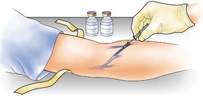
81 Figure 4. Figure 5. Figure 6. Figure 7. 73

82 Figure 8. Muscle Biopsies. Percutaneous muscle biopsies (approximately mg) will be obtained from the vastus lateralis of each participant s thigh (Figures 9-12). Samples will be extracted under local anesthesia of 1% Lidocaine from the middle portion of the muscle at the midway between the patella and the greater trochanter of the femur by Darryn Willoughby, Ph.D. or Mike Spillane, M.S.Ed. For each biopsy, muscle tissue will be extracted from the same location by using the previous incision and depth markings on the needle. First, the participant will lie supine or assume a comfortable reclining position on a sterilized table. Once the extraction point is identified, the area will be shaved clean of leg hair, washed with an antiseptic soap and cleaned with rubbing alcohol. In addition, the biopsy site will further be cleansed by swabbing the area with Betadine (fluid antiseptic) and then draped. A small area of the skin approximately 2 cm in diameter will be anesthetized with a 1.0 ml subcutaneous injection of Lidocaine. Once anesthetized, a scalpel point will be used to produce the initial biopsy site by making an incision approximately 1 cm in length through the skin, subcutaneous fat, and fascia. Due to the localized effects of the anesthetic, the participant should feel no pain during this process. The biopsy needle will be advanced into the incision approximately 1 cm and during this part of the procedure the subject may feel pressure to the thigh area. Once the muscle sample has been obtained, pressure will be immediately applied and the wound will immediately be bandaged. With the incision being so small bleeding is slight; therefore, only a butterfly bandage is needed to close the incision, which is then covered with a pressure bandage. The needle and scalpel blade will be discarded as hazardous waste in an appropriately-labeled plastic sharps container. The site of the biopsy will be cleaned with a sterile alcohol wipe and gauze. The alcohol wipe and gauze then will be discarded in an appropriately labeled biohazard waste receptacle. The tissue sample will be stored at -80 C for future analyses. Once the local anesthesia has taken effect (approximately 2-3 minutes) the biopsy procedure will take approximately seconds. Written instructions for post-biopsy care will be given to the subjects. The participant will be instructed to leave the bandages on for 24 hours (unless unexpected bleeding or pain occurs) and asked to report back to the lab within 24 hours to have the old bandages removed, the incision inspected and new bandages applied. The participant will be further advised to refrain from vigorous 74


83 physical activity during the first 48 hours post-biopsy. These suggestions will minimize pain and possible bleeding of the area. If needed, the subject may take nonprescription analgesic medication such as acetominophen to relieve pain if needed. However, medications such as aspirin, Advil, Nuprin, Bufferin, or Ibuprophen will be discouraged as these medications may lead to ecchymosis at the biopsy site. Soreness of the area may occur for about 24 hours post-biopsy. Figure 9. Figure 10. Figure 11. Figure 12. Equipment Digital Scale. Total body weight will be determined using a digital scale accurate to ±0.02 kg. The scale is calibrated by placing certified 25-kg weights and balancing the scale. Other than general instructions, special skills are not required to measure body weight. Mercurial Sphygmomanometer. Blood pressure will be assessed by auscultation of the brachial artery using a mercurial sphygmomanometer using standard clinical procedures. Bioelectrical Impedance Analyzer (BIA). The Omron HBF-306 Bioelectrical Impedance Analyzer (Omron Healthcare Inc., Vernon Hills, IL) which measures bioresistance and body composition based on a minute low energy, high frequency current transmitted through the body from surface electrodes embedded in the handles of the unit. The analyzer is calibrated internally to a standard electrical current by pressing the calibration key located on the unit. A trained research assistant will perform this procedure. Dual-Energy X-Ray Absorptiometer (DEXA). Body composition measurements will be determined by qualified personnel (in compliance with State Regulations) using a Hologic Discovery W dual energy x-ray absorptiometer (Waltman, MA). This system segments regions of the body (right arm, left arm, trunk, right leg, and left leg) into three compartments (i.e., bone mass, fat mass, and fat-free/soft tissue mass). Quality control (QC) calibration procedures will be performed on a spine phantom (Hologic 75
84 X-CALIBER Model DPA/QDR-1 anthropometric spine phantom) prior to each testing session. In addition, weekly calibration procedures will be performed on a density step calibration phantom. Resistance Exercise Machines. Maximum strength (1-RM strength) tests will be performed on the bench press, seated row, overhead shoulder press, and knee extension machines (Body Masters, Inc., Rayne, LA). Equipment and testing will be contained within the EBNL. Muscle Biopsy Needle. The muscle biopsy technique will be performed with a 5-mm Bergstrom biopsy needle (shown in photos above). Serum and Muscle Protein Analyses. Blood and muscle samples will also be used to assess hormone profiles spectrophotometrically using enzyme-linked immunoabsorbent assays (ELISA) with a Wallac Victor-1420 micoplate reader Wallac Victor-1420 micoplate reader (Perkin-Elmer Life Sciences, Boston, MA). The assays will be performed at either 405 or 450 nm wavlength against a known standard curve. Participants Recruitment Ten apparently healthy, non-resistance trained [no regular, consistent resistance training (i.e. thrice weekly) for at least one year prior to the onset of the study], men between the ages of will volunteer to participate in the study. Enrollment will be open to men of all ethnicities. A recruitment flyer that will be posted on campus and at area fitness centers is attached. Selection Criteria Participants will not be allowed to participate in the study if they: 1. have been involved in a habitual resistance training program (minimum of 3 hours/week for at least 1 year); 2. use tobacco products; 3. have orthopedic limitations that would limit participation in resistance training; 4. have a known allergy to topical anesthetics; 5. have any known metabolic disorder including heart disease, arrhythmias, diabetes, thyroid disease, or hypogonadism; 6. have a bleeding disorder, history of pulmonary disease, hypertension, hepatorenal disease, musculoskeletal disorders, neuromuscular/neurological 76
85 diseases, autoimmune disease, cancer, peptic ulcers, anemia, or chronic infection (e.g., HIV); 7. are taking any heart, pulmonary, thyroid, anti-hyperlipidemic, hypoglycemic, anti-hypertensive, endocrinologic (e.g,thyroid, insulin, etc), emotional/psychotropic (e.g., Prednisone, Ritalin, Adderall), neuromuscular/neurological, or androgenic medications (anabolic steroids); 8. have taken ergogenic levels of nutritional supplements that may affect muscle mass (e.g., creatine, HMB) or anabolic/catabolic hormone levels (e.g., androstenedione, DHEA, etc) within three months prior to the start of the study. 9. have any absolute or relative contraindication for exercise testing or prescription as outlined by the American College of Sports Medicine; 10. report any unusual adverse events associated with this study that in consultation with Darryn Willoughby, Ph.D. who may recommend removal from the study. Compensation or Incentives Participants completing all familiarization and testing sessions as well as turning in all required materials (i.e., dietary logs) in the study will be paid $100. Participants may receive information regarding results of these tests if they desire. If subjects are Baylor students, they will not receive any academic credit for participating in this study. Potential Risks Participants who meet eligibility criteria will be exposed to a very low level of electrical current that will be passed through each subject s body using a bioelectrical impedance analyzer. This analyzer is commercially available and has been used in the health care/fitness industry as a means to assess body composition and body water for over 20 years. The use of the BIA analyzer has been shown to be safe methods of assessing body composition and total body water and is approved by the FDA. Participants who meet eligibility criteria will be subjected to strength testing sessions involving dynamic muscle contractions. Participants in this study will not be experienced resistance trainers, and will be instructed to only perform the prescribed resistance training protocol throughout the duration of the study. As a result of the exercise protocol, participants will most likely experience short-term muscle fatigue. In addition, they will likely experience muscle soreness in muscles in the upper- and lower-body for up to 24 to 48 hours after exercise. This soreness is normal and should be commensurate with the type of muscle soreness participants may have felt after doing unaccustomed physical activity. Muscle strains/pulls resulting from 1-RM testing and the dynamic exercise protocol are possible. During the familiarization session, participants will be informed of the resistance training program and correct lifting technique for each exercise demonstrated. Therefore, potential injury due to exercise will be minimized since all participants will be instructed on how to adhere 77
86 to correct lifting technique. In addition, only Darryn Willoughby, Ph.D., Pete Grandjean, Ph.D., Brian Leutholtz, Mike Spillane, M.S.Ed, or Neil Schwarz, M.S. will conduct the testing and exercise procedures. Participants will be made aware of the intensity and duration of the expected soreness due to the exercise sessions. However, there are minor risks of muscular pain and soreness associated with the resistance training protocol required in this study which are not uncommon to any exercise program especially for individuals who do not resistance train on a regular basis. Participants will donate about approximately 20 milliliters of venous blood a total of 16 times during the study by way of an intravenous catheter and standard phlebotomy using sterile techniques by an experienced phlebotomist using standard procedures. These procedures may cause a small amount of pain when the needle is inserted into the vein as well as some bleeding and bruising. However, proper pressure will be applied upon removal to reduce bruising. The subject may also experience some dizziness, nausea, and/or faint if they are unaccustomed to having blood drawn. Complications resulting from the muscle biopsy are rare, especially in this case where the biopsy is similar to receiving a routine intramuscular injection. As with the blood draw, however, there is a risk of infection if the subject does not adequately cleanse the area for approximately hours post biopsy. While leaving the butterfly bandage in place, participants will be instructed to cleanse the biopsy area with soap and water every 4-6 hours, pat the area dry and reapply a fresh adhesive bandage. The participant will be instructed to leave the bandages on for 24 hours (unless unexpected bleeding or pain occurs) and asked to report back to the lab within 24 hours to have the old bandages removed, the incision inspected and new bandages applied. The participant will be further advised to refrain from vigorous physical activity with the affected leg for 24 hours after the biopsy. There is a potential risk of an allergic reaction to the Lidocaine. All subjects will be asked if they have known allergies to local anesthetics (e.g. Lidocaine, Xylocaine, etc.) that they may have been previously given during dental or hospital visits. Participants with known allergies to anesthesia medications will not be allowed to participate in the study. Darryn Willoughby, Ph.D. or Mike Spillane, M.S.Ed. will perform all muscle biopsies. Researchers involved in collecting data represent trained, non-physician, exercise specialists. All personnel involved in collecting data will be certified in CPR, which is also a condition to holding these professional certifications. A telephone and automated electronic defibrillator (AED) is located in the laboratory in case of any emergencies and there will be no less than two researchers working with each subject during testing. In the event of any unlikely emergency one researcher will check for vital signs and begin any necessary interventions while the other researcher contacts Baylor s campus police at extension Instructions for emergencies are posted above the phone in the event that any other research investigators are available for assistance. 78
87 Potential Benefits The main benefit that participants may obtain from this study is how the body produces testosterone in response to resistance exercise. In addition, participants may gain insight into how they can enhance muscle mass and performance that typically occurs in conjunction with resistance training as well as improved health profiles. Participants may also gain insight about their health and fitness status from the assessments to be performed. Assessment of Risk The greatest risk associated with participating in this study will likely be from the muscle soreness participants will experience from participating in the resistance exercise protocol. However, the intensity of the exercise protocol will be no more than when individuals engaged heavily in a new or different form of physical activity. Therefore, the potential benefits of subjects participating in this study outweigh the potential risks. Compensation for Illness or Injury Each participant will agree to indemnify and hold harmless Baylor University, its officers, directors, faculty, employees, and students for any and all claims for any injury, damage or loss suffered as a result of participation in this study regardless of the cause of injury, damage, or loss. Confidentiality Information obtained from this research (including questionnaires, medical history, laboratory findings, or physical examination) will be kept confidential to the extent permitted by law. However, according to FDA regulations, records will be open to FDA representatives to review if necessary. This may include questionnaires, medical history, laboratory findings/reports, statistical data, and/or notes taken throughout this study. Records of the research may also be subpoenaed by court order or may be inspected by federal regulatory authorities. Data derived from this study may be used in reports, presentations and publications. Participants in this study will not be individually identified unless they give their written consent. All participants will have a number to identify their results. Only the study personnel will know the subject numbers. Only study personnel will have access to the data. All data will be stored in a locked cabinet in the Exercise and Biochemistry Laboratory and only Darryn Willoughby, Ph.D. will have access to the key. Additionally, that confidentiality will be maintained by assigning code numbers to the files, limiting access to data to research assistants, locking cabinets that store data, and providing passwords to limit access to computer files to authorized personnel only. All evidence of primary data will be stored for exactly three years after the completion of the study. At this time data will be destroyed in a manner that instills complete privacy to all participants of the study. Analyzed muscle and blood samples will be 79
88 discarded in an appropriately-labeled biohazard waste disposal container. However, unused muscle and blood samples will be kept in a locked freezer for no longer than one year. If any subsequent analysis occurs with the samples, they will be re-coded to further instill confidentiality. Data Presentation & Publication Data will be presented at an appropriate scientific conference (e.g., American College of Sports Medicine, Experimental Biology, etc.) and published in a peer reviewed scientific journal (e.g., Medicine & Science in Sport and Exercise, Journal of Applied Physiology, etc.). Statement on Conflict of Interest Funding for this study will be provided by the Exercise and Biochemical Nutrition Laboratory of Baylor University. Researchers involved in collecting data in this study have no financial or personal interest in the outcome of results or sponsors. 80
89 Table 1. Overview of Research Design Visit 1 (Familiarizatio n and Entry) Testing Session 1 (Visit 2) 24 Hour Follow- Up (Visit 3) Testing Session 2 (Visit 4) 24 Hour Follow-Up (Visit 5) Explanation of Study Procedures Heart Rate and Blood Pressure Heart Rate and Blood Pressure Heart Rate and Blood Pressure Heart Rate and Blood Pressure Demographic and Health History Form Blood Collection Muscle Biopsy Blood Collection Muscle Biopsy Urine Collection Blood Collection Urine Collection Blood Collection Activity Form Diet Log Analysis Muscle Biopsy Muscle Biopsy General Exam to Determine Qualifications to Participate in Study. Resistance Exercise Session Diet Log Analysis Resistance Exercise Session Informed Consent Form. Determination of Height and Body Weight. Determination of Resting Heart Rate and Blood Pressure Body Composition Assessment. Muscle Strength Assessments 81
90 BAYLOR UNIVERSITY Department of Health, Human Performance, & Recreation Informed Consent Form Title of Investigation: Resistance Mediated Effects of Endogenous Elevations in Testosterone on Exercise-Induced Skeletal Muscle Androgen Receptor Signaling and DNA Binding in Men Principal Investigator: Darryn S. Willoughby, Ph.D. Department of HHPR, Baylor University Co-investigators: Pete Grandjean, Ph.D. Department of HHPR, Baylor University Brian Leutholtz, Ph.D. Department of HHPR, Baylor University Mike Spillane, M.S.Ed. Department of HHPR, Baylor University Neil Schwarz, M.S.Ed. Department of HHPR, Baylor University Sponsors: University) Exercise and Biochemical Nutrition Lab (Baylor Rationale: Resistance exercise stimulates skeletal muscle growth which results from protein accumulation within muscle fibers, thus promoting muscle fiber growth. There are a number of physiological mechanisms known to contribute to this process such as intrinsic factors within skeletal muscle, immune/inflammatory, and endocrine (hormonal). Of the endocrine factors, a number of studies have shown the ability of resistance exercise to increase the endogenous level of testosterone. Testosterone is an androgenic hormone with robust growth-promoting capabilities in skeletal muscle due to its ability to increase muscle protein synthesis. Being a fat-soluble hormone, testosterone will bind with its androgen receptor within skeletal muscle. Upon binding, this hormone-receptor complex migrates into the nucleus where it binds to a specific sequence within various muscle-specific genes, thereby enhancing gene expression. Resistance exercise affects androgen signaling via several mechanisms. First, resistance exercise elevates endogenous testosterone concentrations. Elevations in testosterone due to resistance exercise are known to increase muscle strength following long-term training. Second, muscle contraction increases muscle androgen 82
91 receptor content due to enhancing androgen receptor gene expression. Third, evidence indicates that elevations in endogenous testosterone following resistance exercise could feasibly increase muscle androgen receptor content independently of the influence of muscle contraction. This process of androgen signaling represents an important target for research aimed at promoting muscle growth and/or counteracting the muscle atrophy characteristic of aging, inactivity/immobilization, and various diseases. Therefore, the purpose of this study is to determine the influence of elevated endogenous circulating testosterone levels on skeletal muscle testosterone, androgen receptor protein, and myofibrillar protein concentration, as well as the mrna expression of the androgen receptor. Description of the Study: I will be one of 10 apparently healthy, non-resistance-trained males between the ages 18 to 30 who will participate in this study. I understand that I will be required to visit the laboratory five times during the course of the study in the following manner: visit 1 = entry/familiarization session, visit 2 = testing/resistance exercise session 1, visit 3 = 24 hour follow-up for session 1, visit 4 = testing/resistance exercise session 2, visit 5 = 24 hour follow-up for session 2. Relative to the 2 testing sessions, I will perform a resistance exercise session involving the knee extension exercise on two occasions separated by three weeks. One session will constitute the control session and be preceded by rest and the other will constitute the experimental session and preceded by a bout of high-volume, moderate-intensity upper-body resistance exercise using short rest periods. At each of the 5 visits, I will have my heart rate and blood pressure assessed. At visits 2 5, I will also have blood and muscle samples obtained. During an initial familiarization session (visit 1), I will be informed of the requirements of the study and sign an informed consent statement in compliance with the Human Subjects Guidelines of Baylor University and the American College of Sports Medicine. A trained individual will examine me to determine if I am qualified to participate in this study. If I am cleared to participate in the study, I will be familiarized to the testing procedures and will then undergo assessments for body composition and muscle strength. This session will take approximately 60 minutes to complete. Once I complete the familiarization session, I will be scheduled for the first resistance exercise session (visit 2). During the familiarization session, I understand that I will have my maximum muscle strength determined on the bench press, seated row, overhead shoulder press and knee extension exercises. I will warm up by completing 5 to 10 repetitions with a very light weight and then complete 3 to 5 repetitions with a heavier weight. The weight will then be increased conservatively, and I will attempt to lift the weight for one repetition. If the lift is successful, I will rest for 2 minutes before attempting the next weight increment. This procedure will be continued until I fail to complete the lift. My maximum strength will be recorded as the maximum weight I am able to lift for one repetition. In addition, I will have my body composition (body fat and muscle mass) determined using dual-energy x-ray absorptiometry (DEXA). The DEXA body composition test will involve me lying down on my back in a comfortable position in a pair of shorts/t-shirt. A low dose of radiation will then scan my entire body for 83
92 approximately 6 minutes. Radiation exposure from the DEXA is approximately 1.5 mr per scan. This is similar to the amount of natural background radiation I would receive in one month while living in Waco, TX. The maximal permissible x-ray dose for non-occupational exposure is 500 mr per year. Total radiation dose will be less than 5 mr for the entire study. Following the familiarization session, I will be instructed to refrain from exercise for 48 hours and fast for 8 hours prior to each resistance exercise session. I will be provided with a dietary analysis form that I am to complete for 4 days prior to each resistance exercise testing session. Once I report to the lab for each testing session, I will turn in my dietary analysis form. I understand that I will be required to participate in a 2 separate testing sessions (visits 2 & 4) consisting of the knee extension exercise where I will perform 5 sets of 5 repetitions at 90% - 95% of my maximum strength. However, for one session I will perform 4 sets of 10 repetitions using 80% of my maximum strength on the bench press, seated row, and overhead shoulder press exercises immediately before the knee extension exercise. During the study, I understand that I will provide two urine samples prior to each exercise session using a standard collection protocol for the purpose of accessing my hydration status prior to exercise, as under-hydration can affect my testosterone responses to exercise. I understand that if my hydration status is not acceptable that my testing session will be cancelled and re-scheduled for a later date. I understand that I will donate about 20 milliliters (4 teaspoons) of venous blood from a vein in my arm by way of an intravenous catheter inserted into my arm using sterile techniques by an experienced phlebotomist using standard procedures. This procedure may cause a small amount of pain when the needle is inserted into the vein as well as some bleeding and bruising, and will remain in place until the end of the testing process. However, proper pressure will be applied upon removal to reduce bruising. I understand that I may also experience some dizziness, nausea, and/or faint if I am unaccustomed to having blood drawn. I understand that personnel who will be inserting the intravenous catheter and taking my blood are experienced in phlebotomy (procedures to take blood samples) and are qualified to do so under guidelines established by the Texas Department of Health and Human Services. The process of inserting the catheter and blood draws at each sampling point will take about 5 minutes and I will be required to leave the catheter in place for the duration of the testing session. Throughout the testing, the catheter will be securely placed in my arm; however, during the 3 hours following resistance exercise I may read or participate in other sedentary activities. At visits 2 and 4 (resistance exercise/testing sessions 1 and 2) I will have blood obtained through the catheter at 8 different time points. However, 24 hours after each session (visits 3 and 5) I will have blood sampled through a standard venipuncture. In addition to the blood draws, I will undergo the muscle biopsy. I understand that I will have the biopsy location identified on the thigh (opposite thigh for the second 84
93 exercise session). The biopsy area will be shaved clean of leg hair, washed with antiseptic soap and cleaned with rubbing alcohol. In addition, the biopsy site will be further cleansed by swabbing the area with Betadine (fluid antiseptic). I understand that a small area of the cleaned skin approximately 2 cm in diameter will be anesthetized with a 1.0 ml subcutaneous injection of the topical anesthetic Lidocaine. Once the local anesthesia has taken effect (approximately 2-3 minutes) the biopsy procedure will only take seconds. Once anesthetized, I understand that a scalpel point will be used to make an incision approximately 1 cm in length through the skin. Due to the localized effects of the anesthetic, however, I should feel no pain during this process. At this point, I understand that the biopsy needle will be advanced into the incision approximately 1 cm and during this part of the procedure I may feel pressure in my thigh area. Once the muscle sample has been obtained, pressure will be immediately applied to the incision. With the incision being so small bleeding is slight; therefore, only a butterfly bandage is needed to close the incision, which will then be covered with a pressure bandage. I understand that I will be provided verbal and written instructions for post-biopsy care. I understand that I will be instructed to leave the butterfly bandage in place for 72 hours. However, I understand that I will be asked to report back to the lab 24 hours after the biopsy (unless unexpected bleeding or pain occurs) to have the old bandage removed, the incision inspected and new bandages applied, and that I will also report back to the lab for the same reason at 48 hours after the biopsy. I will be further advised to refrain from vigorous physical activity with my leg during the first 24 hours post-biopsy. I understand that if I feel it necessary I may take a non-prescription analgesic medication such as acetominophen to relieve pain if needed and that some soreness of the area may occur for about 24 hours after the biopsy. I will also be advised to avoid such medications such as aspirin, Advil, Bufferin, Nuprin, or Ibuprofen as they may lead to bruising at the biopsy site. I understand that I will be asked to undergo the muscle biopsy procedure on 8 separate occasions throughout the study. I understand that when I report to the laboratory for the two testing/resistance exercise sessions on visits 2 and 4, I will turn in my 4-day dietary records. In addition, I will have my heart rate and blood pressure determined, and will also provide urine, blood, and muscle samples. I understand that if clinically significant side effects are reported from my participation in the study, I will be referred to discuss the problem with Darryn Willoughby, Ph.D. Upon his discretion, I may be referred to discuss the matter with my primary care physician to determine whether any medical treatment is needed and/or whether I can continue in the study. I understand that if I fail to report my progress and health status to the research assistant I may be removed from the study. I agree to do my best to: 1) follow the instructions outline by the investigators; 2) show up to all scheduled testing times; and 3) put forth my best effort as instructed. I agree not to take any other nutritional supplements or performance enhancing aids during this study (i.e. vitamins/minerals, creatine, HMB, androstenedione, DHEA, etc). In addition, I agree not to take any non-medically prescribed medications and to report any medication that is prescribed for me to take during this study. I understand 85
94 that if I take any nutritional supplements or medications during the course of the study that I will be removed from the study. Exclusionary Criteria I understand that in order to participate in the study, a trained individual will examine me to determine whether I qualify to participate. I understand that I will not be allowed to participate in this study if: 1.) I have any known metabolic disorder including heart disease, arrhythmias, diabetes, thyroid disease, or hypogonadism; 2.) I have a history of pulmonary disease, hypertension, liver or kidney disease, musculoskeletal disorders, neuromuscular or neurological diseases, autoimmune disease, cancer, peptic ulcers, or anemia; 3.) I am taking any heart, pulmonary, thyroid, anti-hyperlipidemic, hypoglycemic, anti-hypertensive, endocrinologic (ie, thyroid, insulin, etc), psychotropic, neuromuscular/neurological, or androgenic medications; 4.) I have any bleeding disorders; 5.) I have any chronic infections (e.g., HIV); 6) I have a known allergic reaction to topical anesthetics. I have reported all nutritional supplements, medically prescribed drugs, and non-medically prescribed drugs that I am presently taking. I have reported whether I have had any prior allergic reactions to topical anesthetics. I have completed medical history questionnaires and am not aware of any additional medical problems that would prevent me from participating in this study. I agree to report all changes in medical status, nutritional and/or pharmacological agents (drugs) that I take during the course of the investigation to Darryn Willoughby, Ph.D. ( ). I understand that if I experience any unexpected problems or adverse events from participating in this study I may be referred to discuss the problem with my primary care physician to determine whether any medical treatment is needed and/or whether I can continue in the study. Risks and Benefits I understand that there are minor risks of muscular pain and soreness associated with the resistance training protocol required in this study which are not uncommon to any exercise program especially for individuals who do not resistance train on a regular basis. On 16 separate occasions during this study, I understand that I will have approximately 4 teaspoons (20 milliliters) of blood drawn from a vein in my forearm. On 14 of these occasions, blood will be obtained using an in-dwelling venous catheter, whereas on 2 other occasions it will involve a standard blood draw using a sterile needle. All blood sampling will be performed by an experienced phlebotomist following an 8-hour fast. This procedure may cause a small amount of pain when the needle is inserted into my vein as well as some bleeding and bruising. I may also experience some dizziness, nausea, and/or faint if I am unaccustomed to having blood drawn. 86
95 On 8 separate occasions during this study (4 at each testing/exercise session), I understand that I will undergo a muscle biopsy in which a small sample of muscle will be obtained from the thigh of my exercised leg. I understand that Darryn Willoughby, Ph.D. or Mike Spillane, M.S.Ed. will perform all of the biopsies and that a local anesthetic (Lidocaine) will be injected into the skin of my thigh prior to the biopsy, which will help prevent any pain and discomfort during the procedure. I understand that I will have a small incision made in my skin and a biopsy needle introduced 1 cm into the incision. I also understand that the incision is so small that it will not require any stitches and will be simply closed with a butterfly bandage and then covered with an adhesive bandage (band-aid). After the anesthetic wears off within 3-4 hours, I understand that the sensation at the biopsy site is comparable to that of a bruise and may persist for hours after the procedure. I understand that I am required to inform the study investigators if I have had any prior allergic reactions to anesthesia (e.g. while in the hospital or during a dental visit). Alternative Treatments This is not a medical treatment. Therefore, if medical treatment is needed, I must obtain treatment for any medical problem I might have from my personal physician. Costs and Payments If I am a Baylor University student, I will not receive any academic credit for participating in this study. I understand that if I am an intercollegiate scholarship athlete I may not be eligible to receive payment to participate in this study. Eligible participants will be paid $100 for completing the familiarization and experimental testing sessions. I also understand that I will be given free blood assessments during the course of the study as described above and may receive information regarding results of these tests if I desire. New Information Any new information obtained during the course of this research that may affect my willingness to continue participation in this study will be provided to me. In addition, I will be informed of any unusual/abnormal clinical findings in which medical referral to my personal physician may be warranted. If I desire, I may request that this information be provided to my physician. Confidentiality I understand that any information obtained about me in this research, including medical history, laboratory findings, or physical examination will be kept confidential to the extent permitted by law. However, I understand in order to ensure that FDA regulations are being followed, it may be necessary for a representative of the FDA to review my records from this study which may include medical history, laboratory findings/reports, statistical data, and/or notes taken about my participation in this study. In addition, I understand that my records of this research may be subpoenaed 87
96 by court order or may be inspected by federal regulatory authorities. I understand that data derived may be used in reports, presentations, and publications. However, I will not be individually identified unless my consent is granted in writing. Additionally, that confidentiality will be maintained by assigning code numbers to my files, limiting access to data to research assistants, locking cabinets that store data, and providing passwords to limit access to computer files to authorized personnel only. I understand that once blood and muscle samples are analyzed that they will be discarded. Right to Withdrawal I understand that I am not required to participate in this study and I am free to refuse to participate or to withdraw from the study at any time. Further, that my decision to withdraw from the study will not affect my care at this institution or cause a loss of benefits to which I might be otherwise entitled. If there is concern about my medical safety, I may be referred to seek medical attention. Compensation for Illness or Injury I understand that if I am injured as a direct result of taking part in this study, I should consult my personal physician to obtain treatment. I understand that the cost associated with the care and treatment of such injury will be the responsibility of me or my insurance carrier. In some cases, insurers may not reimburse claims submitted for a research-related injury resulting from medical procedures or treatments performed as part of a research study. I understand that Baylor University, the investigator s institutions, and the grant sponsor have not budgeted funds to compensate me for injury or illness that may result from my participation in this study and thus will not be accountable for illness or injury acquired during the course of this study. However, I may be referred to my personal physician if any clinically significant medical/psychological findings are observed during the course of this study. I agree to indemnify and hold harmless Baylor University, its officers, directors, faculty, employees, and students for any and all claims for any injury, damage or loss I suffer as a result of my participation in this study regardless of the cause of my injury, damage or loss. Statement on Conflict of Interest I understand that this study is funded by the Exercise and Biochemical Nutrition Laboratory at Baylor University, and that the researchers involved in collecting data in this study have no financial or personal interest in the outcome of results or sponsors. 88
97 Voluntary Consent I certify that I have read this consent form or it has been read to me and that I understand the contents and that any questions that I have pertaining to the research have been, or will be answered by Darryn Willoughby, Ph.D. (principal investigator, Department of Health, Human Performance & Recreation, 120 Marrs McLean Gymnasium, Baylor University, phone: ) or one of the research associates. My signature below means that I am at least 18 years of age and that I freely agree to participate in this investigation. I understand that I will be given a copy of this consent form for my records. If I have any questions regarding my rights as a research subject in this study, I may contact Dr. Michael Sherr (Chair, Baylor University Committee for Protection of Human Subjects in Research), School of Social Work, Baylor University, One Bear Place #97320, Waco, TX , (254) Date Subject's Signature I certify that I have explained to the above individual the nature and purpose of the potential benefits and possible risks associated with participation in this study. I have answered any questions that have been raised and have witnessed the above signature. I have explained the above to the volunteer on the date stated on this consent form. Date Investigator's Signature 89
98 Part 5: Informed Consent Form Checklist When using humans as subjects in research you must obtain their informed consent. Check each of the following items as they appear on your Informed Consent Form and include this checklist with your protocol: x_(a) A statement explaining the purpose of the research. x_(b) A statement of the expected duration of the subject's participation. x_(c) A description of the procedures to be followed. x_(d) A description of any reasonable foreseeable risks or discomforts to the subject, including invasion of privacy. x_(e) A description of any benefits resulting from the research, either to the subject or to others. x_(f) A statement that informs subject of his/her right not to be a subject in a research project that is also a teaching exercise. x_(g) A statement informing subject about how his/her anonymity will be guarded; i.e., that their confidentiality will be protected by assigned code numbers, by limitations of who has access to data, by data storage in locked cabinets, by locked computer files, etc. x_(h) A statement that the subject's participation is voluntary, and that his/her refusal to participate will involve no penalty or loss of benefits to which the subject is otherwise entitled, and that the subject may discontinue participation at any time without penalty or loss of benefits to which the subject is otherwise entitled. _na _(i) A disclaimer, if applicable, regarding the use of the Internet to collect data. x_(j) For research involving more than minimal risk, an explanation regarding the availability of any compensation or any medical treatments if injury occurs (if applicable, see OHRP Reports). x_(k) If written informed consent is required, a place for the subject to sign and date the form and a statement that a copy of the signed consent form will be given to the subject for his/her records. _na_(l) If the subject is a minor, a statement of parental responsibility in consenting to the child's participation in the study with a place for the parent to sign and date the form in addition to the participant's signature. x_(m) Include a short summary of your expertise related to this research proposal. x_(n) The name, address, and telephone number of the principal investigator of the research project, 90
99 and his/her affiliation with Baylor University. If the principal investigator is a graduate student, the name and telephone number of the faculty advisor is also required. x_(o) A statement informing subject that inquiries regarding his/her rights as a subject, or any other aspect of the research as it relates to his/her participation as a subject, can be directed to Baylor's University Committee for Protection of Human Subjects in Research. The chairman is Dr. Matt Stanford, Professor Psychology and Neuroscience, PO Box 97334, Waco, Texas , phone number


100 Want to get Paid to Workout? Non-Resistance-Trained Men Needed for a Weight Lifting Study Researchers in the Exercise & Biochemical Nutrition Lab at Baylor University are recruiting 10 healthy, non-resistancetrained men between the ages of to participate in a study designed to evaluate the effects of their own endogenous testosterone on the effects of the androgen receptor and markers of muscle protein synthesis capacity. Participants will be required to engage in 2 resistance exercise sessions separated by 3 weeks. Participants will be required to undergo strength and body composition testing, and to also submit to providing blood samples and muscle biopsies. Eligible subjects will receive $100 for completing the study and free muscle strength and body fat testing. For more information contact: Exercise & Biochemical Nutrition Lab Department of HHPR Rena Marrs McLean Gymnasium Room Darryn_Willoughby@baylor.edu 92
The Effects of Chronic Creatine Supplementation on Performance and Body Composition of Female Athletes. by Megan Brenner
 The Effects of Chronic Creatine Supplementation on Performance and Body Composition of Female Athletes by Megan Brenner Thesis submitted to the Faculty of Virginia Polytechnic Institute and State University
Reproductive DHT Analyte Information
 Reproductive DHT Analyte Information - 1 - DHT Introduction Dihydrotestosterone (DHT) together with other important steroid hormones such as testosterone, androstenedione (ASD) and dehydroepiandrosterone
Skin metabolism of steroid hormones as endogenous compounds?
 Skin metabolism of steroid hormones as endogenous compounds? Van Luu-The Department of Molecular Medicine Laval University Québec, Canada This work has been supported by L Oréal Research Steroid hormones
ab Testosterone ELISA Kit
 Version 10 Last updated 29 January 2018 ab108666 Testosterone ELISA Kit A competitive immunoenzymatic assay for the quantitative measurement of Testosterone in serum, plasma and urine. This product is
MSD 96-Well MULTI-ARRAY and MULTI-SPOT Human Granulocyte Colony Stimulating Factor (hg-csf) Ultrasensitive Assay
 Ultrasensitive Assay") MSD 96-Well MULTI-ARRAY and MULTI-SPOT Human Granulocyte Colony Stimulating Factor (hg-csf) Ultrasensitive Assay Summary This assay measures Human Granulocyte Colony Stimulating Factor (G-CSF) in a 96-well
MSD 96-Well MULTI-ARRAY and MULTI-SPOT Human Granulocyte Colony Stimulating Factor (hg-csf) Ultrasensitive Assay Summary This assay measures Human Granulocyte Colony Stimulating Factor (G-CSF) in a 96-well
The Science of. NUTRICULA Longevity Journal
 32 December, 2011 The Science of 33 NUTRICULA Longevity Journal As men age, there is often a decline in libido and sexual function. This decline frequently interferes in intimacy within romantic relationships,
Androgenes and Antiandrogenes
 Androgenes and Antiandrogenes Androgens The androgens are a group of steroids that have anabolic and/or masculinizing effects in both males and females. Testosterone [tess-toss-terone], the most important
F7 (Human) Chromogenic Activity Assay Kit
 Chromogenic Activity Assay Kit") F7 (Human) Chromogenic Activity Assay Kit Catalog Number KA0971 96 assays Version: 02 Intended for research use only www.abnova.com Table of Contents Introduction... 3 Background... 3 Principle of the
F7 (Human) Chromogenic Activity Assay Kit Catalog Number KA0971 96 assays Version: 02 Intended for research use only www.abnova.com Table of Contents Introduction... 3 Background... 3 Principle of the
RayBio Human vwf ELISA Kit
 RayBio Human vwf ELISA Kit Catalog #: ELH-vWF User Manual Last revised April 15, 2016 Caution: Extraordinarily useful information enclosed ISO 13485 Certified 3607 Parkway Lane, Suite 100 Norcross, GA
Catabolism in Skeletal Muscle The Phosphagen System
 Catabolism in Skeletal Muscle The Phosphagen System Overview of ATP Regeneration Anaerobic vs Aerobic Metabolism Creatine Kinase Reaction Adenylate Kinase Reaction Purine Nucleotide Cycle Creatine Phosphate
biosensis Rat Fibronectin ELISA Kit Protocol
 biosensis Rat Fibronectin ELISA Kit Protocol Catalog No: BEK-2017-2P For quantitative detection of rat Fibronectin in cell culture supernatants, serum, and citrate, heparin, or EDTA plasma samples only
biosensis Human Lipocalin-2/NGAL ELISA Kit Protocol
 biosensis Human Lipocalin-2/NGAL ELISA Kit Protocol Catalog No: BEK-2141-2P For quantitative detection of human Lipocalin-2 in cell culture supernatants, serum, and heparin treated plasma, saliva, and
ab Androstenedione ELISA Kit
 ab108672 Androstenedione ELISA Kit Instructions for Use A competitive immunoenzymatic assay for the quantitative measurement of Androstenedione in serum and plasma (citrate). This product is for research
RayBio Human Adiponectin ELISA Kit
 RayBio Human Adiponectin ELISA Kit Catalog #: ELH-Adiponectin User Manual Last revised December 5, 2018 Caution: Extraordinarily useful information enclosed ISO 13485 Certified 3607 Parkway Lane, Suite
TESTOFEN. Anabolic & Androgenic Activity GENCOR PACIFIC, INC. Fenugreek Extract standardized for FENUSIDE TM. Copyright 2005 by Gencor Pacific, Inc.
 GENCOR PACIFIC, INC. 920E. Orangethorpe Avenue, Suite B, Anaheim, CA. 92801 Ph: 714.870.8723 714.870.8724 efax: 732.875.0306 drjit@gencorpacific.com manu@gencorpacific.com www.gencorpacific.com TESTOFEN
Relationship between Aerobic Training and Testosterone Levels in Male Athletes
 Relationship between Aerobic Training and Testosterone Levels in Male Athletes Siu Yuen Ng Biology 493 13 th December, 2010 Abstract Salivary testosterone levels of 11 athletes and 15 non athletes were
WADA Technical Document TD2014EAAS. Endogenous Anabolic Androgenic Steroids Measurement and Reporting
 Endogenous Anabolic Androgenic Steroids Measurement and Reporting 1.0 Introduction The purpose of this Technical is to harmonize the approaches to the measurement and reporting of endogenous anabolic androgenic
Nutrition, supplements, and exercise
 Nutrition, supplements, and exercise Walter R. Frontera, MD, PhD Professor and Chair Department of Physical Medicine and Rehabilitation Vanderbilt University School of Medicine And Medical Director of
biosensis Human IGF-II, Insulin-like growth factor II, Somatomedin-A ELISA Kit Protocol
 biosensis Human IGF-II, Insulin-like growth factor II, Somatomedin-A ELISA Kit Protocol Catalog No: BEK-2029-1P For quantitative detection of human IGF-II in cell culture supernatants, cell lysates, tissue
biosensis Human TNFα/Cachectin/TNFSF2 ELISA Kit Protocol
 biosensis Human TNFα/Cachectin/TNFSF2 ELISA Kit Protocol Catalog No: BEK-2100-1P For quantitative detection of human TNFα in cell culture supernatants, serum, and heparin, EDTA or citrate treated plasma
Dietary supplements and nutrition in sports and exercices performance
 Dietary supplements and nutrition in sports and exercices performance Nutrition for endurance sports The most likely contributors to fatigue during an endurance exercise are dehydration and carbohydrates
TESTOFEN HUMAN CLINICAL TRIAL GENCOR PACIFIC, INC. Copyright 2006 by Gencor Pacific, Inc.
 GENCOR PACIFIC, INC. 920 E. Orangethorpe Avenue, Suite B, Anaheim, CA 92801 Ph: 714.870.8723 714.870.8724 efax: 732.875.0306 drjit@gencorpacific.com gita@gencorpacific.com www.gencorpacific.com TESTOFEN
biosensis Human Soluble Tumor Necrosis Factor receptor type II (stnfrii) ELISA Kit Protocol
 ELISA Kit Protocol") biosensis Human Soluble Tumor Necrosis Factor receptor type II (stnfrii) ELISA Kit Protocol Catalog No: BEK-2103-2P For quantitative detection of human soluble TNFRII (stnfrii) in human cell culture supernatants,
biosensis Human Soluble Tumor Necrosis Factor receptor type II (stnfrii) ELISA Kit Protocol Catalog No: BEK-2103-2P For quantitative detection of human soluble TNFRII (stnfrii) in human cell culture supernatants,
RayBio Human TNF-alpha ELISA Kit
 RayBio Human TNF-alpha ELISA Kit Catalog #: ELH-TNFa User Manual Last revised April 15, 2016 Caution: Extraordinarily useful information enclosed ISO 13485 Certified 3607 Parkway Lane, Suite 100 Norcross,
TREATMENT OPTIONS FOR MALE HYPOGONADISM
 TREATMENT OPTIONS FOR MALE HYPOGONADISM Bruce Biundo, RPh, FACA PCCA Pharmacy Consulting Department Updated July 2012 Hypogonadism in men is primarily a state involving lower than expected levels of testosterone,
biosensis Mouse CXCL10/IP-10 ELISA Kit Protocol
 biosensis Mouse CXCL10/IP-10 ELISA Kit Protocol Catalogue No: BEK-2124-2P TABLE OF CONTENTS I Materials provided...2 II Equipment required but not supplied...2 III Technical hints....2 IV Storage of kit
LEUCINE. - A major driving force for Muscle Protein Synthesis
 LEUCINE - A major driving force for Muscle Protein Synthesis An article by Professor Don MacLaren, 2016. Leucine is one of the 9 essential amino acids that are required to be ingested by the body since
Rat myeloperoxidase-antineutrophil cytoplasmic antibody(mpo-anca) ELISA Kit
 ELISA Kit") Rat myeloperoxidase-antineutrophil cytoplasmic antibody(mpo-anca) ELISA Kit Catalog Number. CSB-E08675r For the quantitative determination of rat myeloperoxidase-antineutrophil cytoplasmic antibody(mpo-anca)
Rat myeloperoxidase-antineutrophil cytoplasmic antibody(mpo-anca) ELISA Kit Catalog Number. CSB-E08675r For the quantitative determination of rat myeloperoxidase-antineutrophil cytoplasmic antibody(mpo-anca)
Pre-Lab. 1. What do people mean when they say teenagers have raging hormones?
 Name: Period: Date: You ve got MALE! Hormonal Control of Male Reproductive Processes This simulation explores how sex determination works. You will learn about the impact of testosterone concentration
MODULE #8 - Lesson 3
 MODULE #8 - Lesson 3 21st Century Man: Testosterone and Andropause Module 8 - Lesson 3 21st Century Man: Testosterone and Andropause Testosterone and andropause are 2 pressing issues for men in the 21st
biosensis Mouse Vascular endothelial growth factor A/VEGF-A/VEGF-164/VEGF-1/VEGF- 120/VEGF-2 ELISA Kit Protocol
 biosensis Mouse Vascular endothelial growth factor A/VEGF-A/VEGF-164/VEGF-1/VEGF- 120/VEGF-2 ELISA Kit Protocol Catalog No: BEK-2110-1P For quantitative detection of mouse VEGF-A (VEGF164&VEGF120) in mouse
LIFETIME FITNESS HEALTHY NUTRITION. UNIT 2 Lesson 14 FLEXIBILITY LEAN BODY COMPOSITION
 LIFETIME FITNESS HEALTHY NUTRITION MUSCULAR STRENGTH AEROBIC ENDURANCE UNIT 2 Lesson 14 FLEXIBILITY LEAN BODY COMPOSITION MUSCULAR ENDURANCE Created by Derek G. Becher B.P.E., B. Ed., AFLCA Resistance
Ergogenic Aids 1. Ergogenic Aids. Caffeine
 Ergogenic Aids 1 Dr David Jenkins School of Human Movement Studies The University of Queensland Ergogenic Aids drug derived from a number of plants 2004 removed from WADA Prohibited List possible mechanisms
biosensis Rat Interleukin-1 beta, IL-1β ELISA Kit Protocol
 biosensis Rat Interleukin-1 beta, IL-1β ELISA Kit Protocol For the quantitative detection of rat IL-1β in cell culture supernatants, serum, heparin or EDTA treated plasma samples, and cell homogenates
ab IgG1 Human ELISA Kit
 ab100548 IgG1 Human ELISA Kit Instructions for Use For the quantitative measurement of Human IgG1 in serum and plasma This product is for research use only and is not intended for diagnostic use. Version
biosensis Mouse Brain-derived neurotrophic factor (BDNF) ELISA Kit Protocol
 ELISA Kit Protocol") biosensis Mouse Brain-derived neurotrophic factor (BDNF) ELISA Kit Protocol Catalog No: BEK-2003-2P For quantitative detection of mouse BDNF in cell culture supernatants, cell lysates, serum, and citrate,
biosensis Mouse Brain-derived neurotrophic factor (BDNF) ELISA Kit Protocol Catalog No: BEK-2003-2P For quantitative detection of mouse BDNF in cell culture supernatants, cell lysates, serum, and citrate,
RayBio Human IFN alpha/beta R2 ELISA Kit
 RayBio Human IFN alpha/beta R2 ELISA Kit Catalog #: ELH-IFNabR2 User Manual Last revised April 15, 2016 Caution: Extraordinarily useful information enclosed ISO 13485 Certified 3607 Parkway Lane, Suite
Human ABCD1 ELISA KIT
 Human ABCD1 ELISA KIT Cat. No.:DEIA8716 Pkg.Size:96T Intended use The Human ABCD1 ELISA KIT is for the quantitative detection of human ABCD1 in serum and plasma. General Description ABCD1, also known as
26-G Keewaydin Drive, Salem, NH P: (800) F: (603)
 F: (603)") Dihydrotestosterone ELISA For the quantitative determination of DHT in serum. For Research Use Only. Not for Use in Diagnostic Procedures. Catalog Number: 11-DHTHU-E01 Size: 96 wells Version: 4.1 August
Dihydrotestosterone ELISA For the quantitative determination of DHT in serum. For Research Use Only. Not for Use in Diagnostic Procedures. Catalog Number: 11-DHTHU-E01 Size: 96 wells Version: 4.1 August
ANDROSTENEDIONE SALIVA ELISA Direct immunoenzymatic determination of Androstenedione in saliva
 DCM027-9 Ed. 03/2012 ANDROSTENEDIONE SALIVA ELISA Direct immunoenzymatic determination of Androstenedione in saliva for routine analysis IVD LOT See external label = 96 tests REF DKO027 INTENDED USE Eagle
biosensis Mouse Interleukin-1 beta (IL-1β) ELISA Kit Protocol
 ELISA Kit Protocol") biosensis Mouse Interleukin-1 beta (IL-1β) ELISA Kit Protocol Catalog Number: BEK-2151-1P For quantitative detection of mouse IL-1β in cell culture supernatant, cell lysates, and serum and hepain or EDTA
biosensis Mouse Interleukin-1 beta (IL-1β) ELISA Kit Protocol Catalog Number: BEK-2151-1P For quantitative detection of mouse IL-1β in cell culture supernatant, cell lysates, and serum and hepain or EDTA
biosensis Rat IGF-1/Somatomedin/Insulin-like growth factor ELISA Kit Protocol
 biosensis Rat IGF-1/Somatomedin/Insulin-like growth factor ELISA Kit Protocol Catalog No: BEK-2150-1P For quantitative detection of rat IGF-1 in cell culture supernatants, cell and tissue homogenates,
Updates on Anti-doping and TUE Management in Paralympic Sport
 International Paralympic Committee Updates on Anti-doping and TUE Management in Paralympic Sport Matthew Fedoruk, Ph.D. March 15, 2018 PyeongChang 2018 IPC Medical / Sports Science Committee Workshops
Donald W. Morrish, MD, PhD, FRCPC Presented at Mountain Man: Men's Health Conference, May Terry s Testosterone
 Focus on CME at University of Alberta ADAM: Dealing with the Decline Donald W. Morrish, MD, PhD, FRCPC Presented at Mountain Man: Men's Health Conference, May 2005 We now know there is a decline in total
Female testosterone level chart
 Female testosterone level chart The Borg System is 100 % Female testosterone level chart Mar 23, 2015. Male, Female. Age: T Level (ng/dl):, Age: T Level (ng/dl):. 0-5 mo. 75-400, 0-5 mo. 20-80. 6 mos.-9
National Institute for Public Health and the Environment Annual CRL workshop 22 October Update on natural Hormone studies
 2008 Annual CRL workshop 22 October 2008 Update on natural Hormone studies Natural hormone studies: update Possible approaches - C12/C13 ratio: can result in proof of abuse - Determination of intact esters
CHEMILUMINESCENCE ENZYME IMMUNOASSAY (CLIA) TESTOSTERONE. Testosterone. Cat #
 TESTOSTERONE. Testosterone. Cat #") DIAGNOSTIC AUTOMATION, INC. 21250 Califa Street, Suite 102 and 116, Woodland Hills, CA 91367 Tel: (818) 591-3030 Fax: (818) 591-8383 onestep@rapidtest.com technicalsupport@rapidtest.com www.rapidtest.com
DIAGNOSTIC AUTOMATION, INC. 21250 Califa Street, Suite 102 and 116, Woodland Hills, CA 91367 Tel: (818) 591-3030 Fax: (818) 591-8383 onestep@rapidtest.com technicalsupport@rapidtest.com www.rapidtest.com
RayBio Human BDNF ELISA Kit
 RayBio Human BDNF ELISA Kit Catalog #: ELH-BDNF User Manual Last revised April 15, 2016 Caution: Extraordinarily useful information enclosed ISO 13485 Certified 3607 Parkway Lane, Suite 100 Norcross, GA
MSD 96-Well MULTI-ARRAY CRP Assay
 MSD 96-Well MULTI-ARRAY CRP Assay The following assay protocol has been optimized for analysis of C-reactive protein (CRP) in human serum and plasma samples. MSD Materials Storage Read Buffer T (4X), with
RayBio Mouse bfgf ELISA Kit
 RayBio Mouse bfgf ELISA Kit Catalog #: ELM-bFGF User Manual Last revised April 15, 2016 Caution: Extraordinarily useful information enclosed ISO 13485 Certified 3607 Parkway Lane, Suite 100 Norcross, GA
Growth Hormone & Somatotropin are an Ergogenic Aid
 Growth Hormone & Somatotropin are an Ergogenic Aid BPK 312 MARCH 28 2017 MICHAEL MORKOS PAUL SOURIAL DEL INGVALDSON Table of Contents 1. Hypothesis 2. Clinical Use 3. Mechanism of Action 4. Growth hormone
Recitation question # 05
 Recitation and Lab # 05 The goal of this recitations / labs is to review material related to the CV and respiratory lectures for the second test of this course. Info required to answer this recitation
ab FGF basic (FGF2) Human ELISA Kit
 Human ELISA Kit") ab99979 - FGF basic (FGF2) Human ELISA Kit Instructions for Use For the quantitative measurement of Human FGF basic in serum, plasma, and cell culture supernatants. This product is for research use only
ab99979 - FGF basic (FGF2) Human ELISA Kit Instructions for Use For the quantitative measurement of Human FGF basic in serum, plasma, and cell culture supernatants. This product is for research use only
ab99968 Adiponectin Human ELISA Kit
 ab99968 Adiponectin Human ELISA Kit Instructions for Use For the quantitative measurement of Human Adiponectin in serum, plasma and cell culture supernatants. This product is for research use only and
CHEMILUMINESCENCE IMMUNOASSAY (CLIA) Testosterone
 Testosterone") DIAGNOSTIC AUTOMATION, INC. 21250 Califa Street, Suite 102 and 116, Woodland Hills, CA 91367 Tel: (818) 591-3030 Fax: (818) 591-8383 onestep@rapidtest.com technicalsupport@rapidtest.com www.rapidtest.com
DIAGNOSTIC AUTOMATION, INC. 21250 Califa Street, Suite 102 and 116, Woodland Hills, CA 91367 Tel: (818) 591-3030 Fax: (818) 591-8383 onestep@rapidtest.com technicalsupport@rapidtest.com www.rapidtest.com
Sue Marty, Ph.D., D.A.B.T. The Dow Chemical Company TERA Endocrine Workshop April 23, 2013
 Sue Marty, Ph.D., D.A.B.T. The Dow Chemical Company TERA Endocrine Workshop April 23, 2013 Agenda Value of WoE/MoA determinations in EDSP Information used to examine potential endocrine activity and/or
Associate Professor Geoff Braatvedt
 Associate Professor Geoff Braatvedt Endocrinologist Diabetologist and Physician Green Lane and Auckland City Hospitals Auckland 14:00-14:55 WS #145: Approach to Low Testosterone Values 15:05-16:00 WS #157:
Bt-Cry2Ab and Bt-Cry3Bb1 Multi-trait ELISA Kit DAS ELISA for the detection of Bt-Cry2Ab and Bt-Cry3Bb1 proteins Catalog number: PSM 26800
 List of contents Lot number Item 480 wells 4800 wells Antibody-coated 96-well microtiter plates 5 strip 50 solid Alkaline phosphatase enzyme conjugate, concentrated 0.550 ml 5 x 1.1 ml Peroxidase enzyme
MI Androgen Deficiency Hypogonadism
 MI Androgen Deficiency Hypogonadism WADA TUE Expert Group John A Lombardo, MD October 2014, Columbus, Ohio USA Hypothalamic-Pituitary-Gonadal Axis / 2 Hypogonadism/Androgen Deficiency Clinical syndrome:
ab Sonic Hedgehog Human ELISA Kit
 ab100639 Sonic Hedgehog Human ELISA Kit Instructions for Use For the quantitative measurement of Human Sonic Hedgehog in serum, plasma and cell culture supernatants. This product is for research use only
Issues. What is a low testosterone? Who needs testosterone therapy? Benefits/adverse effects of testosterone replacement Treatment options
 Male Hypogonadism Jauch Symposium Waterloo, IA May 17, 2013 Janet A. Schlechte, M.D. Disclosure of Financial Relationships Janet A. Schlechte, M.D. has no relationships with any proprietary entity producing
Protein: Nutrient Timing & Distribution MATT CARLIN & MELANIE MARSHALL
 Protein: Nutrient Timing & Distribution MATT CARLIN & MELANIE MARSHALL Introduction: Nutrient Timing Strategy: maximize exercise-induced muscular adaptations and facilitate repair of damaged tissue Pre-
ab HB EGF Human ELISA Kit
 ab100531 HB EGF Human ELISA Kit Instructions for Use For the quantitative measurement of Human HB EGF in serum, plasma, and cell culture supernatants. (Human HB EGF concentration is pretty low in normal
ab IL-5 (Interleukin-5) Mouse ELISA Kit
 Mouse ELISA Kit") ab100711 IL-5 (Interleukin-5) Mouse ELISA Kit Instructions for Use For the quantitative measurement of mouse IL-5 in serum, plasma and cell culture supernatants. This product is for research use only and
ab100711 IL-5 (Interleukin-5) Mouse ELISA Kit Instructions for Use For the quantitative measurement of mouse IL-5 in serum, plasma and cell culture supernatants. This product is for research use only and
EVERYTHING YOU NEED TO KNOW ABOUT CREATINE
 [@charlotteclarkeuk] EVERYTHING YOU NEED TO KNOW ABOUT CREATINE T here s are a lot of myths and misconceptions surrounding creatine. Some sources question its safety, suggesting that creatine may cause
Bt-Cry1Ac & Bt-Cry2A Multi-trait ELISA Kit DAS ELISA for the detection of Bt-Cry1Ac & Bt-Cry2A proteins Catalog number: PSM 14900
 List of contents Lot number Item 480 wells 4800 wells Antibody-coated 96-well microtiter plates 5 strip 50 solid Alkaline phosphatase enzyme conjugate, concentrated 0.550 ml 5 x 1.1 ml Peroxidase enzyme
Buy The Complete Version of This Book at Booklocker.com:
 Maintain youthful libido, sexual function, cardiovascular, bone, muscle, overall health. The Gentleman's Guide to the Fountain of Youth Buy The Complete Version of This Book at Booklocker.com: http://www.booklocker.com/p/books/2056.html?s=pdf
biosensis Rat Glial cell line-derived neurotrophic factor/gdnf total /ATF ELISA Kit Protocol
 biosensis Rat Glial cell line-derived neurotrophic factor/gdnf total /ATF ELISA Kit Protocol Catalog No: BEK-2020-1P For quantitative detection of rat GDNF in cell culture supernatants, cell lysates, tissue
Testosterone ELISA Kit
 Testosterone ELISA Kit Catalog Number KA3396 96 assays Version: 03 Intended for research use only www.abnova.com Table of Contents Introduction... 3 Background... 3 Principle of the Assay... 3 General
Secrets of Abang Sado : Effects of testosterone therapy. Azraai Nasruddin
 + Secrets of Abang Sado : Effects of testosterone therapy Azraai Nasruddin + Testosterone Testosterone : Steroid hormone - Made primarily by the testicles in males - Small amounts produced by the adrenal
ab VEGF Human ELISA Kit
 ab100663 VEGF Human ELISA Kit Instructions for Use For the quantitative measurement of Human VEGF in cell lysates and tissue lysates. This product is for research use only and is not intended for diagnostic
Complete. Male Hormones SAMPLE PATIENT. Patient: Order Number: Age: 47 Sex: M MRN:
 Patient: Age: 47 Sex: M MRN: SAMPLE PATIENT Order Number: 81200334 Completed: September 20, 2007 Received: September 20, 2007 Collected: September 20, 2007 Complete Male Hormones Patient: SAMPLE PATIENT
HYPNOS. - A quality pre-sleep protein. Casein. An article by Professor Don Maclaren, 2017
 HYPNOS - A quality pre-sleep protein An article by Professor Don Maclaren, 2017 Many studies have focused on the benefits of whey protein to stimulate muscle protein synthesis (MPS) as well as to attenuate
Training Tip of the Week. MILK: It does the body builder good!
 Training Tip of the Week MILK: It does the body builder good! Believe it or not, milk has a great combination of the right nutrients that helps an athlete recover after a heavy competition or work-out.
Monitoring Hormone Therapy Mark Newman, M.S. President of Precision Analytical Inc.
 Mr Mark Newman Monitoring Hormone Therapy Mark Newman, M.S. President of Precision Analytical Inc. 2 Avoiding Pitfalls in (B)HRT Monitoring and Picking the Right Lab Test Objectives: Outline how expected
RESPIRATORY REGULATION DURING EXERCISE
 RESPIRATORY REGULATION DURING EXERCISE Respiration Respiration delivery of oxygen to and removal of carbon dioxide from the tissue External respiration ventilation and exchange of gases in the lung Internal
Reproductive. Androgens Analytes Information
 Reproductive Androgens Analytes Information - 1 - Androgens Introduction Androgens are a group of C 19 steroids. They are responsible for masculinization of the genital tract as well as the development
Miles To Go Before I Sleep How Much Mileage Is Enough? By Jason R. Karp, M.S.
 Miles To Go Before I Sleep How Much Mileage Is Enough? By Jason R. Karp, M.S. As featured in the May 2007 issue of Running Times Magazine I recently finished reading the book, How to Think Like Einstein.
Monitoring of performance an training in rowers
 Monitoring of performance an training in rowers Jaak Jürimäe Demands of the Sport High maximal oxygen consumption (VO 2max ); Ability to perform at a high percentage of VO 2max for the duration of event;
Homeostasis and Negative Feedback Concepts and Breathing Experiments 1
 Homeostasis and Negative Feedback Concepts and Breathing Experiments 1 I. Homeostasis and Negative Feedback Homeostasis refers to the maintenance of relatively constant internal conditions. For example,
Development of a Clinical Research Method for the Measurement of Testosterone. Dihydrotestosterone
 Development of a Clinical Research Method for the Measurement of Testosterone and Dihydrotestosterone Martijn van Faassen, Maarten van den Noort and Ido Kima University Medical Center, Groningen, Netherlands
Adverse effects of anabolic androgenic steroids abuse on gonadal function, glucose homeostasis and cardiovascular function
 Adverse effects of anabolic androgenic steroids abuse on gonadal function, glucose homeostasis and cardiovascular function Associate Professor Caroline Kistorp, Ph.D Department of Internal Medicine, Herlev
Muscular Factors Muscular Factors
 2014 IDEA World Fitness Convention Running Secrets to Success: Skills and Drills for Trainers Jason Karp, Ph.D. Run-Fit.com 2011 IDEA Personal Trainer of the Year Cardiovascular Factors Cardiac output
HGH for Sale Natural Anti-Aging Human Growth Hormone
 HGH for Sale Natural Anti-Aging Human Growth Hormone Human growth hormone is one of the hottest supplement trends on the market, and now you can purchase top-quality HGH to be delivered right to your home!
What is the difference with Whey, Casein, BCAA's, Glutamine, NO products?
 Charles Glass - Mr. World / IFBB PRO Senior Executive Vice President Personal Trainers Association (PROPTA) PROPTA Master Trainer about Recov Bipeptides This is the best protein supplement I ever tried
KNG1 (Human) ELISA Kit
 ELISA Kit") KNG1 (Human) ELISA Kit Catalog Number KA1040 96 assays Version: 07 Intended for research use only www.abnova.com Table of Contents Introduction... 3 Background... 3 Principle of the Assay... 3 General
KNG1 (Human) ELISA Kit Catalog Number KA1040 96 assays Version: 07 Intended for research use only www.abnova.com Table of Contents Introduction... 3 Background... 3 Principle of the Assay... 3 General
Pco2 *20times = 0.6, 2.4, so the co2 carried in the arterial blood in dissolved form is more than the o2 because of its solubility.
 Physiology, sheet #9 Oxygen, is first dissolved in the plasma and the cytosol of the rbc, we have around blood constitutes 7% of our body weight, oxygen, in the capillaries is present in the rbc s and
Athletics Australia Transgender Policy
 Athletics Australia Transgender Policy Policy Transgender Policy Eligibility of Athletes who have undergone sex reassignment to compete in women s competition. Related Policies Member Protection Policy,
Testosterone Topical/Buccal/Nasal
 BENEFIT APPLICATION Testosterone Topical/Buccal/Nasal DRUG POLICY Benefit determinations are based on the applicable contract language in effect at the time the services were rendered. Exclusions, limitations
Proviron. Proviron Functions & Traits: (Mesterolone)
") Proviron (Mesterolone) Proviron represents one of the oldest anabolic androgenic steroids on the market. A product of the giant pharmaceutical company Schering, it would first appear in 1934. Officially
Proviron (Mesterolone) Proviron represents one of the oldest anabolic androgenic steroids on the market. A product of the giant pharmaceutical company Schering, it would first appear in 1934. Officially
Human Factor Xa Chromogenic Activity Kit
 AssaySense Human Factor Xa Chromogenic Activity Kit Assaypro LLC 3400 Harry S Truman Blvd St. Charles, MO 63301 T (636) 447-9175 F (636) 395-7419 www.assaypro.com For any questions regarding troubleshooting
ab IL-4 (Interleukin-4) Mouse ELISA Kit
 Mouse ELISA Kit") ab100710 IL-4 (Interleukin-4) Mouse ELISA Kit Instructions for Use For the quantitative measurement of mouse IL-4 in cell lysatse and tissue lysates. This product is for research use only and is not intended
ab100710 IL-4 (Interleukin-4) Mouse ELISA Kit Instructions for Use For the quantitative measurement of mouse IL-4 in cell lysatse and tissue lysates. This product is for research use only and is not intended
Author of: The Six-Pack Diet Plan: The Secrets to Getting Lean Abs and a Rock-Hard Body Permanently
 Interview with Rehan Jalali, President, Supplement Research Foundation Author of: The Six-Pack Diet Plan: The Secrets to Getting Lean Abs and a Rock-Hard Body Permanently Background Information: According
CspB ELISA Kit DAS ELISA for the detection of CspB protein Catalog number: PSP 35500
 List of contents Lot number Item 480 wells 4800 wells Antibody-coated 96-well microtiter plates 5 strip 50 solid Peroxidase enzyme conjugate, concentrated 0.550 ml 5 x 1.1 ml RUB6 enzyme conjugate diluent
Meat proteins. Levels of complexity. Levels of complexity Myofibrils C, H, O, N. Amino acids. Myofibrils. Muscle fiber.
 Meat proteins Levels of complexity C, H, O, N Amino acids Actin Myosin Thin filaments Thick filaments Myofibrils Levels of complexity Myofibrils Muscle fiber Primary bundle Secondary bundle Muscle Changes
MONKEY ASSIMILATE STUDY 1
 MONKEY ASSIMILATE STUDY 1 PROCEDURES FOR STUDY Six healthy, lean, adult males volunteered for this study. None of the participants were following any particular protein-rich dietary regime, muscle-toning
EFFECTS OF REVIVOGEN SCALP THERAPY ON TESTOSTERONE METABOLISM IN RECONSTRUCTED HUMAN EPIDERMIS
 IN VITRO BIOLOGICAL TESTING BIOalternatives The state-of-the-art laboratory Proposal n : AD070315C-2 Study n : AD070315B EFFECTS OF REVIVOGEN SCALP THERAPY ON TESTOSTERONE METABOLISM IN RECONSTRUCTED HUMAN
ab TCA-3 (CCL1) Mouse ELISA Kit
 Mouse ELISA Kit") ab155460 TCA-3 (CCL1) Mouse ELISA Kit Instructions for Use For the quantitative measurement of mouse TCA-3 (CCL1) in cell culture supernatants, plasma and serum. This product is for research use only and
ab155460 TCA-3 (CCL1) Mouse ELISA Kit Instructions for Use For the quantitative measurement of mouse TCA-3 (CCL1) in cell culture supernatants, plasma and serum. This product is for research use only and
ull file at
 02 Student: 1. The life span for humans is: A. 75-85 years B. 85-95 years C. 95-105 years D. 105-115 years E. 115-120 years 2. Which of these characteristics is common among centenarians? A. they have
NGAL Rapid ELISA Kit. NGAL Rapid ELISA Kit. For Research Use Only Not for use in diagnostic procedures KIT 037. Diagnostics
 NGAL Rapid ELISA Kit EN For Research Use Only Not for use in diagnostic procedures KIT 037 Diagnostics Revision: NR2012-04RUO Please read these instructions carefully APPLICATION For the in-vitro determination