High Frequency Oscillatory Ventilator (HFOV) Module
|
|
|
- Kenneth Shelton
- 6 years ago
- Views:
Transcription


1 High Frequency Oscillatory Ventilator (HFOV) Module
2 HIGH FREQUENCY OSCILLATORY VENTILATION INTRODUCTION Premature infants and those of low-birth-weight receiving positive pressure ventilation are vulnerable to Pulmonary Injury Sequence (PIS) of prematurity. PIS may begin when the infant, who lacks surfactant, initiates a tidal volume breath. In the immature lung, distal alveoli may prematurely collapse because of the lack of surfactant. The opening pressure required to expand these non-compliant areas is significant. Spontaneously breathing infants may not be able to generate the opening pressures required to inflate distal alveoli. Historically, when mechanical ventilation was initiated, tidal volumes of 10 15cc/kg of ideal body weight were used to ventilate the patient. These volumes are approximately 75 to 150% of the volumes that patients generate during spontaneous ventilation. While conventional ventilation usually provides adequate gas exchange, potentially irreversible lung damage may result. Damaging peak airway pressures, circulatory depression, and pulmonary air leaks are potential complications. DEVELOPMENT OF THE RESPIRATORY SYSTEM The basic respiratory system appears by about the fourth week of gestation. Most systems are developed by about weeks of gestation. The pulmonary system however, continues to develop throughout gestation and the lung does not reach maturity until about 8 years of age. Lung compliance (e.g. lung distensibility) is dependent on the production of surfactant, the product of Type II pneumocytes. Surfactant is synthesized and secreted by Type II alveolar pneumocytes contained within the alveolar walls. Surfactant is 90% phospholipids and 10% proteins. The phospholipids fraction is primarily lecithin. Other phospholipids, including sphingomyelin, make up a smaller portion of surfactant. Surfactant reduces surface tension at the air-liquid interface of the terminal airways and alveoli. This helps maintain alveolar stability and prevents atelectasis. At 18 weeks of gestational age, surfactant can be detected in the amniotic fluid. It is generally agreed that the lung is mature enough to support spontaneous ventilation at about weeks of gestational age. Infants, who are born before 34 weeks of gestational age and require mechanical ventilation, are at risk for lung injury. PATHOPHYSIOLOGY OF LUNG INJURY In general, the majority of lung injuries can be placed into one of two categories. The two categories arediffuse Alveolar Disease (DAD) or Air Leak Syndrome (ALS). A term that seems to encompass both DAD and ALS is Pulmonary Injury Sequence. Pulmonary Injury Sequence (PIS) of prematurity is a continuation of a disease that includes Respiratory Distress Syndrome (RDS), Air leak Syndrome (ALS), Pulmonary Interstitial Emphysema (PIE), and Bronchopulmonary Dysplasia (BPD). ALS/RDS can occur with conditions such as pneumonia, sepsis, meconium aspiration, aspiration of stomach contents, and inhalation injury due to toxic chemicals, oxygen toxicity and chest trauma. Patients who develop ALS/RDS frequently develop respiratory failure. As ALS/RDS becomes more severe, the ability to oxygenate and ventilate decreases secondary to intrapulmonary shunting and ventilation-perfusion mismatch. In an attempt to improve ventilation and oxygenation, clinicians increase tidal volume, peep, and/or FiO2. By increasing tidal volumes on lungs with significantly reduced compliance, the chances for volutrauma also increase. When FiO2 is increased to what is considered toxic levels, (>50%), toxins such as free oxygen radicals are released further exacerbating the lung injury.
3 RDS is an acute inflammatory response in which the lung is not uniformly injured. The diffuse manner in which the lung is affected causes airway resistance and compliance changes. A product of airway resistance and compliance is a phenomenon known as a time constant. A time constant is the time required to make a step change in airway pressure equilibrated through the lungs. Lung regions that have either an increase in airway resistance or compliance will have a long time constant. Conversely, regions with decreased airway resistance or compliance will have a short time constant. This is significant when mechanically ventilating a patient with lung disease. A patient with BPD will have time constants that are long (slow alveoli) because of increased resistance. In RDS, time constants are very short (fast alveoli) because of low compliance. Bronchopulmonary Dysplasia (BPD) is a chronic lung disease (CLD) that develops in preterm and term neonates treated with oxygen and positive pressure ventilation. The frequency of BPD is dependent on the definition used and varies significantly between NICUs and geographic regions. BPD is generally defined as a patient with an oxygen requirement at 28 days of age to maintain a PaO2 > 50 mm Hg and radiographic changes. The pathophysiology of BPD is multifactorial. The alveolar stage of lung development in the human is from about 36 week s gestation to 18 months with most alveoli developing within 5 to 6 months of term birth. Mechanical ventilation and oxygen interferes with alveolar and vascular development in the preterm infant. Pulmonary interstitial emphysema (PIE) is an iatrogenic pulmonary condition of the premature infant with immature lungs. PIE occurs almost exclusively with mechanical ventilation. The pressure used to keep the alveoli open may also rupture the alveoli duct. This usually occurs at the junction of the bronchiole and alveolar duct. Consequently, air escapes into the pulmonary interstitium, lymphatics and venous circulation. The pathophysiology of PIE is fairly straight forward. Immature lungs are underdeveloped and lack an adequate amount of surfactant to keep the alveolar ducts and early alveoli open on inspiration and expiration. This results in a decrease in surface area to accommodate gas exchange and results in widespread atelectasis. This in turn leads to inadequate transfer of carbon dioxide and oxygen across the alveoli into the pulmonary vascular. Air leaks in the immature lung are unique when compared to that of the mature lung. In adults, the alveoli are relatively distensible and the surrounding airway, which is surrounded with cartilage and muscle, is relatively stiff. In infants with immature lungs, the opposite is true. With infants, the most common area for rupture is at the junction of the distal airway and the atelectatic alveoli. This is the region of greatest strain from airway overdistension. Air leaks are the most frequent life threatening complication for mechanically ventilated patients. ALS includes Pulmonary Interstitial Emphysema (PIE), pneumothorax, pneumopericardium, pneumomediastinum, and pneumoperitoneum. Patients who have either Diffuse Alveolar Disease or Air Leak Syndrome present a unique set of challenges for mechanical ventilation. The challenge is to facilitate gas exchange without inflicting further damage to the lung. High Frequency Ventilation is one tool for meeting this challenge. HIGH FREQUENCY VENTILATION There are several variations of high frequency ventilation. They are generally classified as High Frequency Positive Pressure Ventilation, High Frequency Jet Ventilation, and High Frequency Oscillatory Ventilation. High Frequency Oscillatory Ventilation (HFOV) is the most common form of high frequency ventilation used today. HFOV is created when an oscillatory waveform is superimposed on a bias flow of gas at a respiratory rate greater than 60 breaths per minute. HFOV is unique for two reasons. First, the expiratory phase is active. The oscillating device that generates the high-pressure waveform is a reciprocating piston, which generates a negative expiratory pressure. This negative expiratory pressure promotes emptying of the lung. Second, the
4 delivered volume during HFOV is less than the anatomic dead space (1-2cc/kg ideal body weight). HFOV uses a very rapid respiratory rate; usually more than four times the normal rate, and small tidal volumes. Respiratory rates range from 3 to 15 Hertz. One hertz is 60 breaths per cycle. So if a patient is on a hertz of 10, they are receiving 600 breaths per minute. With tidal volumes that are less than physiologic deadspace, how does gas exchange occur? With conventional ventilation, a bulk flow of gas enters the airway with each inspiration. Passive diffusion occurs and the exchange of CO2 and O2 occurs at the end of expiration. That said, how does gas exchange occur with HFOV? Here are four theories that are generally accepted. 1. Convection Streaming: During the inspiratory phase of high frequency ventilation, a pulse is created that moves central molecules further down the airway than those molecules that remain on the periphery of the airway. 2. Pendeluft Effect: The Pendeluft Effect is the mechanism in which over distended alveoli contribute to the inflation of the under inflated alveoli. This is important because of the variance of time constants seen in ARDS. The more compliant alveoli will empty into the less compliant alveoli during exhalation. During inspiration, less compliant alveoli empty into the more compliant alveoli. This theory is generally accepted to be as one of the most significant theories when defining gas distribution and the success of HFOV. 3. Augmented Dispersion (Taylor Dispersion): Taylor dispersion is the theory of gas traveling in a tube. Gas that flows through a tube has a parabolic front (laminar flow). This occurs at the front of the column of gas or along the sides of the column. All the branching and complex structure of the lungs, which leads to turbulence or disruption in the laminar flow, causes the mixing of gases. 4. Cardiogenic Mixing: Cardiogenic mixing may augment gas exchange via an increase in turbulence in those parts of the lung contiguous to the heart. Lung units that border the heart are alternately compressed and then inflate as the heart beats. This leads to an increase in gas exchange. The HFOV circuit is a simple concept in theory: gas flow is administered via a continuos positive airway pressure circuit. This flow is manufactured by gas coming in contact with a diaphragm that alternately pushes and pulls gas through the circuit. The initial high pressures (105 cm H2O for 3100A and 130 cm H2O for 3100B) of the amplitude pressure waves in the proximal airway are rapidly dampened by the conducting airway so the alveoli encounter a very low amplitude wave. This is called the low pass filter effect. Peak pressures slowly decrease from the proximal to the distal airway while the mean airway pressure remains constant. Although these ventilators are capable of generating oscillatory peak-to-peak pressures at the proximal endotracheal tube attachment point as high as 130 cm H2O; no such pressures are developed in the trachea. This is because the respiratory system impedance (which the endotracheal tube is the dominant element) greatly diminishes these high frequency pressure waves and distorts their waveform into a nearly triangular pattern. For instance, using 15 Hz and a compliance of 1ml/cm H2O, the approximate losses are: 90% 2.5 mm ETT 80% 3.5 mm ETT 60% 4.5 mm ETT 47% 5.5 mm ETT 34% 6.5 mm ETT With a larger endotracheal tube, the distal pressures are greater and the reduction in PaCO2 is improved.
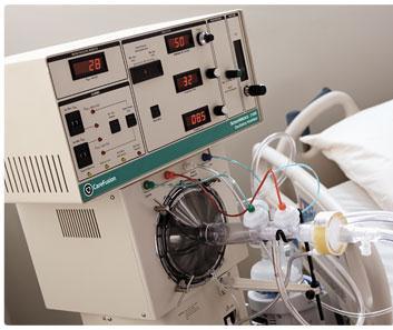
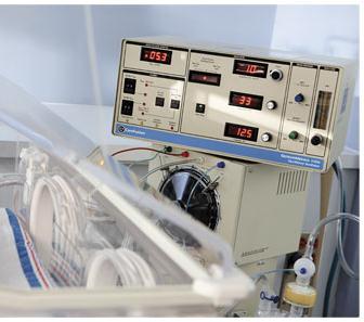
5 INDICATIONS FOR USE OF HIGH FREQUENCY OSCILLATORY VENTILATION There are several indications for the use of High Frequency Oscillatory Ventilation. The two most common indications are Air Leak Syndrome and Acute Respiratory Distress Syndrome. Some general considerations for transitioning a patient from conventional ventilation to high frequency ventilation include a FiO2 >60%, a peep >10 cm H2O, an OI >16, and the inability to maintain a ph >7.25. There are specific disease processes that also merit consideration for initiating HFOV. Patients with Diffuse Alveolar Disease (DAD) may benefit from management by HFOV. The strategy for managing these patients is to maintain a mean airway pressure high enough to recruit and distend collapsed alveoli. This recruitment and distention is intended to reverse the V/Q mismatch and decreases the need for toxic levels of oxygen. Patients with Diffuse Alveolar Disease have time constants in the lungs that vary from region to region. By maintaining a constant mean airway pressure, using small tidal volumes, and limiting pressure fluctuations, volutrauma and shearing injury is minimized. Patients who have Air Leak Syndrome (ALS) may benefit from HFOV. Using a low lung volume strategy, adequate ventilation can be accomplished. By limiting tidal volumes to that above critical opening pressures, and by minimizing pressure fluctuations, the insult to the damaged region of the lung is decreased. Mean airway pressures should be kept as low as possible while maintaining adequate blood oxygen levels. Patients with Respiratory Syncytial Virus (RSV), Status Asthmaticus, or other disease with obstruction to flow are generally not placed on HFOV. These diseases require a long expiratory time and with respiratory rates as high as 15 Hz (900 breaths per minute), the expiratory time is inadequate and air trapping may result. However, patients who develop pneumonia secondary to RSV have been managed quite well on HFOV. Patients born with Lung Hypoplasia Syndrome often benefit from the use of HFOV. Pulmonary Hypoplasia is a relatively common abnormality of lung development and is often associated with congenital diaphragmatic hernia (CDH). Patients benefit because with a single lung, the surface area of lung that participates in gas exchange is reduced by half if not more. The developed lung may be compressed by a mediastinal shift. This shift may cause a compression of the lung, reduce blood flow through the pulmonary veins, and may cause a decrease in venous return. By using MAPs that are fairly low and increasing the MAP in small increments, optimal lung volume can be achieved without extensive pressure increases thus preventing airleaks. Hypoplastic lungs, which have a decrease in the vascular cross-section, develop persistent pulmonary hypertension (PPHN). There are multiple possible causes of PPHN but the management from a pulmonary point of view is basically the same. First, myocardial function must be optimized. Once that is done, the patient may be tried on Nitric Oxide prior to placing a patient on HFOV. If the patient does not respond to or has a minimal response to Nitric Oxide and conventional ventilation, the patient is placed on HFOV for the purpose of optimizing lung volume. It is a common practice to leave the patient on Nitric Oxide while on HFOV. Once optimal lung volume is achieved, the patient should be hyperventilated and hyperoxygenated to promote relaxation of the smooth muscle in the pulmonary vascular bed. Accomplishing this should decrease pulmonary vascular resistance. 3100A and 3100B HIGH FREQUENCY OSCILLATORY VENTILATION The 3100B was developed to allow for HFOV on patients over 35 kg. Both the 3100A and B consists of 6 subsystems, external air/oxygen blender, and an external humidifier.
6 The 6 subsystems are: 1. Pneumatic Logic and Control 2. Patient circuit a. The circuit for the B is longer and more flexible 1. Oscillator Subsystem 2. Airway pressure monitor 3. Electronic control and alarms 4. Electrical supply COMPARISON OF THE 3100A AND 3100B 3100A 3100B MAP 49 cmh2o 59 cmh2o Bias Flow 40 LPM 60 LPM Max Delta P 105 cmh2o 130 cmh2o Safety Dump Press 50 cmh2o 60 cmh2o Safety Dump Alarm Delay No Delay 1.5 sec delay Min Airway Pressure Limit >20% High Paw alarm set 5 cmh2o MAP Pressure Limit Manual-approximately cmh2o Automatic Piston Centering Manual Automatic Cooling Gas Flow 10 LPM 25 LPM Circuit Length 31 inches 51 inches Visual Max set Red LED; non-latching Red LED, latching
7 CONTROLS AND INDICATORS This section will deal with the control knobs and indicators on both the 3100A and B. With a few exceptions, the functions of the control knobs are the same for both ventilators. Bias Flow Bias Flow: controls and indicates the rate of continuous flow of humidified gas. This gas supplies fresh gas that aids in flushing the CO2. Bias flow is also a limiting factor for setting mean airway pressure. The maximum available flow is 40 LPM for the 3100 A and 60 LPM for the 3100 B.
8 Power/Delta P Mean Pressure Limit Mean Pressure Adjust Power/Delta P: determines the amount of power that is driving the oscillator piston back and forth. The knob scale is a 10-turn locking dial that is not calibrated in % power but is marked for purposes of establishing reference points. This knob has control of the displacement of the oscillator piston and the oscillatory pressure Delta P. Mean Pressure Limit: controls the limit above which proximal mean airway pressure cannot be increased. The approximate range is 10 to 45cmH2O and is set manually approximately 2cm H2O above the mean. The 3100B mean pressure limit is internally set at 5cmH2O above the mean airway pressure and requires no intervention by the clinician. Mean Pressure Adjust: this adjusts the mean airway pressure by controlling the resistance of the Paw Control valve. When adjusted, the control fixes the mean pressure at the ETT/patient connection. Changes to Frequency, % I time, Power, Piston Centering, and adding water to the humidification chamber may affect the mean airway pressure. When setting the power on the 3100A, the goal is to achieve chest wiggle from the clavicles to the umbilicus. With the 3100B, the goal is to achieve chest wiggle from the clavicles to mid-thigh.

9 Amplitude % Inspiratory Time Frequency Amplitude: pressure gradient between peak inspiratory and peak expiratory pressures within the oscillatory circuit. Note: The gradient in the airways is significantly less than the pressure gradient in the circuit. For patients on the 3100B, the amplitude should be set 20 above the PaCO2. % Inspiratory Time: determines the amount of time the piston is in the inspiratory position. When the clinician changes the % Inspiratory Time, it changes the starting position of the piston to begin inspiration. A % Inspiratory Time of 33% gives an I:E ratio of 1:2. A % Inspiratory Time of 50% gives an I:E ration of 1:1. Since this control affects the shape of the waveform, changing % Inspiratory Time will affect the Mean Airway Pressure and the Delta P. Frequency: sets the oscillator frequency in Hertz. One Hertz is 60 cycles and is measured in cycles per minute. An example of this is a hertz of 5 is 300 cycles per minute. Both A and B have a range of 3 to 15 Hertz.
10 Mean Airway Pressure Piston Centering Set Max Paw Set Min Paw Alarm Silence Reset Start/Stop Mean Airway Pressure: displays the Paw on a digital meter in cmh20 Set Max Paw: this determines the level at which the Max Paw Exceeded Warning Alarm is activated. The 3100B does not have a manual alarm to set; it is internal and is preset at 5 cmh2o above the set Paw. Set Min Paw: this determines the level at which the Min Paw Exceeded Warning Alarm is activated. Alarm silence: silences alarms for 45 seconds Reset: this momentary push button resets all Safety Alarms and the Power Failure alarm. Certain conditions must first be corrected before the reset and restart of the oscillator will occur. Start/Stop: this changes the oscillator between enable and disable..
11 Paw>50 cmh2o Paw <20% of Set Max Paw : Paw>50 cmh2o: this indicates that the preset Safety alarm has been activated. On the 3100B, this alarm is set at 60 cmh2o. When this alarm is activated, both the 3100A and 3100B will automatically shut down. When this occurs, bias flow continues, the dump valve opens and holds the airway pressure near the current atmospheric pressure. The only way to reset this alarm is to correct the problem and push the reset button. Paw <20% of Set Max Paw : This alarm is activated when the Paw level is equal to 20% of the thumbwheel setting. When this alarm is activated, both the 3100A and 3100B will automatically shut down the oscillator. When this occurs, bias flow continues, the dump valve opens, and atmospheric pressure is held. The 3100B alarm is internally set to alarm at 5 cmh2o above the Set Max Paw. Additionally, there is no delay in the dump valve opening with the 3100A. With the 3100B, there is a 1.5 second delay. After the 1.5 second delay, the 3100B will reset itself whereas the 3100A does not.

12 Piston Centering Control On/ Off Switch Piston Centering Control: Determines the center position of the oscillator piston. Power Switch: Turns power on to the 3100A and B system.
13 TROUBLE SHOOTING THE 3100A AND 3100B Conditions Possible Causes Possible Remedies Displayed Paw > 50cmH2O 1. Patient at high Paw and 1. Bias flow rate possibly Alarm spontaneously breathing insufficient, re-adjust Paw usinghigher flow 2. Obstruction in exp limb 2. Replace patient circuit 3. Obstruction in pressure 3. Replace patient circuit sense line 4. Interference from radio 4. Remove source of transmitter interference Displayed Paw < Set 1. Patient spontaneously 1. Bias Flow rate possibly Min thumbwheel alarm breathing insufficient, re-adjust Paw using higher flow 2. Improper setting of 2. Change setting thumbwheel switch 3.Improper setting of Paw adjust or flow meter 3. Change setting 4. Patient circuit temp drop 4. Check and correct circuit temperature 5. Improper setting of Paw 4. Change setting limit 6. Leak in patient circuit 6. Eliminate leak or replace circuit 7. Cap diaphragm leak 7. Replace cap diaphragm 8. Interference from a radio 8. Remove source of transmitter interference Displayed Paw > Set Max 1. Patient spontaneously 1. Bias Flow rate possibly Paw thumbwheel alarm breathing insufficient, re-adjust Paw using higher flow 2. Improper setting of 2. Change setting thumbwheel switch 3.Obstruction of exp limit 3. Replace the patient circuit 4. Obstruction of pressure 4. Replace the Patient circuit sense line 5. Patient circuit temperature 5. Check and correct circuit rise temperature 6. Interference from a radio 6. Remove the source transmitter of interference
14 TROUBLE SHOOTING THE 3100A AND 3100B cont. Conditions Possible Causes Possible Remedies Displayed Paw < 20cmH2O 1. Improper setting of 1. Change setting Set Max Thumbwheel Alarm thumbwheel switch 2. Improper setting of Paw 2. Change Setting adjust or flow meter 3. Improper setting of Paw 3. Change Setting limit 4. Leak in humidifier or 4. Eliminate leak or patient circuit, including replace circuit patient disconnect 5. Cap diaphragm leak 5. Replace cap diaphragm 6. Interference from a 6. Remove source of radio transmitter interference 7. Open water trap stopcock 7. Close water trap stopcock Oscillator stopped with 1. Power setting too low 1. Bias Flow rate possibly no other alarm occuring and P is less than insufficient, re-adjust 6 cm H2O Paw using higher flow 2. Oscillator piston not 2. Change setting centered 3.Oscillator failure 3. Remove from service Source Gas Low Alarm 1. Input pressure less than 1. Check input gas line 30 psi, either from blender or cooling gas 2. Input filter needs 2. Replace filters replacement 3. Flow restriction in gas 3. Replace the patient circuit supply lines 4. Internal leak 4. Call Sensormedics Battery Low Alarm 1. Battery voltage less 1. Replace battery than optimal 2. Battery disconnected 2. Properly reconnect battery Oscillator Overheated 1. No cooling gas flow 1. Assure cooling gas supply Alarm is attached 2. Oscillator overheated 2. Check cooling gas flow for due to poor cooling gas blocked filter element or flow restricted supply hose replace if necessary 3. Oscillator overheated to 3. Readjust centering while mis-centered at very monitoring Paw high P setting 4. Oscillator overheated due 4. Call Sensormedics to mechanical failure of oscillator subsystem
15 TROUBLE SHOOTING THE 3100A AND 3100B cont. Failure During Checkout Conditions Possible Causes Possible Remedies Reset/ Power Failure 1. AC power removed from 1. Check line cord system or main power interuption 2. Internal power failure 2. To start oscillator after correcting problem, apply power to system, press and hold "Reset" to establish Paw, and then press Stop/ Start switch Failure to meet Patient 1. Leak in patient circuit 1. Eliminate leak or replace Circuit Calibration or humidifier connections patient circuit 2. Improper flow meter 2. Set flow meter to 20 lpm setting sighting on center of ball 3. Open water trap 3. Close water trap stopcock stopcock 4. Internal Leak or 4. Call Sensormedics maladjustment Failure of ventilator 1. Incorrect Patient Circuit 1. Perform Patient Circuit Performance Check- calibration calibration Paw out of range (low) 2. Center of flow meter not 2. Adjust flow to center of used to make 20 lpm ball adjustment 3. Incorrect altitude range 3. Use appropriate altitude being used range for your facility 4. Oscillator not centered 4. Center piston using properly centering control 5. Internal failure 5. Call Sensormedics Failure of ventilator 1. Incorrect Patient Circuit 1. Perform Patient Circuit Performance Check - calibration Paw out of range (high) 2. Center of flow meter ball 2. Adjust flow to center of not used to make 20 lpm ball adjustment 3. Incorrect altitude range 3. Use appropriate altitude being used for your facility 4. Oscillator not centered 4. Center piston properly properly using centering control 5. Paw set with Paw limit 5. Correct settings and not Paw adjust 6. Internal failure 6. Call Sensormedics Failure of Ventilator 1. Bias flow tubing from 1. Use bias flow tubing Performance Check- humidifier to circuit has supplied with circuit and P out of range (low) been cut to less than 30 ", do not shorten or tubing not supplied with patient circuit being used 2. Oscillator not centered 2. Center piston properly properly using centering control 3. Power not set at 6 3. Set power to 6 4. Compression 4. Bypass humidifier for characteristics of performance check, then humidifier allowing reattach P to drop 5. Internal failure 5. Call Sensormedics
16 TROUBLE SHOOTING THE 3100A AND 3100B cont. Unexplained Operation Conditions Possible Causes Possible Remedies Failure of Ventilator 1. Oscillator not centered 1. Center piston Performance check - P 2. Oscillator not warmed up 2. Allow Oscillator to warm out of range (High) for 5 minutes 3. Incorrectaltitude range 3. Use appropriate altitude being used range for your facility 4. Internal failure 4. Call Sensormedics Oscillator shuts down and 1. Set Max Thumbwheel set 1. Reset the thumbwheel to lower Dump valve opens during to high. Paw > 20% set setting closer to target Paw Operation max 2. Drastic change in Paw 2. Re- establish Paw and over- aggressive control make any small change using the Paw adjustments to Paw using adjust flow meter adjust 3. ETT has become 3. Reconnect ETT disconnected 4. Radio frequency 4. Locate and distance interference offending devise Oscillator will not restart 1. Set max thumbwheel 1. Reset thumbwheel to lower After temporary control set too high setting closer to target Paw Disconnect (such as 2. To restart oscillator, Paw 2. Reduce Max thumbwheel for routine suctioning) must first be >20% of setting temporary until set max, but in order to oscillator starts. If it still achieve Paw >20% of set will not start, reduce power oscillator must be on and increase Paw to target level using flow meter and Paw control valve- then increase power while keeping Paw on target by adjusting flow meter or Paw control valve down Paw unstable- jumps by 1. Water collecting at Paw 1. Adjust circuit height for 2-3 cm H2O adjust valve better draining 2. Patient spontaneously 2. Bias flow rate possibly breathing insufficient, re-adjust Paw using higher flow 3. Worn or defective cap 3. Replace cap diaphragms diaphragm 4. Internal failure 4. Call Sensormedics


17 ASSEMBLING THE PATIENT CIRCUIT Connect the patient circuit body to the bellows/water trap assembly. Snap the three identical cap/diaphragm assemblies on to the three valve bodies located on the patient circuit body.


18 Note: Be sure to check the diaphragm assemblies to ensure there are no leaks. 2. Attach the assembled patient circuit to the face of the oscillator compartment using the four T-handle quarter turn fasteners.

19 Attach the three color-coded tubes to their corresponding valve caps, following the color-coding scheme: Color of Line Attaches To 1. Green Paw Control Valve 2. Blue Paw Limit Valve 3. Red Dump Valve 4. Clear Paw Sensing Port Note: The differing lengths and color coding of the tubes and the physical arrangement of the valves within the Patient circuit minimizes the possibility of cross connection. The colored lines are not disposable

20 Insert the humidifier temperature probe in the tapered-opening near the patient Y. PATIENT CIRCUIT CALIBRATION PROCEDURE On the side of both the 3100A and 3100B ventilator, is Patient Circuit Calibration Procedure that is to be performed before a patient is placed on the 3100A or 3100B High Frequency Ventilator. This check is to be done off patient.

21 VENTILATOR PERFOMANCE CHECKS On the top of both the 3100A and 3100B ventilator, is a Ventilator Performance Check that is to be performed before a patient is placed on the 3100A or 3100B High Frequency Ventilator. This check is to be done off patient.

22 START-UP PROCEDURES A Quick-Checklist label for pre-patient hook-up verification is attached to the ventilator. An example for both the 3100A and 3100B is below.

23 TASKS AND CONSIDERATIONS FOR INITIATING HFOV When a patient is being considered for HFOV, there are several tasks and considerations to be kept in mind when initiating HFOV. Labs: An ABG should be drawn prior to and approximately 30 minutes after initiating HFOV. A blood gas should be drawn within 1 hour of all changes and when clinically indicated. Radiology: A chest x-ray should be taken prior to initiation if possible. A follow-up chest x-ray should be taken 1 hour after initiation of HFOV. The clinician should look for a more expanded chest, usually to 9 ribs. Ideally, a cxr should be obtained after changes to the mean airway pressure. Routine cxr s are recommended every 6 hours for the first 24 hours and then every day. Changes in chest radiographs may not be appreciated for at least 4-6 hours to allow a patient time to fully recruit collapsed alveoli.
24 Sedation: Patients should be sedated to the point of being comfortable and not fighting the ventilator. Paralytic agents are not necessarily indicated unless previously in use. If a paralytic was in use prior to initiating HFOV, it should be discontinued, as tolerated by the patient. 3100B HIGH FREQUENCY OSCILLATORY VENTILATION PATIENT SET UP 1. Perform circuit calibration. Note: Remember that this is done off the patient 2. Perform Ventilator performance check. Note: Remember that this is done off the patient 3. Patient set up: A. Adjust Bias Flow to 30 lpm initially B. Set the MAP adjust knob to achieve the desired MAP, usually 5cmH2O above the mean on conventional ventilation, per physician orders 1. Set MAP alarm limit to +5cmH20 above MAP C. Set the Frequency to physician s orders, usually 3-6 Hz D. Set Inspiratory time to 33% E. Set the Power to achieve amplitude 20 above the PaCO2. Depending on the size of the patient, power is set in a range of 4-9. F.. Adjust the Set Min alarm to 3cmH20 below the MAP and readjust as needed 1. Depress and hold the Restart Button until the MAP increases to >5cmH20. Press the Start/Stop to start the piston. Note: Prior to connecting the ventilator, suction the patient well. Place the patient on the oscillator. Set the MAP to 40 cmh2o for 40 seconds, and then start the ventilator. Immediately assess the Chest Wiggle and adjust the power setting to achieve optimal wiggle. Chest Wiggle for patients on the 3100B is a visual vibration between the clavicles and upper thigh. The power required to achieve optimal chest wiggle is the baseline measurement. G. Start with a FiO2 of 100% and wean as tolerated H. Adjust the MAP according to the O2 saturation I. Obtain an ABG approximately 30 minutes after initiating HFOV. 1. To improve ventilation, increase the P in increments of 5cmH20. If the P is 3 times greater than the Paw, decrease frequency by 1 Hz to decrease CO2 and allow for the decrease in P. 2. To improve oxygenation, optimal lung volume must be maintained. Optimal lung volume is reached once you see stable O2sats. Once the desired saturation is obtained, decrease FiO2 until it is <50%. J. Approximately 1-2 hours after initiating HFOV, obtain a CXR. The diaphragm should be approximately at the 9 th rib on the right side. 3100A HIGH FREQUENCY OSCILLATORY VENTILATION PATIENT SETUP 1. As with all patients, perform circuit calibration. Note: Remember that this is done off the patient 1. As with all patients, perform ventilator performance check. Note: Remember that this is done off the patient. 2. Patient set up: A. Adjust Bias Flow to 20 lpm initially
25 B. Set the MAP adjust knob to achieve the desired MAP per physician orders 1. High Volume Strategy: RDS, PPHN a. MAP usually 2-3cmH2O above MAP on conventional ventilation a. Start with an FiO2 of 100%. Once optimal lung volume has been achieved, start weaning the FiO2 2. Low Volume Strategy: Pulmonary Interstitial Emphysema a. MAP usually set at or below MAP used on conventional ventilation a. Gradually decrease the MAP by 1cmH2O Every 15 minutes as tolerated, accepting ph > 7.25 and PaCO2 < 65 torr Note: Diaphragmatic Hernia and MAS do not fall into either category and must be managed accordingly C. Set the power to obtain an amplitude equal to the PIP on conventional ventilation per physicians to: 1. Make adjustments until good chest wiggle and optimal PaCO2 is achieved D. Set the frequency for infants per physician orders to: 1. weighing < 750 grams 15Hz 2. weighing > 750 grams 10 Hz E. Inspiratory time should always be set at 33% to avoid air trapping unless otherwise ordered by a PICU attending or fellow. MANAGEMENT OF HIGH FREQUENCY OSCILLATORY VENTILATOR A. Respiratory Care Practitioner Responsibilities 1.. Check orders a. Orders should be rewritten, by the ICU Fellow or Attending, every 24 hours b. Make sure that the settings are appropriate for the patient 2. Perform patient assessment per CMCD Patient Assessment Guidelines 3. Chest X-ray a. Verify chest expansion by counting the number of ribs. At CMCD, chest expansion is considered adequate if the diaphragm is at the level of the 9 th rib. 1. Chest expansion is one of the clinical indicators for adequate Paw. a. Assess the position of the ETT. It should be comfortably above the carina. 4. Equipment a. Thoroughly inspect ventilator circuit b. Verify that all wheels are locked and secured c. Verify that circuit is stable and secure d. Verify that there is adequate water in the humidifier and that humidification is adequate. 1. Note that if the water level is low in the humidification chamber, this will affect the Mean Airway Pressure e. Calibrate and insure proper operation of the O2 analyzer f. Verify that there is a back up oscillator. If not, notify the Team Leader for that unit immediately. 5. Ventilator Checks are done every hour a. Assess patient circuit for proper gas exhaust. b. Verify water level in the humidification chamber.
26 c. Drain the water from the bellows and the 60cc syringe 1. Do not empty the syringe completely. Leave approximately 1/4 of water in the syringe to seal and prevent a leak. d. Perform a patient assessment in compliance with CMCD Guidelines e. Assess for adequate chest wiggle. 1. Patients on the 3100A: clavicle to umbilicus 2. Patients on the 3100B: clavicle to mid-thigh f. Check recent lab results 6. Lab and Chest X-ray frequency a. Arterial blood gases to 60 minutes following initiation of HFOV 2. Q2H for the first 8 hours following initiation 3. Q4H per physician order for the duration of time on HFOV 4. 1 hour after changes made to HFOV settings b. Chest X-ray minutes to 1 hour following initiation of HFOV 2. Q12H for the following 24 hours 3. Qam for duration of time on HFOV 4. When clinically indicated 5. Following changes in MAP 7. Invasive and Non-Invasive monitoring a. Placement of any invasive monitoring device; Art line, CVP, or Swann Ganz Catheter should be done prior to initiation of HFOV a. Continuous Cardiac Monitoring and Pulse oximetry 8. Blood pressure and urine output should be assessed with each ventilator check to assure that both are adequate and that no acute change has occurred 9. Head ultrasounds a. Should be done on patients with a open fontanel prior to initiation of HFOV. b. Follow up ultrasounds should be done when clinically indicated.. B. High Frequency Oscillatory Ventilation Management Guidelines 1. Ventilation Management a. Decreasing CO2 is accomplished primarily by adjusting 2the Delta P. Monitor adjustments in Delta P by watching chest wiggle. b. If maximum Delta P is unable to improve ventilation, decreasing the frequency (Hertz) will increase the displacement of the piston increasing tidal volumes. a. If these two adjustments are not successful, consider increasing the bias flow. This may lead to an increase in the CO2 washout. Note: Ensure that there is an adequate airleak around the ETT. If the cuff is inflated, deflate the cuff until an airleak can be heard. In extreme cases, the patient may need to be reintubated with a smaller tube to facilitate an adequate airleak. With patients on the 3100B, deflate the cuff on the ETT until MAP decreases by 5 cmh2o. At this point, increase the MAP back to ordered parameter. a. If elevated PaCO2 persists, an adjustment to % Inspiratory Time may be indicated. An increase of approximately 10% may be obtained by increasing the I Time % to 50%.
27 Note that this is not a common practice at CMCD and must be done only with a physician order and observed by either the PICU Fellow or Attending. 2. Oxygenation Management a. Oxygenation is a primary function of Peak Airway Pressure (Paw). Paw is usually set 10 to 30% higher than that of conventional ventilation. a. Proper inflation or Paw is reflected by a chest x-ray that reveals expansion to 9 ribs above the diaphragm. b. The clinician should be aware of the disease process that is being managed. Higher Paw should be used when the patient has Diffuse Alveolar Disease (DAD). The theory is that higher Paw s will recruit and distend collapsed alveoli. Patients who have Air Leak Injury (ALI) should be maintained on a minimal Paw and oxygenation managed with increasing FiO2. Higher Paw may cause additional injury or exacerbation of the injury to the lung. c. Increasing the FiO2 is another way of improving oxygenation. Once the desired improvement in oxygenation is achieved, the clinician should wean the FiO2 to a level of 50% or less as the patient tolerates. Once a level of <50% is achieved, weaning the Paw may be appropriate. C. High Frequency Treatment Strategies Clinical Indicators Therapeutic Intervention Rationale FiO2 above 60% High PaCO2 with: PaO2 = OK Increase Delta P Achieve optimal PaO2 PaO2 = low Increase Paw, Delta P, FiO2 Improves O2 delivery PaO2 = high Increase Delta P, Decrease FiO2 Decrease FiO2 to minimize O2 exposure Normal PaCO2 with: PaO2 = OK No action No action PaO2 = low Increase Paw, FiO2 Improves O2 delivery PaO2 = high Decrease FiO2 Decrease FiO2 to minimize O2 exposure Low PaCO2 with: PaO2 = OK Decrease Delta P Achieve optimal PaO2 PaO2 = low Increase Paw/FiO2 decrease Delta P Improve O2 delivery PaO2 = high Decrease FiO2, Delta P Decrease FiO2 to minimize O2 exposure FiO2 below 60% High PaO2 with: PaO2 = OK Increase Delta P Achieve optimal PaCO2 PaO2 = low I ncrease FiO2
28 Delta P Improve PaO2 PaO2 = high Increase Delta P decrease Paw Decrease Paw to decrease Normal PaO2 with: PaO2 = OK No Action No Action PaO2 = low Increase FiO2 Improves PaO2 PaO2 = high Decrease Paw, FiO2 Reduces PaO2 Low PaCO2 with: PaO2 = okay Decrease Delta P Achieve optimal PaCO2 PaO2= low Increase FiO2, decrease Delta P Improves PaO2 PaO2 = high Decrease Paw, Delta P Achieve optimal PaO2 and PaO2 PaO2 D. Suctioning: Indications and Proper Technique 1. Indications a. Acute increase in Delta P b. Decreased chest wiggle and increased Delta P c. Sudden or acute drop in SpO2 2. Proper Technique a. Patients on HFOV should be suctioned only as needed. It is strongly recommended that a closed suction system be used when suctioning a patient on HFOV. Patients may de-recruit and collapse alveoli if disconnected from HFOV. b. Suction Technique 1. Pre oxygenate with 100% FiO2 Manually ventilate patient with 100% oxygen with ambu bag 1. Pass the catheter quickly and return patient to ambu bag. 2. Repeat suctioning if needed. 3. When finished suctioning, perform a Sustained Maximal Inflation c. Sustained Maximal Inflation 1. For patients on the 3100A, connect patient to ventilator and increase MAP 2 cmh2o above set ventilator MAP a. Restart ventilator b. Once O2 saturations return to normal levels, decrease MAP to previous settings 2. With the 3100B, connect patient to ventilator and increase MAP to 40 for 40 seconds. a. Restart ventilator and decrease MAP to previous settings as tolerated E. Sedation and Paralysis At CMCD, patients are routinely sedated when being ventilated on HFOV. The decision to medically paralyze a patient is physician driven. Each attending has their preference. 1. Indications for sedation may include the following: a. tachycardia b. hypertension
29 c. increased Delta P a. pressure limiting ventilator b. spontaneous breathing c. increased agitation 2. Indications for paralyzing may include the following: a. inability to adequately ventilate or oxygenate b. pressure limiting ventilator c. patient safety F. Positioning Studies in adults have shown that positioning patients, who have ARDS, prone is an effective way of increasing Functional Residual Capacity (FRC) thus improving oxygenation. Currently, a multi-center study is under way to determine if the pediatric population will benefit from being placed prone. If the patient has no other injuries that contraindicate them being placed prone, the therapist may want to discuss placing the patient prone with the physician prior to initiating HFOV. Patients who are left supine may benefit from having the head of the bed raised slightly to help keep the contents of the abdominal cavity from placing pressure on the diaphragm. Again, make sure that there are no contraindications for raising the head of the bed. Weaning from High Frequency Oscillatory Ventilation The goals for using HFOV when treating patients with acute lung disease is to minimize ventilator induced lung injury while achieving nontoxic oxygen levels. Weaning from high frequency is a slow process that allows sufficient time for the changes to be representative of the patient's pulmonary status. Weaning from HFOV should be done in a systematic manner that prevents the lung from de-recruiting. The following steps for weaning from HFOV are guidelines only. They represent the practice of Critical Care Services at CMCD, information found in a review of current professional articles, and recommendations found in various research studies. Weaning FiO2 Attempt to wean the FiO2 to a nontoxic level (<50%) while maintaining a SpO2 of >90%. If the patient does not maintain an adequate saturation at an FiO2 of < 60%, no additional attempts at weaning should be made until the patient can tolerate them. Weaning Paw If the patient is able to maintain a SpO2 >90% on an FiO2 50%, obtain a physician s order and start a gradual reduction in Paw of.5-3 cmh20 Q4-6 hours as tolerated. If weaning is overly aggressive, lung volumes may drop below closing pressures and desaturation may occur. Increasing the Paw 1-2 cmh2o above the previous Paw level may not be sufficient to re-recruit collapsed alveoli. Weaning Power When the ph is normalized and the PaCO2 is within acceptable limits, the power/delta P should be weaned before the frequency. There seems to be no agreed upon value or amount to decrease the power by. Remember that small changes in the power can make a significant change in ventilation. Close attention should be paid to the patient's blood gas values. Note: When weaning the power, ensure there is adequate chest wiggle with the changes. Weaning Frequency When adequate chest wiggle is achieved with reduction of power, the frequency may be weaned. The clinician must remember that frequency is inversely related to tidal volume. To wean the tidal volume, increase the
30 frequency. This will reduce the size of the tidal volume. Close attention should be paid to the patient's blood gas values. As with weaning conventional ventilation, it is advisable to wean one parameter at a time. Multiple changes may cloud the issue and the clinician may not be able to distinguish which change lead to the clinical change. HFOV weaning difficulties are usually evidenced by restlessness, increased work of breathing, fluctuations in mean airway pressure, and a decrease in saturation. Once a patient has demonstrated that he is ready to convert to conventional mechanical ventilation, the following settings may be considered as a starting point. These are merely suggested settings. The final decision for ventilator settings is either the PICU fellow or attending. Transitioning to Conventional Ventilation When a patient is ready to transition to conventional ventilation, many of the same precautions taken when placing a patient on HFOV need to be employed. Much like weaning power, there seems to be no firm agreement on what the Paw needs to be on conventional ventilation. The literature varies from as low as 8 cmh2o to a high of 24 cmh2o. The physicians at CMCD will determine what level of Paw they want. A suggested mode of ventilation is SIMV Pressure Control. Current literature suggest setting the pressure control level to achieve a delivered tidal volume of 6 8 ml/kg of ideal body weight. The same literature also suggest that a pressure support level be set to achieve a delivered tidal volume on a spontaneous breath of 3 5 ml/kg of ideal body weight. A respiratory rate of is a good suggested starting rate when transitioning to conventional ventilation. An arterial blood gas should be obtained 30 to 60 minutes after transfer to conventional ventilation to further guide ventilator adjustments. Keep in mind that different CCS attending physicians at CMCD have different philosophies of how to manage patients requiring HFOV. The information provided in the above sections is meant to be used as clinical guidelines only.
31 REFERENCES 1. Patricia Hynes-Gay, R.N., Rod MacDonald, R.R.T. (2001) Using High-Frequency Oscillatory Ventilation to Treat Adults With Acute Respiratory Distress Syndrome. Critical Care Nurse, Vol 21, No. 5, October 2001: Steve Derdak, D.O. (2001) High Frequency Oscillatory Ventilation; Clinical Management Strategies For Adult Patients. L1856 Revision A, November 2001: Ray Higginson, RN, B.N. (2002) High Frequency Oscillatory Ventilation. Chest Medicine On-Line, September Sherry E, Courtney, M.D., David J Durand, M.D., Jeanette M. Asselin, RRT, M.S., Mark L. Hudak, M.D., Judy L. Aschner, M.D., and Craig T. Shoemaker, M.D. (2002) High-Frequency Oscillatory Ventilation Verses Conventional Mechanical Ventilation for Very Low Birth Weight Infants. N Engl J Med, Vol 347, No. 9, August 29, 2002: Operator s manual, SensorMedics, Model 300B High Frequency Oscillatory Ventilator, SensorMedics Corporation, Saui Ranch Parkway, Yorba Linda, Ca Fedora, M., Klimovic, M., Domink, P., and Nekvasil, R., (2001) The Influence of An Early Application of High Frequency Oscillatory Ventilation on The Outcome In Paediatric Acute Respiratory Distress Syndrome. Scripta Medica 74 (4), October, 2001: Ruben D. Restrepo, M.D., RRT, Stephen Dickson, RRT, (2002) Low Lung Inflation Strategy and High Frequency Oscillatory Ventilation. RT June/July Jerry A. Krishnan, M.D., and Roy G. Bower, M.D. (2000) High Frequency Ventilation for Acute Lung Injury and ARDS. Chest. 2000; 118: Curtis Caldwell, RN, CNNP (2002) High Frequency Ventilation. Chest Medicine On-Line, September, Catherine A Connors, RN, BSN, CCRN, and Cathy Rosenthal-Dichter, RN, MN, CCRN. (1997) Pediatrics: Components of Breathing: Pediatric Ventilatory Challenges. Critical Care Nurse Volume 17 Number 1, NICU Ventilation Management, Protocol-CHEC Guidelines Department of Pediatrics, Affilliated with the University Of Ottawa and 12. Children s Hospital of Eastern Ontario 13. Stephen Derdak, Sangeeta Mehata, Thomas E Stewart, Terry Smith, Mark Rogers, Timothy G. Buchman, Brian Carlin, Stuart Lowson, John Granton, and the Multicenter Oscillatory Ventilation for Acute Respiratory Distress Syndrome Trial (MOAT) Study Investigators. (2002) High Frequency Oscillatory Ventilation for Acute Respiratory Distress Syndrome in Adults, A Randomized, Controlled Trial. Am J Respir Crit Care Med Volume 166, May 2002, Donald Null, M.D., and Noah Perlman, BS, RRT (1999) High Frequency Oscillatory Ventilation: Disease Specific Clinical Management Strategies. 15. Critical Care Review SensorMedics Corporation Robert Harwood. Intrauterine development and comparative respiratory anatomy. In Kacmarek, R.M., Mack, C., and Dimas, S. (1990). The Essentials Of Respiratory Care, 3 rd Edition, St Louis: Mosby-Year Book 17. Operator s Manual, SensorMedics, Model 3100A High Frequency Oscillatory Ventilator, SensorMedics Corporation, Saui Ranch Parkway, Yorba Linda, Ca Abhay J Bhatt, M.D. Assistant Professor, Department of Pediatrics, Division of Newborn Medicine, University of Mississippi Medical Center, emedicine, May 29, Beverly P Wood, MD, MS Professor, Departments of Radiology and Pediatrics, Division of Medical Education, Keck School of Medicine, 20. University of Southern California, emedicine, October 30, William Driscoll, DO, Assistant Professor, Department of Neonatology, Winthrop University hospital, State University of New York Stony Brook 22. School of Medicine, emedicine, February 7, Neil R MacIntyre, MD, Duke University Medical Center, Mechanical Ventilation Strategies for Lung Protection. Presentation at Conference May 1999, Wisconsin Society for Respiratory Care 24. Steven E. Sittig, RRT, Neonatal Mechanical Ventilation Support. AARC Times, April 1999, Georgopoulos D, Mitrouska I, Markopoulou K, Patakas D, Anthonisen NR, Effects of breathing patterns on mechanically ventilated patients with 26. Chronic obstructive pulmonary disease and dynamic hyperinflation, Intensive Care Med Nov: 21(11):880-6
 http://www.priory.com/cmol/hfov.htm INTRODUCTION The vast majority of patients who are admitted to an Intensive Care Unit (ICU) will need artificial ventilation (Jones et al 1998). The usual means through
3100A Competency Exam
 NAME DATE (Circle the appropriate answer) 3100A Competency Exam 1. Of the following, which best describes the mechanics of ventilation used by the 3100A? a. Active inspiration with passive exhalation b.
QUICK REFERENCE GUIDE
 cm H O 2 cm H O 2 cm HO 2 PSI cm H O 2 ON OFF UPPER LIMIT LOWER LIMIT UPPER LIMIT LOWER LIMIT LIFE PULSE HIGH-FREQUENCY VENTILATOR QUICK REFERENCE GUIDE 01388-08.11 MONITOR PIP JET VALVE ALARMS READY SILENCE
PART EIGHT HIGH FREQUENCY PERCUSSIVE OSCILLATION (HFPOV )
") PART EIGHT HIGH FREQUENCY PERCUSSIVE OSCILLATION (HFPOV ) Note: For maximal comparative understanding, FIRST read PART SEVEN which defines the concept of High Frequency Oscillatory Ventilation (HFOV).
PART EIGHT HIGH FREQUENCY PERCUSSIVE OSCILLATION (HFPOV ) Note: For maximal comparative understanding, FIRST read PART SEVEN which defines the concept of High Frequency Oscillatory Ventilation (HFOV).
Initiation and Management of Airway Pressure Release Ventilation (APRV)
") Initiation and Management of Airway Pressure Release Ventilation (APRV) Eric Kriner RRT Pulmonary Critical Care Clinical Specialist Pulmonary Services Department Medstar Washington Hospital Center Disclosures
Initiation and Management of Airway Pressure Release Ventilation (APRV) Eric Kriner RRT Pulmonary Critical Care Clinical Specialist Pulmonary Services Department Medstar Washington Hospital Center Disclosures
VENTILATORS PURPOSE OBJECTIVES
 VENTILATORS PURPOSE To familiarize and acquaint the transfer Paramedic with the skills and knowledge necessary to adequately maintain a ventilator in the interfacility transfer environment. COGNITIVE OBJECTIVES
Mechanical Ventilation
 PROCEDURE - Page 1 of 5 Purpose Scope Physician's Order Indications Procedure Mechanical Artificial Ventilation refers to any methods to deliver volumes of gas into a patient's lungs over an extended period
Operating Instructions for Microprocessor Controlled Ventilators
 Page 1 of 5 Operating Instructions for Microprocessor Controlled Ventilators Purpose Audience Scope Physician's Order To provide guidelines for the procedure for the use of microprocessor controlled ventilators.
HIGH FREQUENCY JET VENTILATION (HFJV): EQUIPMENT PREPRATION
: EQUIPMENT PREPRATION") POLICY The physician orders High Frequency Jet Ventilation (HFJV). The Respiratory Therapist in discussion with the physician will determine blood gas targets and ventilation settings for the treatment
POLICY The physician orders High Frequency Jet Ventilation (HFJV). The Respiratory Therapist in discussion with the physician will determine blood gas targets and ventilation settings for the treatment
Volume Diffusion Respiration (VDR)
") Volume Diffusion Respiration (VDR) A therapy with many uses Jeffrey Pietz, MD April 15, 2016 VDR ventilation has been used to treat patients with: ARDS Meconium Aspiration Burn and Inhalation Injury RDS
Volume Diffusion Respiration (VDR) A therapy with many uses Jeffrey Pietz, MD April 15, 2016 VDR ventilation has been used to treat patients with: ARDS Meconium Aspiration Burn and Inhalation Injury RDS
OPEN LUNG APPROACH CONCEPT OF MECHANICAL VENTILATION
 OPEN LUNG APPROACH CONCEPT OF MECHANICAL VENTILATION L. Rudo Mathivha Intensive Care Unit Chris Hani Baragwanath Aacademic Hospital & the University of the Witwatersrand OUTLINE Introduction Goals & Indications
carefusion.com CareFusion Yorba Linda, CA CareFusion
 CareFusion 22745 Savi Ranch Parkway Yorba Linda, CA 92887 800.231.2466 toll-free 714.283.2228 tel 714.283.8493 fax CareFusion Germany 234 GmbH Leibnizstrasse 7 97204 Hoechberg Germany +49 931 4972-0 tel
Bunnell LifePulse HFV Quick Reference Guide # Bunnell Incorporated
 Bunnell Incorporated n www.bunl.com n 800-800-4358 (HFJV) n info@bunl.com 436 Lawndale Drive n Salt Lake City, Utah 84115 n intl 801-467-0800 n f 801-467-0867 Bunnell LifePulse HFV Quick Reference Guide
EMS INTER-FACILITY TRANSPORT WITH MECHANICAL VENTILATOR COURSE OBJECTIVES
 GENERAL PROVISIONS: EMS INTER-FACILITY TRANSPORT WITH MECHANICAL VENTILATOR COURSE OBJECTIVES Individuals providing Inter-facility transport with Mechanical Ventilator must have successfully completed
AUTOVENT 4000 VENTILATOR
 OVERVIEW AUTOVENT 4000 Only properly trained and approved Escambia County Bureau of Public Safety Paramedics are to use the AutoVent 4000 ventilator manufactured by LSP to transport patients already on
excellence in care Procedure Management of patients with difficult oxygenation. For Review Aug 2015
 Difficult Oxygenation HELI.CLI.12 Purpose This procedure describes the processes and procedures for a lung protective strategy in the mechanical ventilation of patients that are difficult to oxygenate
Difficult Oxygenation Distribution: Sydney X Illawarra X Orange X
 HELICOPTER OPERATING PROCEDURE HOP No: C/12 Issued: May 2011 Page: 1 of 5 Revision No: Original Difficult Oxygenation Distribution: Sydney X Illawarra X Orange X TRIM No: 09/300 Document No: D10/9973 X
Basics of Mechanical Ventilation. Dr Shrikanth Srinivasan MD,DNB,FNB,EDIC Consultant, Critical Care Medicine Medanta, The Medicity
 Basics of Mechanical Ventilation Dr Shrikanth Srinivasan MD,DNB,FNB,EDIC Consultant, Critical Care Medicine Medanta, The Medicity Overview of topics 1. Goals 2. Settings 3. Modes 4. Advantages and disadvantages
B. A clinical emergency exists in which a profound hypoxia is determined to be present.
 I. Subject: Oxyhood-Oxygen Therapy for Neonates II. Policy: Oxygen therapy by oxyhood shall be initiated upon a physician's order by nurses and Respiratory Therapy personnel trained in the principles of
How does HFOV work? John F Mills MBBS, FRACP, M Med Sc, PhD Neonatologist Royal Children s Hospital. Synopsis
 How does HFOV work? John F Mills MBBS, FRACP, M Med Sc, PhD Neonatologist Royal Children s Hospital Synopsis Definition of an oscillator Historical perspective Differences between HFOV and CMV Determinants
Capnography in the Veterinary Technician Toolbox. Katie Pinner BS, LVT Bush Advanced Veterinary Imaging Richmond, VA
 Capnography in the Veterinary Technician Toolbox Katie Pinner BS, LVT Bush Advanced Veterinary Imaging Richmond, VA What are Respiration and Ventilation? Respiration includes all those chemical and physical
INTRODUCTION TO BI-VENT (APRV) INTRODUCTION TO BI-VENT (APRV) PROGRAM OBJECTIVES
 INTRODUCTION TO BI-VENT (APRV) PROGRAM OBJECTIVES") INTRODUCTION TO BI-VENT (APRV) INTRODUCTION TO BI-VENT (APRV) PROGRAM OBJECTIVES PROVIDE THE DEFINITION FOR BI-VENT EXPLAIN THE BENEFITS OF BI-VENT EXPLAIN SET PARAMETERS IDENTIFY RECRUITMENT IN APRV USING
INTRODUCTION TO BI-VENT (APRV) INTRODUCTION TO BI-VENT (APRV) PROGRAM OBJECTIVES PROVIDE THE DEFINITION FOR BI-VENT EXPLAIN THE BENEFITS OF BI-VENT EXPLAIN SET PARAMETERS IDENTIFY RECRUITMENT IN APRV USING
Introduction to Conventional Ventilation
 Introduction to Conventional Ventilation Dr Julian Eason Consultant Neonatologist Derriford Hospital Mechanics Inspiration diaphragm lowers and thorax expands Negative intrathoracic/intrapleural pressure
PART SEVEN THE HISTORY AND APPLICATION OF HIGH FREQUENCY OSCILLATORY VENTILATION (HFOV)
") PART SEVEN THE HISTORY AND APPLICATION OF HIGH FREQUENCY OSCILLATORY VENTILATION (HFOV) Reciprocating pistons with an eccentric travel speed, moving to and fro within a cylinder (with a common inlet/outlet),
PART SEVEN THE HISTORY AND APPLICATION OF HIGH FREQUENCY OSCILLATORY VENTILATION (HFOV) Reciprocating pistons with an eccentric travel speed, moving to and fro within a cylinder (with a common inlet/outlet),
QUICK GUIDE: VOLUME GUARANTEE VENTILATION Prematurely Born Infants < Weeks Gestation
 Initial Set-up QUICK GUIDE: VOLUME GUARANTEE VENTILATION Prematurely Born Infants < 32 +0 Weeks Gestation Mode: SIPPV + VG Tidal volume: 4-5 ml/kg Pmax (PIP limit): 25 cmh 2 O PEEP: 5 cmh 2 O & Insp. time
Physiological Basis of Mechanical Ventilation
 Physiological Basis of Mechanical Ventilation Wally Carlo, M.D. University of Alabama at Birmingham Department of Pediatrics Division of Neonatology wcarlo@peds.uab.edu Fine Tuning Mechanical Ventilation
Principles of mechanical ventilation. Anton van Kaam, MD, PhD Emma Children s Hospital AMC Amsterdam, The Netherlands
 Principles of mechanical ventilation Anton van Kaam, MD, PhD Emma Children s Hospital AMC Amsterdam, The Netherlands Disclosure Research grant Chiesi Pharmaceuticals Research grant CareFusion GA: 27 weeks,
Mechanical Ventilation. Mechanical Ventilation is a Drug!!! is a drug. MV: Indications for use. MV as a Drug: Outline. MV: Indications for use
 Mechanical Ventilation is a Drug!!! Mechanical Ventilation is a drug I am an employee of Philips Healthcare Hospital Respiratory Care Group and they help me pay for my kids education Jim Laging, RRT, RCP
Mechanical Ventilation
 Mechanical Ventilation Chapter 4 Mechanical Ventilation Equipment When providing mechanical ventilation for pediatric casualties, it is important to select the appropriately sized bag-valve mask or endotracheal
RESPIRATORY CARE POLICY AND PROCEDURE MANUAL. a) Persistent hypoxemia despite improved ventilatory pattern and elevated Fl02
 Persistent hypoxemia despite improved ventilatory pattern and elevated Fl02") The University of Mississippi AND PROCEDURE MANUAL Effective Date: June 30, 1990 Revised Date: December 2009 MANUAL CODE Page 1 of 5 PREPARED BY: Respiratory Care Policy and Procedure Review Committee
The University of Mississippi AND PROCEDURE MANUAL Effective Date: June 30, 1990 Revised Date: December 2009 MANUAL CODE Page 1 of 5 PREPARED BY: Respiratory Care Policy and Procedure Review Committee
NOTE: If not used, provider must document reason(s) for deferring mechanical ventilation in a patient with an advanced airway
 for deferring mechanical ventilation in a patient with an advanced airway") APPENDIX: TITLE: Mechanical Ventilator Use REVISED: November 1, 2017 I. Introduction: Mechanical Ventilation is the use of an automated device to deliver positive pressure ventilation to a patient. Proper
APPENDIX: TITLE: Mechanical Ventilator Use REVISED: November 1, 2017 I. Introduction: Mechanical Ventilation is the use of an automated device to deliver positive pressure ventilation to a patient. Proper
The physiological functions of respiration and circulation. Mechanics. exercise 7. Respiratory Volumes. Objectives
 exercise 7 Respiratory System Mechanics Objectives 1. To explain how the respiratory and circulatory systems work together to enable gas exchange among the lungs, blood, and body tissues 2. To define respiration,
RSPT 1060 OBJECTIVES OBJECTIVES OBJECTIVES EQUATION OF MOTION. MODULE C Applied Physics Lesson #1 - Mechanics. Ventilation vs.
 RSPT 1060 MODULE C Applied Physics Lesson #1 - Mechanics OBJECTIVES At the end of this module, the student should be able to define the terms and abbreviations used in the module. draw & explain the equation
Indications for Mechanical Ventilation. Mechanical Ventilation. Indications for Mechanical Ventilation. Modes. Modes: Volume cycled
 Mechanical Ventilation Eric A. Libré, MD VCU School of Medicine Inova Fairfax Hospital and VHC Indications for Mechanical Ventilation Inadequate ventilatory effort Rising pco2 with resp acidosis (7.25)
Neonatal tidal volume targeted ventilation
 Neonatal tidal volume targeted ventilation Colin Morley Retired Professor of Neonatal Medicine, Royal Women s Hospital, Melbourne, Australia. Honorary Visiting Fellow, Dept Obstetrics and Gynaecology,
Lung Volumes and Capacities
 Lung Volumes and Capacities Normally the volume of air entering the lungs during a single inspiration is approximately equal to the volume leaving on the subsequent expiration and is called the tidal volume.
PART ONE CHAPTER ONE PRIMARY CONSIDERATIONS RELATING TO THE PHYSIOLOGICAL AND PHYSICAL ASPECTS OF THE MECHANICAL VENTILATION OF THE LUNG
 PART ONE CHAPTER ONE PRIMARY CONSIDERATIONS RELATING TO THE PHYSIOLOGICAL AND PHYSICAL ASPECTS OF THE MECHANICAL VENTILATION OF THE LUNG POSSIBLE ORIGIN OF THE MECHANICAL VENTILATION OF THE LUNG- The first
Chapter 4: Ventilation Test Bank MULTIPLE CHOICE
 Instant download and all chapters Test Bank Respiratory Care Anatomy and Physiology Foundations for Clinical Practice 3rd Edition Will Beachey https://testbanklab.com/download/test-bank-respiratory-care-anatomy-physiologyfoundations-clinical-practice-3rd-edition-will-beachey/
5/9/2017. Definition & Description. HFOV Rationale, Physiology, & Applications. Definition & Description. Rationale. High Frequency Ventilation Types
 High Frequency Oscillatory Ventilation Learning Objectives Explain the indications, rationale & monitoring for HFOV Explain the effects of adjusting HFOV ventilator controls Arthur Jones EdD, RRT This
High Frequency Ventilation. Neil MacIntyre MD Duke University Medical Center Durham NC USA
 High Frequency Ventilation Neil MacIntyre MD Duke University Medical Center Durham NC USA High frequency ventilation Concept of ventilator induced lung injury and lung protective ventilatory strategies
APRV: Moving beyond ARDSnet
 APRV: Moving beyond ARDSnet Matthew Lissauer, MD Associate Professor of Surgery Medical Director, Surgical Critical Care Rutgers, The State University of New Jersey What is APRV? APRV is different from
Disclosures. The Pediatric Challenge. Topics for Discussion. Traditional Anesthesia Machine. Tidal Volume = mls/kg 2/13/14
 2/13/14 Disclosures Optimal Ventilation of the Pediatric Patient in the OR Consulting Draeger Medical Jeffrey M. Feldman, MD, MSE Division Chief, General Anesthesia Dept. of Anesthesiology and Critical
VENTILATION STRATEGIES FOR THE CRITICALLY UNWELL
 VENTILATION STRATEGIES FOR THE CRITICALLY UNWELL Dr Nick Taylor Visiting Emergency Specialist Teaching Hospital Karapitiya Senior Specialist and Director ED Training Clinical Lecturer, Australian National
RESPIRATORY PHYSIOLOGY RELEVANT TO HFOV
 RESPIRATORY PHYSIOLOGY RELEVANT TO Physiology of CMV 1 What is? Ventilation using a relatively high continuous distending pressure at the airway opening, around which an oscillatory wave is generated to
Advanced Ventilator Modes. Shekhar T. Venkataraman M.D. Professor Critical Care Medicine and Pediatrics University of Pittsburgh School of Medicine
 Advanced Ventilator Modes Shekhar T. Venkataraman M.D. Shekhar T. Venkataraman M.D. Professor Critical Care Medicine and Pediatrics University of Pittsburgh School of Medicine Advanced modes Pressure-Regulated
Virginia Beach EMS. Oxylator EMX. Debra H. Brennaman, RN, MPA, NREMT-P
 Virginia Beach EMS Oxylator EMX Debra H. Brennaman, RN, MPA, NREMT-P Oxylator EMX Overview Patient responsive oxygen powered resuscitation / ventilation device intended to provide emergency ventilatory
Test Bank for Pilbeams Mechanical Ventilation Physiological and Clinical Applications 6th Edition by Cairo
 Test Bank for Pilbeams Mechanical Ventilation Physiological and Clinical Applications 6th Edition by Cairo Link full download: http://testbankair.com/download/test-bank-for-pilbeams-mechanicalventilation-physiological-and-clinical-applications-6th-edition-by-cairo/
How Ventilators Work. Chapter 3
 How Ventilators Work Chapter 3 To care for a ventilator patient, you need to know: The various functions of the ventilator used How the ventilator interacts with the patient How changes in lung condition
Mechanical Ventilation
 Mechanical Ventilation Understanding Modes Rob Chatburn, RRT-NPS, FAARC Research Manager Respiratory Therapy Cleveland Clinic Associate Professor Case Western Reserve University 1 Overview Characteristics
Respiratory Pulmonary Ventilation
 Respiratory Pulmonary Ventilation Pulmonary Ventilation Pulmonary ventilation is the act of breathing and the first step in the respiratory process. Pulmonary ventilation brings in air with a new supply
birth: a transition better guidelines better outcomes the birth experience a challenging transition the fountains of life: 2/8/2018
 better guidelines better outcomes neonatal resuscitation Anne G. Wlodaver, MD neonatology OU medical center the birth experience a challenging transition birth requires major and sudden transitions some
Automatic Transport Ventilator
 Automatic Transport Ventilator David M. Landsberg, MD, FACP, FCCP, EMT-P Luke J. Gasowski, RRT, NPS, ACCS, CCP-C, FP-C Christopher J. Fullagar, MD, FACEP, EMT-P Stan Goettel, MS, EMT-P Author credits /
MEDICAL EQUIPMENT IV MECHANICAL VENTILATORS. Prof. Yasser Mostafa Kadah
 MEDICAL EQUIPMENT IV - 2013 MECHANICAL VENTILATORS Prof. Yasser Mostafa Kadah Mechanical Ventilator A ventilator is a machine, a system of related elements designed to alter, transmit, and direct energy
PERFORMANCE EVALUATION #34 NAME: 7200 Ventilator Set Up DATE: INSTRUCTOR:
 PERFORMANCE EVALUATION #34 NAME: 7200 Ventilator Set Up DATE: 1. **Identify and name the filters on the 7200ae. 2. **Explain how each filter is sterilized. 3. **Trace the gas flow through the ventilator
Mechanical Ventilation. Which of the following is true regarding ventilation? Basics of Ventilation
 Mechanical Ventilation Jeffrey L. Wilt, MD, FACP, FCCP Associate Professor of Medicine Michigan State University Associate Program Director MSU-Grand Rapids Internal Medicine Residency Which of the following
UNDERSTANDING NEONATAL WAVEFORM GRAPHICS. Brandon Kuehne, MBA, RRT-NPS, RPFT Director- Neonatal Respiratory Services
 UNDERSTANDING NEONATAL WAVEFORM GRAPHICS Brandon Kuehne, MBA, RRT-NPS, RPFT Director- Neonatal Respiratory Services Disclosures Purpose: To enhance bedside staff s knowledge of ventilation and oxygenation
SLE5000 Infant Ventilator with HFO
 SLE5000 Infant Ventilator with HFO When the smallest thing matters SLE5000 - The Total Solution for Infant Ventilation Shown on optional roll stand. Ventilator may be mounted either way round on stand.
Trust Guideline for Neonatal Volume Guarantee Ventilation (VGV)
") A Clinical Guideline For Use in: By: For: Division responsible for document: Key words: Name of document author: Job title of document author: Name of document author s Line Manager: Job title of author
A Clinical Guideline For Use in: By: For: Division responsible for document: Key words: Name of document author: Job title of document author: Name of document author s Line Manager: Job title of author
Introduction. Respiration. Chapter 10. Objectives. Objectives. The Respiratory System
 Introduction Respiration Chapter 10 The Respiratory System Provides a means of gas exchange between the environment and the body Plays a role in the regulation of acidbase balance during exercise Objectives
Neonatal Assisted Ventilation. Haresh Modi, M.D. Aspirus Wausau Hospital, Wausau, WI.
 Neonatal Assisted Ventilation Haresh Modi, M.D. Aspirus Wausau Hospital, Wausau, WI. History of Assisted Ventilation Negative pressure : Spirophore developed in 1876 with manual device to create negative
RESPIRATORY PHYSIOLOGY, PHYSICS AND PATHOLOGY IN RELATION TO ANAESTHESIA AND INTENSIVE CARE
 Course n : Course 3 Title: RESPIRATORY PHYSIOLOGY, PHYSICS AND PATHOLOGY IN RELATION TO ANAESTHESIA AND INTENSIVE CARE Sub-category: Intensive Care for Respiratory Distress Topic: Pulmonary Function and
The aim of this guideline is to describe the indications and procedure for using high flow nasal prong oxygen
 High flow nasal prong HFNP oxygen therapy o Introduction & aim o Indications o Contraindications o Management equipment setup patient management o Weaning o Complications Introduction Humidified high flow
The Crossvent 2i+ 2. Ventilator Concept (brief theory of operation and features)
") The Crossvent 2i+ 1. How is this ventilator classified 2. Ventilator Concept (brief theory of operation and features) -Your Two Choices with this Ventilator 3. An overview of the device (an in-service)
The Crossvent 2i+ 1. How is this ventilator classified 2. Ventilator Concept (brief theory of operation and features) -Your Two Choices with this Ventilator 3. An overview of the device (an in-service)
PART TWO CHAPTER 4 THE RAMIFICATIONS OF INTRAPULMONARY PERCUSSIVE VENTILATION (IPV ) and associated MECHANICAL INTRATHORACIC VESICULAR PERISTALSIS
 and associated MECHANICAL INTRATHORACIC VESICULAR PERISTALSIS") PART TWO CHAPTER 4 THE RAMIFICATIONS OF INTRAPULMONARY PERCUSSIVE VENTILATION (IPV ) and associated MECHANICAL INTRATHORACIC VESICULAR PERISTALSIS A DISCUSSION In 1979 F. M. Bird conceived the technology
PART TWO CHAPTER 4 THE RAMIFICATIONS OF INTRAPULMONARY PERCUSSIVE VENTILATION (IPV ) and associated MECHANICAL INTRATHORACIC VESICULAR PERISTALSIS A DISCUSSION In 1979 F. M. Bird conceived the technology
Mechanical Ventilation of the Patient with ARDS
 1 Mechanical Ventilation of the Patient with ARDS Dean Hess, PhD, RRT, FAARC Assistant Professor of Anesthesia Harvard Medical School Assistant Director of Respiratory Care Massachusetts General Hospital
Mechanical Ventilation. Flow-Triggering. Flow-Triggering. Advanced Concepts. Advanced Concepts in Mechanical Ventilation
 Mechanical Ventilation Advanced Concepts in Mechanical Ventilation Flow-Triggering Trigger = the variable that causes the vent to begin the inspiratory phase Common triggers 1-2- 3- Effort required to
VT PLUS HF performance verification of Bunnell Life-Pulse HFJV (High Frequency Jet Ventilator)
") VT PLUS HF performance verification of Bunnell Life-Pulse HFJV (High Frequency Jet Ventilator) VT PLUS HF provides a special mode for evaluating the performance of high frequency ventilators while connected
VT PLUS HF performance verification of Bunnell Life-Pulse HFJV (High Frequency Jet Ventilator) VT PLUS HF provides a special mode for evaluating the performance of high frequency ventilators while connected
6 th Accredited Advanced Mechanical Ventilation Course for Anesthesiologists. Course Test Results for the accreditation of the acquired knowledge
 6 th Accredited Advanced Mechanical Ventilation Course for Anesthesiologists Course Test Results for the accreditation of the acquired knowledge Q. Concerning the mechanics of the newborn s respiratory
Collin County Community College. Lung Physiology
 Collin County Community College BIOL. 2402 Anatomy & Physiology WEEK 9 Respiratory System 1 Lung Physiology Factors affecting Ventillation 1. Airway resistance Flow = Δ P / R Most resistance is encountered
Principles of Mechanical Ventilation: A Graphics-Based Approach
 Principles of Mechanical Ventilation: A Graphics-Based Approach Steven M. Donn, MD, FAAP Professor of Pediatrics Neonatal-Perinatal Medicine C.S. Mott Children s Hospital University of Michigan Health
Technical Data and Specifications
 Technical Data and Specifications INTENDED USE Ventilator designed to provide Invasive and Non-invasive ventilation for the critical care management of adult, pediatric and neonate-infant (including premature)
Medical Instruments in the Developing World
 2.2 Ventilators 2.2.1 Clinical Use and Principles of Operation Many patients in an intensive care and the operating room require the mechanical ventilation of their lungs. All thoracic surgery patients,
Completed downloadable Test Bank for Pilbeams Mechanical Ventilation Physiological and Clinical Applications 5th Edition by Cairo
 Completed downloadable Test Bank for Pilbeams Mechanical Ventilation Physiological and Clinical Applications 5th Edition by Cairo Link full download: http://testbankcollection.com/download/pilbeams-mechanicalventilation-physiological-and-clinical-applications-5th-edition-test-bank-cairo
4/18/12 MECHANISM OF RESPIRATION. Every Breath You Take. Fun Facts
 Objectives MECHANISM OF RESPIRATION Dr Badri Paudel Explain how the intrapulmonary and intrapleural pressures vary during ventilation and relate these pressure changes to Boyle s law. Define the terms
Selecting the Ventilator and the Mode. Chapter 6
 Selecting the Ventilator and the Mode Chapter 6 Criteria for Ventilator Selection Why does the patient need ventilatory support? Does the ventilation problem require a special mode? What therapeutic goals
SLE4000. Infant Ventilator with touch-screen operation. When the smallest thing matters
 SLE4000 Infant Ventilator with touch-screen operation When the smallest thing matters SLE4000 - The Total Solution for Conventional Infant Ventilation SLE is a world leader in the design and manufacture
Chapter 37: Pulmonary Ventilation. Chad & Angela
 Chapter 37: Pulmonary Ventilation Chad & Angela Respiratory Structures Basic Structures of Respiration Nasal/Oral Cavities Larynx Trachea Bronchi Secondary Bronchi Bronchioles Alveoli Mechanics of Ventilation
AQUADEX FLEXFLOW SYSTEM QUICK REFERENCE GUIDE
 AQUADEX FLEXFLOW SYSTEM QUICK REFERENCE GUIDE AQUADEX FLEXFLOW SYSTEM DISCLAIMER The quick reference guide is not intended to replace CHF Solutions, Inc. Aquadex FlexFlow Direction For Use (DFU). Always
Respiration (revised 2006) Pulmonary Mechanics
 Pulmonary Mechanics") Respiration (revised 2006) Pulmonary Mechanics PUL 1. Diagram how pleural pressure, alveolar pressure, airflow, and lung volume change during a normal quiet breathing cycle. Identify on the figure the
Respiration (revised 2006) Pulmonary Mechanics PUL 1. Diagram how pleural pressure, alveolar pressure, airflow, and lung volume change during a normal quiet breathing cycle. Identify on the figure the
Flight Medical presents the F60
 Flight Medical presents the F60 Reliable Ventilation Across the Spectrum of Care Adult & Pediatric Pressure/Volume Control Basic/Advanced Modes Invasive/NIV High Pressure/Low Flow O2 Up to 12 hours batteries
Presentation Overview. Monitoring Strategies for the Mechanically Ventilated Patient. Early Monitoring Strategies. Early Attempts To Monitor WOB
 Monitoring Strategies for the Mechanically entilated Patient Presentation Overview A look back into the future What works and what may work What s all the hype about the WOB? Are ventilator graphics really
What is an Optimal Paw Strategy?
 What is an Optimal Paw Strategy? A Physiological Rationale Anastasia Pellicano Neonatologist Royal Children s Hospital, Melbourne Acute injury sequence Barotrauma Volutrauma Atelectotrauma Biotrauma Oxidative
Physiological based management of hypoxaemic respiratory failure
 Physiological based management of hypoxaemic respiratory failure David Tingay 1. Neonatal Research, Murdoch Children s Research Institute, Melbourne 2. Neonatology, Royal Children s Hospital 3. Dept of
Respiratory Physiology Gaseous Exchange
 Respiratory Physiology Gaseous Exchange Session Objectives. What you will cover Basic anatomy of the lung including airways Breathing movements Lung volumes and capacities Compliance and Resistance in
Standards and guidelines for care and management of patients requiring oxygen therapy.
 PURPOSE Standards and guidelines for care and management of patients requiring oxygen therapy. STANDARDS Ongoing management of oxygen therapy requires a prescriber s order. The order must specify oxygen
Unit II Problem 4 Physiology: Diffusion of Gases and Pulmonary Circulation
 Unit II Problem 4 Physiology: Diffusion of Gases and Pulmonary Circulation - Physical principles of gases: Pressure of a gas is caused by the movement of its molecules against a surface (more concentration
Invasive mechanical ventilation:
 Invasive mechanical ventilation definition mechanical ventilation: using an apparatus to facilitate transport of oxygen and CO2 between the atmosphere and the alveoli for the purpose of enhancing pulmonary
PICU Resident Self-Study Tutorial The Basic Physics of Oxygen Transport. I was told that there would be no math!
 Physiology of Oxygen Transport PICU Resident Self-Study Tutorial I was told that there would be no math! INTRODUCTION Christopher Carroll, MD Although cells rely on oxygen for aerobic metabolism and viability,
Organis TestChest. Flight Simulator for Intensive Care Clinicians
 Organis TestChest Flight Simulator for Intensive Care Clinicians Organis TestChest Critical Care challenges Training on ventilation modes with simulation is crucial for patient safety The equipment and
ONLINE DATA SUPPLEMENT. First 24 hours: All patients with ARDS criteria were ventilated during 24 hours with low V T (6-8 ml/kg
 APPENDIX 1 Appendix 1. Complete respiratory protocol. First 24 hours: All patients with ARDS criteria were ventilated during 24 hours with low V T (6-8 ml/kg predicted body weight (PBW)) (NEJM 2000; 342
Boards and Beyond: Pulmonary
 Boards and Beyond: Pulmonary A Companion Book to the Boards and Beyond Website Jason Ryan, MD, MPH i ii Table of Contents Pulmonary Anatomy 1 Treatment of COPD/Asthma 45 Pulmonary Physiology 4 Pneumonia
Lung recruitment maneuvers
 White Paper Lung recruitment maneuvers Assessment of lung recruitability and performance of recruitment maneuvers using the P/V Tool Pro Munir A Karjaghli RRT, Clinical Application Specialist, Hamilton
Operation and Maintenance of the EPV200 Portable Ventilator
 Operation and Maintenance of the EPV200 Portable Ventilator 1 Applications of the EPV200 The EPV200 Portable Ventilator is a gas powered electronically controlled mechanical ventilator, designed to provide
Ventilators. Dr Simon Walton Consultant Anaesthetist Eastbourne DGH KSS Basic Science Course
 Ventilators Dr Simon Walton Consultant Anaesthetist Eastbourne DGH KSS Basic Science Course Objectives Discuss Classification/ terminology Look at Modes of ventilation How some specific ventilators work
Recommendations on Checking Anaesthesia Delivery Systems
 Page 1 of 11 Recommendations on Checking Anaesthesia Delivery Version Effective Date 1 Oct 1992 (reviewed Feb 07, Feb 02) 2 2004 3 Nov 2011 4 Dec 2016 Document No. HKCA T1 v4 Prepared by College Guidelines
HONG KONG COLLEGE OF ANAESTHESIOLOGISTS TECHNICAL GUIDINES RECOMMENDATIONS ON CHECKING ANAESTHESIA DELIVERY SYSTEMS
 RECOMMENDATIONS ON CHECKING ANAESTHESIA DELIVERY SYSTEMS 1. INTRODUCTION An anaesthesia delivery system includes any machine, equipment or apparatus which supplies gases, vapours, local anaesthesia and/or
HOBIT Trial Checklist
 ED INITIAL IDENTIFICATION AND NOTIFICATION OF HOBIT TRIAL PATIENTS HOBIT eligibility met pending head CT results If patient has pacemaker/aicd/insulin pump/other implantable device, verify that the device
This is a pre-copyedited, author-produced PDF of an article accepted for publication in Neonatal Network following peer review. The version of record
 This is a pre-copyedited, author-produced PDF of an article accepted for publication in Neonatal Network following peer review. The version of record [Neonatal Network (August 2013) Vol 32 (4): 246-261.
Objectives. Respiratory Failure : Challenging Cases in Mechanical Ventilation. EM Knows Respiratory Failure!
 Respiratory Failure : Challenging Cases in Mechanical Ventilation Peter DeBlieux, MD, FAAEM, FACEP LSUHSC University Hospital Pulmonary and Critical Care Medicine Emergency Medicine pdebli@lsuhsc.edu Objectives
Mechanical ven3la3on. Neonatal Mechanical Ven3la3on. Mechanical ven3la3on. Mechanical ven3la3on. Mechanical ven3la3on 8/25/11. What we need to do"
 8/25/11 Mechanical ven3la3on Neonatal Mechanical Ven3la3on Support oxygen delivery, CO2 elimination" Prevent added injury, decrease ongoing injury" Enhance normal development" Mark C Mammel, MD University