Chapter 22. Respiratory System
|
|
|
- Dorothy Dalton
- 6 years ago
- Views:
Transcription
1 Chapter 22 Respiratory System
2 Breathing all our body processes directly or indirectly require ATP ATP synthesis requires oxygen and produces carbon dioxide drives the need to breathe to take in oxygen, and eliminate carbon dioxide the respiratory system consists of a system of tubes that delivers air to the lung oxygen diffuses into the blood, and carbon dioxide diffuses out respiratory and cardiovascular systems work together to deliver oxygen to the tissues and remove carbon dioxide considered jointly as cardiopulmonary system disorders of lungs directly effect the heart and vise versa respiratory system and the urinary system collaborate to regulate the body s acid base balance 22-2
3 Respiration Respiration has three meanings: 1. ventilation of the lungs (breathing) 1. the exchange of gases between the air and blood, and between blood and the tissue fluid 1. the use of oxygen in cellular metabolism 22-3
4 Functions of Respiratory System O2 and CO2 exchange between blood and air speech and other vocalizations sense of smell affects ph of body fluids by eliminating CO2 affects blood pressure by synthesis of vasoconstrictor, angiotensin II breathing creates pressure gradients between thorax and abdomen that promote the flow of lymph and venous blood breath-holding helps expel abdominal contents during urination, defecation, and childbirth (Valsalva maneuver) 22-4
5 Principal Organs of Respiratory System nose, pharynx, larynx, trachea, bronchi, lungs incoming air stops in the alveoli millions of thin-walled, microscopic air sacs exchanges gases with the bloodstream through the alveolar wall, and then flows back out conducting division of the respiratory system those passages that serve only for airflow no gas exchange nostrils through major bronchioles respiratory division of the respiratory system consists of alveoli and other gas exchange regions upper respiratory tract in head and neck nose through larynx lower respiratory tract organs of the thorax trachea through lungs 22-5

6 Organs of Respiratory System Copyright The McGraw-Hill Companies, Inc. Permission required for reproduction or display. Nasal cavity Posterior nasal aperture Hard palate Soft palate Epiglottis Nostril Pharynx Larynx Esophagus Trachea Left lung Right lung Left main bronchus Lobar bronchus Segmental bronchus Pleural cavity Pleura (cut) Diaphragm Figure 22.1 nose, pharynx, larynx, trachea, bronchi, lungs 22-6
7 The Nose functions of the nose warms, cleanses, and humidifies inhaled air detects odors in the airstream serves as a resonating chamber that amplifies the voice 22-7
8 Nasal Cavity nasal fossae right and left halves of the nasal cavity nasal septum divides nasal cavity roof and floor of nasal cavity ethmoid and sphenoid bones form the roof hard palate forms floor separates the nasal cavity from the oral cavity and allows you to breathe while you chew food 22-8
9 Nasal Cavity vestibule beginning of nasal cavity small dilated chamber just inside nostrils Nasal conchae - occupied by three folds of tissue narrowness and turbulence insure that most air contacts mucous membranes cleans, warms, and moistens the air olfactory epithelium detect odors covers a small area of the roof of the nasal fossa and adjacent parts of the septum and superior concha ciliated pseudostratified columnar epithelium with goblet cells immobile cilia to bind odorant molecules 22-9
10 Nasal Cavity respiratory epithelium lines rest of nasal cavity except vestibule ciliated pseudostratified columnar epithelium with goblet cells cilia are motile goblet cells secrete mucus and cilia propel the mucous posteriorly toward pharynx swallowed into digestive tract erectile tissue every 30 to 60 minutes, erectile tissue on one side swells with blood restricts air flow through that fossa most air directed through other nostril and fossa allowing engorged side time to recover from drying preponderant flow of air shifts between the right and left nostrils once or twice an hour 22-10
 Figure 22.")
11 Regions of Pharynx Copyright The McGraw-Hill Companies, Inc. Permission required for reproduction or display. Nasal septum: Perpendicular plate Septal cartilage Vomer Pharynx: Nasopharynx Oropharynx Laryngopharynx (c) Figure 22.3c 22-11
12 Pharynx pharynx (throat) a muscular funnel extending about 13 cm (5 in.) three regions of pharynx nasopharynx receives auditory tubes and contains pharyngeal tonsil 90 downward turn traps large particles (>10µm) oropharynx space between soft palate and epiglottis contains palatine tonsils laryngopharynx nasopharynx passes only air and is lined by pseudostratified columnar epithelium oropharynx and laryngopharynx pass air, food, and drink and are lined by stratified squamous epithelium 22-12
13 Larynx larynx (voice box) cartilaginous chamber about 4 cm (1.5 in.) primary function is to keep food and drink out of the airway has evolved to produce sound epiglottis flap of tissue that guards the superior opening of the larynx at rest, stands almost vertically during swallowing, extrinsic muscles of larynx pull larynx upward tongue pushes epiglottis down to meet it closes airway and directs food to the esophagus behind it 22-13
")
14 Views of Larynx Copyright The McGraw-Hill Companies, Inc. Permission required for reproduction or display. Epiglottis Epiglottis Hyoid bone Hyoid bone Epiglottic cartilage Thyroid cartilage Thyroid cartilage Vestibular fold Vocal cord Cricoid cartilage Arytenoid muscle Trachea (a) Anterior Tracheal cartilage (b) Posterior (c) Median Figure 22.4 a-c 22-14
15 Walls of Larynx walls of larynx are quite muscular deep intrinsic muscles operate the vocal cords superior extrinsic muscles connect the larynx to hyoid bone elevate the larynx during swallowing interior wall has two folds superior vestibular folds play no role in speech close the larynx during swallowing inferior vocal cords produce sound when air passes between them contain vocal ligaments covered with stratifies squamous epithelium best suited to endure vibration and contact between the cords glottis the vocal cords and the opening between them 22-15

16 Endoscopic View of the Larynx Copyright The McGraw-Hill Companies, Inc. Permission required for reproduction or display. Anterior Epiglottis Glottis Vestibular fold Vocal cord Trachea Posterior (a) Phototake Figure 22.5a 22-16
")
17 Action of Vocal Cords Copyright The McGraw-Hill Companies, Inc. Permission required for reproduction or display. Adduction of vocal cords Abduction of vocal cords Thyroid cartilage Cricoid cartilage Anterior Vocal cord Lateral cricoarytenoid muscle Posterior Arytenoid cartilage Corniculate cartilage (a) Posterior cricoarytenoid muscle (c) Base of tongue Epiglottis Vestibular fold Vocal cord Glottis Corniculate cartilage (b) (d) Figure 22.6 a-d 22-17
18 Trachea trachea (windpipe) a rigid tube about 12 cm (4.5 in.) long and 2.5 cm (1 in.) in diameter supported by 16 to 20 C-shaped rings of hyaline cartilage reinforces the trachea and keeps it from collapsing when you inhale opening in rings faces posteriorly towards esophagus trachealis muscle spans opening in rings gap in C allows room for the esophagus to expand as swallowed food passes by contracts or relaxes to adjust air flow 22-18
19 Trachea inner lining of trachea is a ciliated pseudostratified columnar epithelium composed mainly of mucus-secreting cells, ciliated cells, and stem cells mucociliary escalator mechanism for debris removal mucus traps inhaled particles upward beating cilia drives mucus toward pharynx where it is swallowed 22-19

20 Tracheal Epithelium Copyright The McGraw-Hill Companies, Inc. Permission required for reproduction or display. Cilia Goblet cell Figure 22.8 Custom Medical Stock Photo, Inc. 4 µm 22-20
 Trachealis muscle")
21 Lower Respiratory Tract Copyright The McGraw-Hill Companies, Inc. Permission required for reproduction or display. Mucus Larynx Thyroid cartilage Mucociliary escalator Particles of debris Cricoid cartilage Epithelium: Goblet cell Ciliated cell Mucous gland Trachea Cartilage Chondrocytes (b) Trachealis muscle Lobar bronchi Main bronchi Lumen Mucosa Segmental bronchi (a) Hyaline cartilage ring Mucous gland Figure 22.7 a-c (c)

22 Lungs - Surface Anatomy Copyright The McGraw-Hill Companies, Inc. Permission required for reproduction or display. Larynx: Thyroid cartilage Cricoid cartilage Trachea Apex of lung Main bronchi Superior lobe Superior lobar bronchus Costal surface Horizontal fissure Middle lobar bronchus Superior lobe Middle lobe Inferior lobar bronchus Oblique fissure Mediastinal surfaces Inferior lobe Base of lung (a) Anterior view Cardiac impression Inferior lobe Oblique fissure Apex Superior lobe Lobar bronchi Pulmonary arteries Pulmonary veins Hilum Middle lobe Pulmonary ligament Inferior lobe Diaphragmatic surface (b) Mediastinal surface, right lung Figure

23 Thorax - Cross Section Copyright The McGraw-Hill Companies, Inc. Permission required for reproduction or display. Anterior Breast Sternum Ribs Pericardial cavity Heart Left lung Right lung Visceral pleura Aorta Pleural cavity Vertebra Parietal pleura Spinal cord Posterior Ralph Hutchings/Visuals Unlimited Figure
24 Lungs lung costal surface pressed against the ribcage mediastinal surface faces medially toward the heart hilum slit through which the lung receives the main bronchus, blood vessels, lymphatics and nerves right lung shorter than left because the liver rises higher on the right has three lobes superior, middle, and inferior separated by horizontal and oblique fissure left lung taller and narrower because the heart tilts toward the left and occupies more space on this side of mediastinum has indentation cardiac impression has two lobes superior and inferior separated by a single oblique fissure 22-24
25 Bronchial Tree bronchial tree a branching system of air tubes in each lung from main bronchus to 65,000 terminal bronchioles main (primary) bronchi supported by c-shaped hyaline cartilage rings lobar (secondary) bronchi supported by crescent shaped cartilage plates three rt. lobar (secondary) bronchi superior, middle, and inferior one to each lobe of the right lung two lt. lobar bronchi - superior and inferior one to each lobe of the left lung segmental (tertiary) bronchi - supported by crescent shaped cartilage plates 10 on right, and 8 on left bronchopulmonary segment functionally independent unit of the lung tissue 22-25
26 Bronchial Tree all bronchi are lined with ciliated pseudostratified columnar epithelium lamina propria has an abundance of mucous glands and lymphocyte nodules (bronchus-associated lymphoid tissue, BALT) intercept inhaled pathogens all divisions of bronchial tree have a large amount of elastic connective tissue contributes to the recoil that expels air from lungs mucosa also has a well-developed layer of smooth muscle muscularis mucosae which contracts or relaxes to constrict or dilate the airway, regulating air flow pulmonary artery branches closely follow the bronchial tree on their way to the alveoli bronchial artery services bronchial tree with systemic blood arises from the aorta 22-26
27 Bronchial Tree bronchioles lack cartilage 1 mm or less in diameter pulmonary lobule - portion of lung ventilated by one bronchiole terminal bronchioles Final branches of conducting division each terminal bronchiole gives off two or more smaller respiratory bronchioles respiratory bronchioles have alveoli budding from their walls considered the beginning of the respiratory division since alveoli participate in gas exchange divide into 2-10 alveolar ducts end in alveolar sacs grape-like clusters of alveoli arrayed around a central space called the atrium 22-27
28 Path of Air Flow Conducting division nasal cavity > pharynx > larynx > trachea > main bronchus > lobar bronchus > segmental bronchus > bronchiole > terminal bronchiole > respiratory division respiratory bronchiole > alveolar duct > atrium > alveolars 22-28
 1 mm (b) 1")
29 Lung Tissue Copyright The McGraw-Hill Companies, Inc. Permission required for reproduction or display. Bronchiole: Epithelium Smooth muscle Alveoli Terminal bronchiole Pulmonary arteriole Respiratory bronchiole Branch of pulmonary artery Alveolar duct Alveoli Alveolar duct (a) 1 mm (b) 1 mm a: Dr. Gladden Willis/Visuals Unlimited; b: Visuals Unlimited Figure

30 Alveolar Blood Supply Copyright The McGraw-Hill Companies, Inc. Permission required for reproduction or display. Bronchiole Pulmonary arteriole Pulmonary venule Alveoli Alveolar sac Terminal bronchiole Capillary networks around alveoli Respiratory bronchiole Figure 22.12a (a) 22-30
31 Alveoli 150 million alveoli in each lung, providing about 70 m2 of surface for gas exchange cells of the alveolus squamous (type I) alveolar cells thin, broad cells that allow for rapid gas diffusion between alveolus and bloodstream cover 95% of alveolus surface area great (type II) alveolar cells round to cuboidal cells that cover the remaining 5% of alveolar surface repair the alveolar epithelium when the squamous (type I) cells are damaged secrete pulmonary surfactant a mixture of phospholipids and proteins that coats the alveoli and prevents them from collapsing when we exhale alveolar macrophages (dust cells) most numerous of all cells in the lung wander the lumen and the connective tissue between alveoli keep alveoli free from debris by phagocytizing dust particles 100 million dust cells perish each day as they ride up the mucociliary escalator to be swallowed and digested with their load of debris
32 Respiratory Membrane capillaries supplied by the pulmonary artery respiratory membrane the barrier between the alveolar air and blood respiratory membrane consists of: squamous alveolar cells endothelial cells of blood capillary their shared basement membrane important to prevent fluid from accumulating in alveoli gases diffuse too slowly through liquid to sufficiently aerate the blood Capillaries absorb excess liquid extensive lymphatic drainage 22-32

 Great alveolar cell Alveolar macrophage Air Respiratory membrane: Squamous")
33 Alveolus Copyright The McGraw-Hill Companies, Inc. Permission required for reproduction or display. Respiratory membrane Capillary endothelial cell Fluid with surfactant Squamous alveolar cell Lymphocyte (b) Great alveolar cell Alveolar macrophage Air Respiratory membrane: Squamous alveolar cell Shared basement membrane Capillary endothelial cell CO2 O2 Blood (c) Figure b-c 22-33
34 The Pleurae and Pleural Fluid visceral pleura serous membrane that covers lungs parietal pleura adheres to inner surface of the rib cage, and superior surface of the diaphragm pleural cavity space between pleurae normally no room between the membranes, but contains a film of slippery pleural fluid functions of pleurae and pleural fluid reduce friction create pressure gradient lower pressure than atmospheric pressure and assists lung inflation compartmentalization prevents spread of infection from one organ to others 22-34
35 Pulmonary Ventilation breathing (pulmonary ventilation) inspiration (inhaling) and expiration (exhaling) respiratory cycle one complete inspiration and expiration quiet respiration while at rest, effortless, and automatic forced respiration deep rapid breathing, such as during exercise flow of air in and out of lung depends on a pressure difference between air pressure within lungs and outside body breathing muscles change lung volumes and create differences in pressure relative to the atmosphere 22-35
36 Respiratory Muscles diaphragm prime mover of respiration contraction flattens diaphragm and enlarging thoracic cavity and pulling air into lungs relaxation allows diaphragm to bulge upward again, compressing the lungs and expelling air accounts for two-thirds of airflow internal and external intercostal muscles synergist to diaphragm between ribs stiffen the thoracic cage during respiration prevents it from caving inward when diaphragm descends contribute to enlargement and contraction of thoracic cage adds about one-third of the air that ventilates the lungs scalenes synergist to diaphragm quiet respiration holds ribs 1 and 2 stationary 22-36
37 Accessory Respiratory Muscles accessory muscles of respiration act mainly in forced respiration forced inspiration Accessory muscles greatly increase thoracic volume normal quiet expiration an energy-saving passive process achieved by the elasticity of the lungs and thoracic cage as muscles relax, structures recoil to original shape and original (smaller) size of thoracic cavity, results in air flow out of the lungs forced expiration greatly increased abdominal pressure pushes viscera up against diaphragm increasing thoracic pressure, forcing air out 22-37
38 Accessory Respiratory Muscles Valsalva maneuver consists of taking a deep breath, holding it by closing the glottis, and then contracting the abdominal muscles to raise abdominal pressure and pushing organ contents out childbirth, urination, defecation, vomiting 22-38
 Scalenes (fix or elevate ribs 1 2) External intercostals (elevate ribs 2 12, widen thoracic cavity) Pectoralis minor (cut) (elevates ribs 3 5)")
39 Respiratory Muscles Copyright The McGraw-Hill Companies, Inc. Permission required for reproduction or display. Inspiration Sternocleidomastoid (elevates sternum) Scalenes (fix or elevate ribs 1 2) External intercostals (elevate ribs 2 12, widen thoracic cavity) Pectoralis minor (cut) (elevates ribs 3 5) Forced expiration Internal intercostals, interosseous part (depress ribs 1 11, narrow thoracic cavity) Internal intercostals, intercartilaginous part (aid in elevating ribs) Diaphragm (ascends and reduces depth of thoracic cavity) Diaphragm (descends and increases depth of thoracic cavity) Rectus abdominis (depresses lower ribs, pushes diaphragm upward by compressing abdominal organs) External abdominal oblique (same effects as rectus abdominis) Figure
40 Neural Control of Breathing no autorhythmic pacemaker cells for respiration, as in the heart exact mechanism for setting the rhythm of respiration remains unknown breathing depends on repetitive stimuli of skeletal muscles from brain neurons in medulla oblongata and pons control unconscious breathing voluntary control provided by motor cortex inspiratory neurons: fire during inspiration expiratory neurons: fire during forced expiration 22-40
41 Brainstem Respiratory Centers automatic, unconscious cycle of breathing is controlled by three pairs of respiratory centers in the reticular formation of the medulla oblongata and the pons respiratory nuclei in medulla ventral respiratory group (VRG) primary generator of the respiratory rhythm inspiratory neurons in quiet breathing fire for about two seconds expiratory neurons fire for about three seconds allowing inspiratory muscles to relax produces a respiratory rhythm of ~12 breath per minute dorsal respiratory group (DRG) modifies the rate and depth of breathing receives influences from external sources pons pontine respiratory group (PRG) modifies rhythm of the VRG by outputs to both the VRG and DRG adapts breathing to special circumstances such as sleep, exercise, vocalization, and emotional responses 22-41

42 Copyright The McGraw-Hill Companies, Inc. Permission required for reproduction or display. Key Inputs to respiratory centers of medulla Outputs to spinal centers and respiratory muscles Respiratory Control Centers Output from hypothalamus, limbic system, and higher brain centers Pons Pontine respiratory group (PRG) Dorsal respiratory group (DRG) Central chemoreceptors Glossopharyngeal n. Ventral respiratory group (VRG) Vagus n. Medulla oblongata Intercostal nn. Spinal integrating centers Phrenic n. Diaphragm and intercostal muscles Figure Accessory muscles of respiration 22-42
43 Central and Peripheral Input to Respiratory Centers hyperventilation anxiety triggered state breathing is so rapid that it expels CO2 from the body faster than it is produced. blood CO2 levels drop, the ph rises > cerebral arteries constrict central chemoreceptors brainstem neurons that respond to changes in ph of cerebrospinal fluid ph of cerebrospinal fluid reflects the CO2 level in the blood by regulating respiration to maintain stable ph, respiratory center also ensures stable CO2 level in the blood peripheral chemoreceptors located in the carotid and aortic bodies of the large arteries above the heart respond to the O2 and CO2 content and the ph of blood 22-43
44 Central and Peripheral Input to Respiratory Centers stretch receptors found in the smooth muscles of bronchi and bronchioles, and in the visceral pleura respond to inflation of the lungs inflation (Hering-Breuer) reflex triggered by excessive inflation protective reflex that inhibits inspiratory neurons stopping inspiration irritant receptors nerve endings amid the epithelial cells of the airway respond to smoke, dust, pollen, chemical fumes, cold air, and excess mucus trigger protective reflexes such as bronchoconstriction, shallower breathing, breath-holding (apnea), or coughing 22-44

45 Peripheral Chemoreceptors Copyright The McGraw-Hill Companies, Inc. Permission required for reproduction or display. Sensory nerve fiber in glossopharyngeal nerve Carotid body Sensory nerve fibers in vagus nerves Common carotid artery Aortic bodies Aorta Heart Figure
46 Voluntary Control of Breathing voluntary control over breathing originates in the motor cortex of frontal lobe of cerebrum sends impulses down corticospinal tracts to respiratory neurons in spinal cord, bypassing brainstem limits to voluntary control breaking point when CO2 levels rise to a point when automatic controls override one s will 22-46
47 Pressure and Airflow respiratory airflow is governed by the same principles of flow, pressure, and resistance as blood flow the flow of a fluid is directly proportional to the pressure difference between two points the flow of a fluid is inversely proportional to the resistance atmospheric pressure drives respiration the weight of the air above us 760 mm Hg at sea level - 1 atmosphere (atm) lower at higher elevations Boyle s Law at a constant temperature, the pressure of a given quantity of gas is inversely proportional to its volume if the lungs contain a quantity of a gas and the lung volume increases, their internal pressure (intrapulmonary pressure) falls if the pressure falls below atmospheric pressure the air moves into the lungs if the lung volume decreases, intrapulmonary pressure rises if the pressure rises above atmospheric pressure the air moves out of the lungs 22-47
48 Inspiration the two pleural layers, their cohesive attraction to each other, and their connections to the lungs and their lining of the rib cage bring about inspiration when the ribs swing upward and outward during inspiration, the parietal pleura follows them the visceral pleura clings to it by the cohesion of water and it follows the parietal pleura it stretches the alveoli within the lungs the entire lung expands along the thoracic cage as it increases in volume, its internal pressure drops, and air flows in intrapleural pressure the slight vacuum that exists between the two pleural layers about -4 mm Hg drops to -6 mm Hg during inspiration as parietal pleura pulls away some of this pressure change transfers to the interior of the lungs intrapulmonary pressure the pressure in the alveoli drops -3 mm Hg pressure gradient from 760 mm Hg atmosphere to 757 mm Hg in alveoli allows air to flow into the lungs 22-48
49 Inspiration another force that expands the lungs is Charles s Law Charles s Law the given quantity of a gas is directly proportional to its absolute temperature on a cool day, 16 C (60 F) air will increase its temperature by 21 C (39 F) during inspiration inhaled air is warmed to 37 C (99 F) by the time it reaches the alveoli inhaled volume of 500 ml will expand to 536 ml and this thermal expansion will contribute to the inflation of the lungs in quiet breathing, the dimensions of the thoracic cage increase only a few millimeters in each direction enough to increase its total volume by 500 ml. thus, 500 ml of air flows into the respiratory tract 22-49

50 Respiratory Cycle Copyright The McGraw-Hill Companies, Inc. Permission required for reproduction or display. No airflow Atmospheric pressure 760 mm Hg Pleural cavity Intrapulmonary pressure 760 mm Hg Diaphragm Intrapleural pressure 756 mm Hg Ribs swing upward like bucket handles during inspiration. 1 At rest, atmospheric and intrapulmonary pressures are equal, and there is no airflow. 2 Inspiration Ribs swing downward like bucket handles during expiration. 4 Pause Airflow Airflow Intrapleural pressure 4 mm Hg Intrapleural pressure 6 mm Hg Intrapulmonary pressure +3 mm Hg Intrapulmonary pressure 3 mm Hg Diaphragm rises 3 Expiration Diaphragm flattens Rib Rib Rib Rib Sternum Ribs elevated, thoracic cavity expands laterally Sternum Sternum Sternum swings up, thoracic cavity expands anteriorly 2 In inspiration, the thoracic cavity expands laterally,vertically and anteriorly; intrapulmonary pressure drops 3 mm Hg below atmospheric pressure, and air flows into the lungs. Ribs depressed, thoracic cavity narrows Sternum Sternum swings down, thoracic cavity contracts posteriorly 3 In expiration, the thoracic cavity contracts in all three directions; intrapulmonary pressure rises 3 mm Hg above atmospheric pressure, and air flows out of the lungs. Figure
51 Expiration relaxed breathing passive process achieved mainly by the elastic recoil of the thoracic cage recoil compresses the lungs volume of thoracic cavity decreases raises intrapulmonary pressure to about +3 mm Hg air flows down the pressure gradient and out of the lungs forced breathing accessory muscles raise intrapulmonary pressure as high as +30 mmhg massive amounts of air moves out of the lungs 22-51
52 Pneumothorax pneumothorax - presence of air in pleural cavity thoracic wall is punctured inspiration sucks air through the wound into the pleural cavity potential space becomes an air filled cavity loss of negative intrapleural pressure allows lungs to recoil and collapse atelectasis - collapse of part or all of a lung can also result from an airway obstruction 22-52
53 Resistance to Airflow three factors influencing airway resistance diameter of the bronchioles bronchodilation increase in the diameter of a bronchus or bronchiole epinephrine and sympathetic stimulation stimulate bronchodilation increase air flow bronchoconstriction decrease in the diameter of a bronchus or bronchiole histamine, parasympathetic nerves, cold air, and chemical irritants stimulate bronchoconstriction suffocation from extreme bronchoconstriction brought about by anaphylactic shock and asthma pulmonary compliance the ease with which the lungs can expand the change in lung volume relative to a given pressure change compliance reduced by degenerative lung diseases in which the lungs are stiffened by scar tissue surface tension of the alveoli and distal bronchioles surfactant reduces surface tension of water 22-53
54 Alveolar Surface Tension thin film of water needed for gas exchange creates surface tension that acts to collapse alveoli and distal bronchioles pulmonary surfactant produced by the great alveolar cells decreases surface tension by disrupting the hydrogen bonding in water premature infants that lack surfactant suffer from infant respiratory distress syndrome (IRDS) great difficulty in breathing treated with artificial surfactant until lungs can produce own 22-54
55 Measurements of Ventilation spirometer a device that recaptures expired breath and records such variables such as rate and depth of breathing, speed of expiration, and rate of oxygen consumption respiratory volumes tidal volume - volume of air inhaled and exhaled in one cycle during quiet breathing (500 ml) inspiratory reserve volume - air in excess of tidal volume that can be inhaled with maximum effort (3000 ml) expiratory reserve volume - air in excess of tidal volume that can be exhaled with maximum effort (1200 ml) residual volume - air remaining in lungs after maximum expiration (1300 ml) 22-55
 5,000 4,000 Inspiratory reserve volume Vital capacity Inspiratory capacity Tidal volume 3,000 Total lung capacity Expiratory reserve volume 2,000")
56 Lung Volumes and Capacities Copyright The McGraw-Hill Companies, Inc. Permission required for reproduction or display. 6,000 Maximum possible inspiration Lung volume (ml) 5,000 4,000 Inspiratory reserve volume Vital capacity Inspiratory capacity Tidal volume 3,000 Total lung capacity Expiratory reserve volume 2,000 1,000 Maximum voluntary expiration Residual volume Functional residual capacity
57 Respiratory Capacities vital capacity - total amount of air that can be inhaled and then exhaled with maximum effort VC = ERV + TV + IRV (4700 ml) important measure of pulmonary health inspiratory capacity - maximum amount of air that can be inhaled after a normal tidal expiration IC = TV + IRV (3500 ml) functional residual capacity - amount of air remaining in lungs after a normal tidal expiration FRC = RV + ERV (2500 ml) total lung capacity maximum amount of air the lungs can contain TLC = RV + VC (6000 ml) 22-57
58 Respiratory Capacities spirometry the measurement of pulmonary function aid in diagnosis and assessment of restrictive and obstructive lung disorders restrictive disorders those that reduce pulmonary compliance limit the amount to which the lungs can be inflated any disease that produces pulmonary fibrosis black-lung, tuberculosis obstructive disorders those that interfere with airflow by narrowing or blocking the airway make it harder to inhale or exhale a given amount of air asthma, chronic bronchitis emphysema combines elements of restrictive and obstructive disorders 22-58
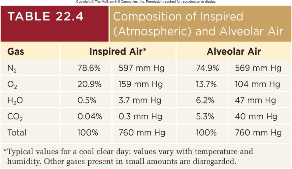
59 Gas Exchange and Transport composition of air 78.6 % nitrogen, 20.9% oxygen, 0.04% carbon dioxide, 0 4% water vapor depending on temperature and humidity, and minor gases argon, neon, helium, methane and ozone 22-59
60 Daltons Law The total atmospheric pressure is the sum of the contributions of the individual gases partial pressure the separate contribution of each gas in a mixture at sea level 1 atm. of pressure = 760 mmhg nitrogen constitutes 78.6% of the atmosphere, thus PN2 = 78.6% x 760 mm Hg = 597 mm Hg PO2 = 20.9% x 760 mm Hg = 159 mm Hg PH2O = 0.5% x 760 mm Hg = 3.7 mm Hg PCO2 = 0.04% x 760 mm Hg = 0.3 mm Hg PN2 + PO2 + PH2O + PCO2 = 760 mmhg
61 Composition of Inspired and Alveolar Air composition of inspired air and alveolar is different because of three influences: 1. air is humidifies by contact with mucous membranes 1. freshly inspired air mixes with residual air left from the previous respiratory cycle 1. alveolar PH2O is more than 10 times higher than inhaled air oxygen is diluted and it is enriched with CO2 alveolar air exchanges O2 and CO2 with the blood PO2 of alveolar air is about 65% that of inspired air PCO2 is more than 130 times higher 22-61
62
63 Alveolar Gas Exchange air in the alveolus is in contact with a film of water covering the alveolar epithelium for oxygen to get into the blood it must dissolve in this water pass through the respiratory membrane separating the air from the bloodstream for carbon dioxide to leave the blood it must pass the other way diffuse out of the water film into the alveolar air gases diffuse down their own concentration gradient until the partial pressure of each gas in the air is equal to its partial pressure in water 22-63
64 Alveolar Gas Exchange Henry s law at the air-water interface, for a given temperature, the amount of gas that dissolves in the water is determined by its solubility in water and its partial pressure in air the greater the PO2 in the alveolar air, the more O2 the blood picks up since blood arriving at an alveolus has a higher PCO2 than air, it releases CO2 into the air at the alveolus, the blood is said to unload CO2 and load O2 unload CO2 and load O2 involves erythrocytes efficiency depends on how long RBC stays in alveolar capillaries 0.25 sec necessary to reach equilibrium at rest, RBC spends 0.75 sec in alveolar capillaries strenuous exercise, 0.3, which is still adequate each gas in a mixture behaves independently one gas does not influence the diffusion of another 22-64
 Oxygen Air Air Time Blood Blood Figure 22.")
65 Alveolar Gas Exchange Copyright The McGraw-Hill Companies, Inc. Permission required for reproduction or display. Air Air Time Blood Blood Initial state Equilibrium state (a) Oxygen Air Air Time Blood Blood Figure Initial state Equilibrium state (b) Carbon dioxide
66 Factors Affecting Gas Exchange pressure gradient of the gases PO2 = 104 mm Hg in alveolar air versus 40 mm Hg in blood PCO2 = 46 mm Hg in blood arriving versus 40 mm Hg in alveolar air hyperbaric oxygen therapy treatment with oxygen at greater than one atm of pressure gradient difference is more, and more oxygen diffuses into the blood treat gangrene, carbon monoxide poisoning at high altitudes the partial pressures of all gases are lower gradient difference is less, and less oxygen diffuses into the blood solubility of the gases CO2 20 times as soluble as O2 equal amounts of O2 and CO2 are exchanged across the respiratory membrane because CO2 is much more soluble and diffuses more rapidly O2 is twice as soluble as N
67 Factors Affecting Gas Exchange membrane thickness - only 0.5 µm thick presents little obstacle to diffusion membrane surface area ml blood in alveolar capillaries, spread thinly over 70 m2 emphysema, lung cancer, and tuberculosis decrease surface area for gas exchange 22-67

68 Concentration Gradients of Gases Copyright The McGraw-Hill Companies, Inc. Permission required for reproduction or display. Expired air Inspired air PO2 116 mm Hg PCO2 32 mm Hg PO2 159 mm Hg PCO2 0.3 mm Hg Alveolar gas exchange Alveolar air O2 loading PO2 104 mm Hg CO2 unloading PCO2 40 mm Hg CO2 Gas transport O2 Pulmonary circuit O2 carried from alveoli to systemic tissues CO2 carried from systemic tissues to alveoli Deoxygenated blood Oxygenated blood PO2 40 mm Hg PCO2 46 mm Hg PO2 95 mm Hg PCO2 40 mm Hg Systemic circuit Systemic gas exchange CO2 O2 O2 unloading CO2 loading Tissue fluid PO2 40 mm Hg PCO2 46 mm Hg Figure
 p ee St ad gr t, d pi ra 40 Redu ced al g rad ient n io us 110 Nor m ff di 158 O2 Ambient PO2 (mm Hg) n ie Air at sea level (1 atm) and gradie nt, slo O d")
69 Ambient Pressure & Concentration Gradients 2,500 Copyright The McGraw-Hill Companies, Inc. Permission required for reproduction or display. Air in hyperbaric chamber (100% O2 at 3 atm) p ee St ad gr t, d pi ra 40 Redu ced al g rad ient n io us 110 Nor m ff di 158 O2 Ambient PO2 (mm Hg) n ie Air at sea level (1 atm) and gradie nt, slo O d 2 iffu sion wer O 2 diffu sion Air at 3,000 m (10,000 ft) Figure Atmosphere Venous blood arriving at alveoli Pressure gradient of O
 Normal Fluid and blood cells in alveoli Alveolar walls thickened by edema (b) Pneumonia Confluent alveoli Figure 22.")
70 Lung Disease Affects Gas Exchange Copyright The McGraw-Hill Companies, Inc. Permission required for reproduction or display. (a) Normal Fluid and blood cells in alveoli Alveolar walls thickened by edema (b) Pneumonia Confluent alveoli Figure (c) Emphysema 22-70

71 Perfusion Adjustments Copyright The McGraw-Hill Companies, Inc. Permission required for reproduction or display. Decreased airflow Reduced PO2 in blood vessels Response to reduced ventilation Result: Blood flow matches airflow Increased airflow Elevated PO2 in blood vessels Response to increased ventilation Vasodilation of pulmonary vessels Vasoconstriction of pulmonary vessels Decreased blood flow Increased blood flow (a) Perfusion adjusted to changes in ventilation Figure 22.22a 22-71

72 Ventilation Adjustments Copyright The McGraw-Hill Companies, Inc. Permission required for reproduction or display. Reduced PCO2 in alveoli Response to reduced perfusion Decreased blood flow Result: Airflow matches blood flow Increased blood flow Elevated PCO2 in alveoli Response to increased perfusion Constriction of bronchioles Dilation of bronchioles Decreased airflow Increased airflow (b) Ventilation adjusted to changes in perfusion Figure 22.22b 22-72
73 Gas Transport gas transport - the process of carrying gases from the alveoli to the systemic tissues and vise versa oxygen transport 98.5% bound to hemoglobin 1.5% dissolved in plasma carbon dioxide transport 70% as bicarbonate ion 23% bound to hemoglobin 7% dissolved in plasma 22-73
74 Oxygen Transport hemoglobin molecule specialized in oxygen transport four protein (globin) portions each with a heme group which binds one O2 to the ferrous ion (Fe2+) one hemoglobin molecule can carry up to 4 O2 oxyhemoglobin (HbO2) O2 bound to hemoglobin deoxyhemoglobin (HHb) hemoglobin with no O2 100 % saturation Hb with 4 oxygen molecules 50% saturation Hb with 2 oxygen molecules 22-74
75 Carbon Monoxide Poisoning carbon monoxide (CO) - competes for the O2 binding sites on the hemoglobin molecule colorless, odorless gas carboxyhemoglobin CO binds to ferrous ion of hemoglobin binds 210 times as tightly as oxygen non-smokers - less than 1.5% of hemoglobin occupied by CO smokers- 10% in heavy smokers atmospheric concentrations of 0.2% CO is quickly lethal 22-75
 in mm Hg 100")
76 Oxyhemoglobin Dissociation Curve Copyright The McGraw-Hill Companies, Inc. Permission required for reproduction or display. 20 O2 unloaded to systemic tissues ml O2 /dl of blood Percentage O2 saturation of hemoglobin Systemic tissues Partial pressure of O2 (PO2) in mm Hg 100 Alveoli Figure relationship between hemoglobin saturation and PO
77 Carbon Dioxide Transport 90% of CO2 is hydrated to form carbonic acid CO2 + H2O H2CO3 HCO3- + H+ then dissociates into bicarbonate and hydrogen ions 5% binds to the amino groups of plasma proteins and hemoglobin to form carbamino compounds chiefly carbaminohemoglobin (HbCO2) carbon dioxide does not compete with oxygen they bind to different moieties on the hemoglobin molecule hemoglobin can transport O2 and CO2 simultaneously 5% is carried in the blood as dissolved gas 22-77
78 Systemic Gas Exchange systemic gas exchange - the unloading of O2 and loading of CO2 at the systemic capillaries CO2 loading CO2 diffuses into the blood carbonic anhydrase in RBC catalyzes CO2 + H2O H2CO3 HCO3- + H+ chloride shift keeps reaction proceeding, exchanges HCO3- for Cl H+ binds to hemoglobin O2 unloading H+ binding to HbO2 reduces its affinity for O2 tends to make hemoglobin release oxygen HbO2 arrives at systemic capillaries 97% saturated, leaves 75% saturated venous reserve oxygen remaining in the blood after it passes through the capillary beds 22-78

79 Systemic Gas Exchange Copyright The McGraw-Hill Companies, Inc. Permission required for reproduction or display. Respiring tissue Capillary blood 7% CO2 Dissolved CO2 gas CO2 + plasma protein Carbamino compounds 23% CO2 HbCO2 CO2 + Hb 70% CO2 CO2 + H2O CAH H2CO3 Chloride shift Cl HCO3 + H+ 98.5% O2 O2 O2 + HHb 1.5% Dissolved O2 gas Figure HbO2+ H+ Key Hb Hemoglobin HbCO2 HbO2 HHb CAH Carbaminohemoglobin Oxyhemoglobin Deoxyhemoglobin Carbonic anhydrase 22-79
80 Alveolar Gas Exchange Revisited reactions that occur in the lungs are reverse of systemic gas exchange CO2 unloading as Hb loads O2 its affinity for H+ decreases, H+ dissociates from Hb and bind with HCO3 CO2 + H2O H2CO3 HCO3- + H+ reverse chloride shift HCO3- diffuses back into RBC in exchange for Cl-, free CO2 generated diffuses into alveolus to be exhaled

81 Alveolar Gas Exchange Copyright The McGraw-Hill Companies, Inc. Permission required for reproduction or display. Alveolar air CO2 Respiratory membrane 7% Capillary blood Dissolved CO2 gas CO2 + plasma protein Carbamino compounds 23% CO2 CO2 + Hb 70% CO2 CO2 + H2O CAH Chloride shift Cl HbCO2 H2 CO3 HCO3 + H+ 98.5% O2 + HHb O2 O2 1.5% Dissolved O2 gas Key Hb Figure HbO2 + H+ HbCO2 HbO2 HHb CAH Hemoglobin Carbaminohemoglobin Oxyhemoglobin Deoxyhemoglobin Carbonic anhydrase 22-81
82 Adjustment to the Metabolic Needs of Individual Tissues hemoglobin unloads O2 to match metabolic needs of different states of activity of the tissues ambient PO2 active tissue has PO2 ; O2 is released from Hb temperature active tissue has temp; promotes O2 unloading Bohr effect active tissue has CO2, which lowers ph of blood ; promoting O2 unloading bisphosphoglycerate (BPG) RBCs produce BPG which binds to Hb; O2 is unloaded Haldane effect rate of CO2 loading is also adjusted to varying needs of the tissues, low level of oxyhemoglobin enables the blood to transport more CO2 body temp (fever), thyroxine, growth hormone, testosterone, and epinephrine all raise BPG and cause O2 unloading» metabolic rate requires oxygen 22-82
 Effect of temperature 40 60 80 100 120 140 PO2 (mm Hg) Figure 22.26a 22-83")
83 Oxygen Dissociation and Temperature Percentage saturation of hemoglobin Copyright The McGraw-Hill Companies, Inc. Permission required for reproduction or display C 20 C C C Normal body temperature (a) Effect of temperature PO2 (mm Hg) Figure 22.26a 22-83
 70 60 ph 7.20 50 40 30 20 10 0 0 (b) Effect of ph 20 40 60 80 PO2 (mm Hg) 100 120 140 Figure 22.")
84 Oxygen Dissociation and ph Copyright The McGraw-Hill Companies, Inc. Permission required for reproduction or display. Percentage saturation of hemoglobin ph ph 7.40 (normal blood ph) ph (b) Effect of ph PO2 (mm Hg) Figure 22.26b Bohr effect: release of O2 in response to low ph 22-84
85 Blood Gases and the Respiratory Rhythm rate and depth of breathing adjust to maintain levels of: ph PCO2 40 mm Hg PO2 95 mm Hg brainstem respiratory centers receive input from central and peripheral chemoreceptors that monitor the composition of blood and CSF most potent stimulus for breathing is ph, followed by CO2, and least significant is O
86 Hydrogen Ions pulmonary ventilation is adjusted to maintain the ph of the brain central chemoreceptors in the medulla oblongata produce about 75% of the change in respiration induced by ph shift yet H+ does not cross the blood-brain barrier very easily CO2 does and in CSF reacts with water and produces carbonic acid dissociates into bicarbonate and hydrogen ions most H+ remains free and greatly stimulates the central chemoreceptors hydrogen ions are also a potent stimulus to the peripheral chemoreceptors which produce about 25% of the respiratory response to ph change 22-86
87 Effects of Hydrogen Ions respiratory acidosis and respiratory alkalosis ph imbalances resulting from a mismatch between the rate of pulmonary ventilation and the rate of CO2 production hyperventilation is a corrective homeostatic response to acidosis blowing off CO2 faster than the body produces it pushes reaction to the left CO2 (expired) + H2O H2CO3 HCO3- + H+ reduces H+ (reduces acid) raises blood ph towards normal 22-88
88 Effects of Hydrogen Ions hypoventilation is a corrective homeostatic response to alkalosis allows CO2 to accumulate in the body fluids faster than we exhale it shifts reaction to the right CO2 + H2O H2CO3 HCO3- + H+ raising the H+ concentration, lowering ph to normal ketoacidosis acidosis brought about by rapid fat oxidation releasing acidic ketone bodies (diabetes mellitus) induces Kussmaul respiration hyperventilation cannot remove ketone bodies, but blowing off CO2, it reduces the CO2 concentration and compensates for the ketone bodies to some degree 22-89
89 Carbon Dioxide indirect effects on respiration through ph as seen previously direct effects CO2 at beginning of exercise may directly stimulate peripheral chemoreceptors and trigger ventilation more quickly than central chemoreceptors 22-90
90 Effects of Oxygen PO2 usually has little effect on respiration chronic hypoxemia, PO2 less than 60 mm Hg, can significantly stimulate ventilation hypoxic drive respiration driven more by low PO2 than by CO2 or ph emphysema, pneumonia high elevations after several days 22-91
91 Chronic Obstructive Pulmonary Disease COPD refers to any disorder in which there is a long-term obstruction of airflow and a substantial reduction in pulmonary ventilation major COPDs are chronic bronchitis and emphysema usually associated with smoking other risk factors include air pollution or occupational exposure to airborne irritants 22-95
92 Chronic Obstructive Pulmonary Disease chronic bronchitis inflammation and hyperplasia of the bronchial mucosa cilia immobilized and reduced in number goblet cells enlarge and produce excess mucus develop chronic cough to bring up extra mucus with less cilia to move it sputum formed (mucus and cellular debris) ideal growth media for bacteria incapacitates alveolar macrophages leads to chronic infection and bronchial inflammation 22-96
93 Chronic Obstructive Pulmonary Disease emphysema alveolar walls break down lung has larger but fewer alveoli much less respiratory membrane for gas exchange lungs fibrotic and less elastic healthy lungs are like a sponge; in emphysema, lungs are more like a rigid balloon air passages collapse obstructs outflow of air air trapped in lungs weaken thoracic muscles spend three to four times the amount of energy just to breathe 22-97
94 Smoking and Lung Cancer lung cancer accounts for more deaths than any other form of cancer most important cause is smoking (15 carcinogens) squamous-cell carcinoma (most common) transformation of bronchial epithelium dividing cells invade bronchial wall, cause bleeding lesions dense swirls of keratin replace functional respiratory tissue 22-99
95 Lung Cancer adenocarcinoma originates in mucous glands small-cell (oat cell) carcinoma least common, most dangerous named for clusters of cells that resemble oat grains originates in primary bronchi, metastasizes quickly to other organs
96 Progression of Lung Cancer 90% originate in primary bronchi tumor invades bronchial wall, compresses airway; may cause atelectasis often first sign is coughing up blood metastasis is rapid; usually occurs by time of diagnosis common sites: pericardium, heart, bones, liver, lymph nodes and brain prognosis poor after diagnosis only 7% of patients survive 5 years
 Healthy lung, mediastinal surface (b) Smoker's lung with carcinoma a: The McGraw-Hill Companies/Dennis Strete, photographer; b: Biophoto")
97 Effect of Smoking Copyright The McGraw-Hill Companies, Inc. Permission required for reproduction or display. Tumors (a) Healthy lung, mediastinal surface (b) Smoker's lung with carcinoma a: The McGraw-Hill Companies/Dennis Strete, photographer; b: Biophoto Associates/Photo Researchers, Inc. Figure a-b
UNIT 9 - RESPIRATORY SYSTEM LECTURE NOTES
 UNIT 9 - RESPIRATORY SYSTEM LECTURE NOTES 9.01 GENERAL FUNCTIONS OF THE RESPIRATORY SYSTEM A. Brings oxygenated air to the alveoli B. Removes air containing carbon dioxide C. Filters, warms, and humidifies
Chapter 16 Respiratory System
 Introduction Chapter 16 Respiratory System The respiratory system consists of tubes that filter incoming air and transport it to alveoli where gases are exchanged. Think pair share: what organs are associated
Chapter 15. Lecture and Animation Outline
 Chapter 15 Lecture and Animation Outline To run the animations you must be in Slideshow View. Use the buttons on the animation to play, pause, and turn audio/text on or off. Please Note: Once you have
Chapter 23. Gas Exchange and Transportation
 Chapter 23 Gas Exchange and Transportation What is air? Mixture of gasses 78.6 % nitrogen 20.9% oxygen 0.04% carbon dioxide 0 4% water vapor depending on temperature and humidity and minor gases argon,
(Slide 1) Lecture Notes: Respiratory System
 Lecture Notes: Respiratory System") (Slide 1) Lecture Notes: Respiratory System I. (Slide 2) The Respiratory Tract A) Major structures and regions of the respiratory Tract/Route INTO body 1) nose 2) nasal cavity 3) pharynx 4) glottis 5)
(Slide 1) Lecture Notes: Respiratory System I. (Slide 2) The Respiratory Tract A) Major structures and regions of the respiratory Tract/Route INTO body 1) nose 2) nasal cavity 3) pharynx 4) glottis 5)
Respiratory System Physiology. Dr. Vedat Evren
 Respiratory System Physiology Dr. Vedat Evren Respiration Processes involved in oxygen transport from the atmosphere to the body tissues and the release and transportation of carbon dioxide produced in
Chapter 23. Gas Exchange and Transportation
 Chapter 23 Gas Exchange and Transportation What is air? Mixture of gasses 78.6 % nitrogen 20.9% oxygen 0.04% carbon dioxide 0 4% water vapor depending on temperature and humidity other minor gases argon,
Chapter 17 The Respiratory System: Gas Exchange and Regulation of Breathing
 Chapter 17 The Respiratory System: Gas Exchange and Regulation of Breathing Overview of Pulmonary Circulation o Diffusion of Gases o Exchange of Oxygen and Carbon Dioxide o Transport of Gases in the Blood
Outline - Respiratory System. Function of the respiratory system Parts of the respiratory system Mechanics of breathing Regulation of breathing
 Respiratory system Function Outline - Respiratory System I. II. III. IV. Respiratory System The function of the respiratory system is to bring in oxygen to the body and remove carbon dioxide. Function
Human Biology Respiratory System
 Human Biology Respiratory System Respiratory System Responsible for process of breathing Works in cooperation with Circulatory system Three types: 1. Internal Respiration 2. External Respiration 3. Cellular
82 Respiratory Tract NOTES
 82 Respiratory Tract NOTES RESPIRATORY TRACT The respiratory tract conducts air to the lungs where gaseous exchange occurs. It is separated into air-conducting and respiratory (where gas exchange occurs)
Physiology of Respiration
 Physiology of Respiration External Respiration = pulmonary ventilation breathing involves 2 processes: inspiration expiration Inspiration an active process involves contraction of diaphragm innervated
2/28/18. Respiratory System. 1 Copyright 2016 by Elsevier Inc. All rights reserved. Introduction. Anatomy. Physiology. Respiratory System
 Introduction Respiratory System Chapter 28 Respiration: We inhale air, extract oxygen from it, exhale air Cardiovascular and respiratory systems work together Failure of either system: - Disruption of
B. Lining - epithelium
 RESPIRATORY SYSTEM Function supply body with oxygen and remove carbon dioxide. This process is called respiration, can be broken down into 4 components. I. Pulmonary Ventilation or breathing movement of
Chapter 22 The Respiratory System
 Chapter 22 The Respiratory System 1 Respiration Pulmonary ventilation (breathing): movement of air into and out of the lungs External respiration: O 2 and CO 2 exchange between the lungs and the blood
Chapter 23: Respiratory System
 Chapter 23: Respiratory System I. Functions of the Respiratory System A. List and describe the five major functions of the respiratory system: 1. 2. 3. 4. 5. II. Anatomy and Histology of the Respiratory
Chapter 13 The Respiratory System
 Chapter 13 The Respiratory System by Dr. Jay M. Templin Brooks/Cole - Thomson Learning Atmosphere Tissue cell External respiration Alveoli of lungs 1 Ventilation or gas exchange between the atmosphere
Chapter 11: Respiratory System Review Assignment
 Name: Date: Mark: / 45 Chapter 11: Respiratory System Review Assignment Multiple Choice = 45 Marks Identify the choice that best completes the statement or answers the question. 1. Which of the following
Respiratory System. Part 2
 Respiratory System Part 2 Respiration Exchange of gases between air and body cells Three steps 1. Ventilation 2. External respiration 3. Internal respiration Ventilation Pulmonary ventilation consists
Respiratory Pulmonary Ventilation
 Respiratory Pulmonary Ventilation Pulmonary Ventilation Pulmonary ventilation is the act of breathing and the first step in the respiratory process. Pulmonary ventilation brings in air with a new supply
Breathing: The normal rate is about 14 to 20 times a minute. Taking in of air is called Inspiration and the forcing out of air is called Expiration.
 Biology 12 Respiration Divisions of Respiration Breathing: entrance and exit of air into and out of the lungs External Respiration: exchange of gases(o2 and CO2) between air (in alveoli) and blood Internal
Physiology Unit 4 RESPIRATORY PHYSIOLOGY
 Physiology Unit 4 RESPIRATORY PHYSIOLOGY In Physiology Today Respiration External respiration ventilation gas exchange Internal respiration cellular respiration gas exchange Respiratory Cycle Inspiration
1. Label a diagram of the respiratory system. Objective sheet 3 Notes
 1. Label a diagram of the respiratory system Objective sheet 3 Notes 2. Functions of the respiratory structures Name Description Function Nasal Cavity Trachea Bronchi (Singular Bronchus) Bronchioles Lungs
The Respiratory System
 Essentials of Human Anatomy & Physiology Elaine N. Marieb Seventh Edition Chapter 13 The Respiratory System Slides 13.1 13.30 Lecture Slides in PowerPoint by Jerry L. Cook Organs of the Respiratory system
Structures of the Respiratory System include:
 Respiratory System Structures of the Respiratory System include: ü Oral Cavity ü Nasal Cavity ü Pharynx ü Epiglottis ü Larynx ü Trachea ü Diaphragm ü Lung ü Bronchus ü Bronchioles ü Alveolus ü Pulmonary
Introduction. Respiration. Chapter 10. Objectives. Objectives. The Respiratory System
 Introduction Respiration Chapter 10 The Respiratory System Provides a means of gas exchange between the environment and the body Plays a role in the regulation of acidbase balance during exercise Objectives
Lesson 9.1: The Importance of an Organ Delivery System
 Lesson 9.1: The Importance of an Organ Delivery System Animals require a continuous supply of oxygen (O 2 ) for cellular respiration, and they must expel carbon dioxide (CO 2 ), the waste product of this
Respiration - Human 1
 Respiration - Human 1 At the end of the lectures on respiration you should be able to, 1. Describe events in the respiratory processes 2. Discuss the mechanism of lung ventilation in human 3. Discuss the
Respiratory system. Premedical - Biology
 Respiratory system Premedical - Biology Composition of atmosphere Nitrogen (N 2 ) 840 ppmv (78.084%) Oxygen (O 2 ) 209,460 ppmv (20.946%) Carbon dioxide (CO 2 ) 387 ppmv (0.0387%) Argon (Ar), Neon (Ne),
Respiratory System Review
 KEY THIS TEST WILL BE COMPLETED IN ONE CLASS PERIOD MONDAY, MARCH 10. 2014 Respiratory System Review Name A. Directions: Fill in the blank with the appropriate vocabulary word or words (several examples
Respiratory System -Training Handout
 Respiratory System -Training Handout Karen L. Lancour National Rules Committee Chairman Life Science FUNCTIONS: Provides oxygen to the blood stream and removes carbon dioxide Enables sound production or
Human Anatomy & Physiology
 Human Anatomy & Physiology 19 Karen Webb Smith Unit Five Respiratory System URLs http://yucky.kids.discovery.com/flash/body/pg000138.html http://www.stemnet.nf.ca/~dpower/resp/exchange.htm #Breathing http://www.emc.maricopa.edu/faculty/farabee/
GAS EXCHANGE & PHYSIOLOGY
 GAS EXCHANGE & PHYSIOLOGY Atmospheric Pressure Intra-Alveolar Pressure Inspiration 760 mm HG at Sea Level (= 1 atm) Pressure due to gases (N2, O2, CO2, Misc.) Pressure inside the alveolus (air sac) Phrenic
Respiratory System. 1. muscular tube lined by mucous membrane 2. throat 3. nasopharynx, oropharynx, laryngopharynx
 I. Functions of the Respiratory System A. gas exchange B. prevent dehydration C. sound D. olfaction E. ph regulation II. Anatomy of the Respiratory System A. Nose 1. external nares vestibule nasal cavity
The Respiratory System. Medical Terminology
 The Respiratory System Medical Terminology The respiratory system is where gas exchange occurs via respiration; inhalation/exhalation. pick up oxygen from inhaled air expels carbon dioxide and water sinus
Be sure you understand these four functions of the respiratory system before you begin this lab.
 Biology 212: Human Anatomy and Physiology II ************************************************************************************************************* RESPIRATORY ANATOMY & VENTILATION *************************************************************************************************************
The Respiratory System
 PowerPoint Lecture Slides prepared by Meg Flemming Austin Community College C H A P T E R 15 The Respiratory System Chapter 15 Learning Outcomes 15-1 15-2 Describe the primary functions of the respiratory
I. Gas Exchange Respiratory Surfaces Respiratory Surface:
 I. Gas Exchange Respiratory Surfaces Respiratory Surface: Common characteristics of respiratory surfaces: a) Moist: allows for the RAPID diffusion of dissolved gasses across its surface. Whereas the respiratory
BIOLOGY 12: UNIT J - CHAPTER 15 - REVIEW WORKSHEET RESPIRATORY SYSTEM
 BIOLOGY 12: UNIT J - CHAPTER 15 - REVIEW WORKSHEET RESPIRATORY SYSTEM A. CHAPTER REVIEW 1. Define the four components of respiration. 2. What happens to the air as it moves along the air passages? What
Respiration. The ins and outs
 Respiration The ins and outs Functions 1. To bring O 2 into the body and transfer it to the blood stream 2. To remove CO 2 Circulation and respiration work together to achieve these functions Why Do We
 Question 1: Define vital capacity. What is its significance? Vital capacity is the maximum volume of air that can be exhaled after a maximum inspiration. It is about 3.5 4.5 litres in the human body. It
Respiration. The resspiratory system
 Respiration The resspiratory system The Alveoli The lungs have about 300 million alveoli, with a total crosssec onal area of 50 70 m2.. Each alveolar sac is surrounded by blood capillaries. The walls of
GASEOUS EXCHANGE 17 JULY 2013
 GASEOUS EXCHANGE 17 JULY 2013 Lesson Description In this lesson we: Discuss what is gaseous exchange? Consider requirements of an efficient gaseous exchange surface. Look at diversity in gas exchange systems.
4/18/12 MECHANISM OF RESPIRATION. Every Breath You Take. Fun Facts
 Objectives MECHANISM OF RESPIRATION Dr Badri Paudel Explain how the intrapulmonary and intrapleural pressures vary during ventilation and relate these pressure changes to Boyle s law. Define the terms
Chapter 13 The Respiratory System
 VI edit Pag 451-499 Chapter 13 The Respiratory System V edit. Pag 459-509 Tissue cell Alveoli of lungs Atmosphere 1 External respiration Ventilation or gas exchange between the atmosphere and air sacs
25/4/2016. Physiology #01 Respiratory system Nayef Garaibeh Rawan Alwaten
 25/4/2016 Physiology #01 Respiratory system Nayef Garaibeh Rawan Alwaten Respiratory System Introduction: - We breath while we are sleeping, talking, working and resting. - Respiratory diseases are abundant
CHAPTER 17 BREATHING AND EXCHANGE OF GASES
 268 BIOLOGY CHAPTER 17 BREATHING AND EXCHANGE OF GASES 17.1 Respiratory Organs 17.2 Mechanism of Breathing 17.3 Exchange of Gases 17.4 Transport of Gases 17.5 Regulation of Respiration 17.6 Disorders of
GASEOUS EXCHANGE IN HUMANS 06 AUGUST 2014
 GASEOUS EXCHANGE IN HUMANS 06 AUGUST 2014 In this lesson we: Lesson Description Look at gaseous exchange in humans in terms of o Ventilation o Inspiration o Expiration o Transport of gases o Homeostatic
1 CHAPTER 17 BREATHING AND EXCHANGE OF GASES https://biologyaipmt.com/
 1 CHAPTER 17 BREATHING AND EXCHANGE OF GASES https://biologyaipmt.com/ CHAPTER 17 BREATHING AND EXCHANGE OF GASES Oxygen (O2) is utilised by the organisms to indirectly break down nutrient molecules like
Animal Systems: The Respiratory System
 Animal Systems: The Respiratory System Tissues, Organs, and Systems of Living Things Cells, Cell Division, and Animal Systems and Plant Systems Cell Specialization Human Systems The Digestive The Circulatory
Then the partial pressure of oxygen is x 760 = 160 mm Hg
 1 AP Biology March 2008 Respiration Chapter 42 Gas exchange occurs across specialized respiratory surfaces. 1) Gas exchange: the uptake of molecular oxygen (O2) from the environment and the discharge of
PARTS AND STRUCTURE OF THE RESPIRATORY SYSTEM
 PARTS AND STRUCTURE OF THE RESPIRATORY SYSTEM Parts of the Respiratory System The RS can be divided into two parts: 1. Respiratory Tract, (path that air follows). Nasal passage Pharynx Larynx Trachea Bronchi,
Respiration. Chapter 39
 Respiration Chapter 39 Impacts, Issues Up in Smoke Smoking immobilizes ciliated cells and kills white blood cells that defend the respiratory system; highly addictive nicotine discourages quitting 39.1
P215 Respiratory System, Part 2
 P15 Respiratory System, Part Gas Exchange Oxygen and Carbon Dioxide constant need for oxygen constant production of carbon dioxide exchange (and movement) lung alveoli pulmonary arteries pulmonary capillaries
Gas Exchange Respiratory Systems
 alveoli gills Gas Exchange Respiratory Systems elephant seals 2008-2009 Why do we need a respiratory system? respiration for respiration Need O 2 in for aerobic cellular respiration make ATP Need CO 2
Gaseous exchange. Grade 11
 z Gaseous exchange Grade 11 z Terminology 1. Breathing 2. Gaseous exchange 3. Diffusion 4. Spongy mesophyll cells 5. Tracheae 6. Gills 7. Alveoli 8. Larynx 9. Diaphragm 10. Endothelium 1. Pleura 2. Squamous
BIOLOGY 12 - RESPIRATION - CHAPTER NOTES
 BIOLOGY 12 - RESPIRATION - CHAPTER NOTES We often think of respiration as just breathing. In fact, breathing is just one part of this physiological process. As biologists, we divide respiration up into
AP Biology. Gas Exchange Respiratory Systems. Gas exchange. Why do we need a respiratory system? Optimizing gas exchange. Gas exchange in many forms
 alveoli Gas Exchange Respiratory Systems gills elephant seals 2008-2009 Why do we need a respiratory system? Need O 2 in food respiration for respiration for aerobic cellular respiration make ATP Need
LECTURE NOTES ANATOMY & PHYSIOLOGY II (A. IMHOLTZ) RESPIRATORY P1 OF 8
 RESPIRATORY P1 OF 8") LECTURE NOTES ANATOMY & PHYSIOLOGY II (A. IMHOLTZ) RESPIRATORY P1 OF 8 I. The main functions of the respiratory system include: a. Exchange of O 2 and CO 2. b. Voice production. c. Regulation of plasma
LECTURE NOTES ANATOMY & PHYSIOLOGY II (A. IMHOLTZ) RESPIRATORY P1 OF 8 I. The main functions of the respiratory system include: a. Exchange of O 2 and CO 2. b. Voice production. c. Regulation of plasma
Assignments for Life Processes(Respiration)
") Assignments for Life Processes(Respiration) 1 Question 1 Why do organisms need food? Organisms need food for obtaining energy to perform the vital functions. Question 2 What is a respiratory substrate?
Assignments for Life Processes(Respiration) 1 Question 1 Why do organisms need food? Organisms need food for obtaining energy to perform the vital functions. Question 2 What is a respiratory substrate?
alveoli Chapter 42. Gas Exchange elephant seals gills AP Biology
 alveoli Chapter 42. Gas Exchange gills elephant seals Gas exchange O 2 & CO 2 exchange exchange between environment & cells provides O 2 for aerobic cellular respiration need moist membrane need high
CHAPTER 3: The respiratory system
 CHAPTER 3: The respiratory system Practice questions - text book pages 56-58 1) When the inspiratory muscles contract, which one of the following statements is true? a. the size of the thoracic cavity
system. and then into the tissues. Diffusion of wastes such as Carbon Dioxide from tissues into blood and out of blood into the lungs.
 Respiratory System 1.Exchange Why do of we gases breathe? into the Think blood of all and the tissues. reasons Diffusion why we of Oxygen need a respiratory into blood from the lungs system. and then into
Respiratory Lecture Test Questions Set 3
 Respiratory Lecture Test Questions Set 3 1. The pressure of a gas: a. is inversely proportional to its volume b. is unaffected by temperature changes c. is directly proportional to its volume d. does not
RESPIRATORY SYSTEM OF RABBIT:
 RESPIRATION The term respiration means exchange of gases (particularly oxygen and carbon dioxide) between an organism and its medium. Physiologically, the term respiration may be defined as a biochemical
2. State the volume of air remaining in the lungs after a normal breathing.
 CLASS XI BIOLOGY Breathing And Exchange of Gases 1. Define vital capacity. What is its significance? Answer: Vital Capacity (VC): The maximum volume of air a person can breathe in after a forced expiration.
Chapter 23: The Respiratory System
 Chapter 23: The Respiratory System I. The Respiratory System: An Introduction, p. 814 Objectives: 1. Describe the primary functions of the respiratory system. 2. Explain how the delicate respiratory exchange
AP Biology. Chapter 42. Gas Exchange. Optimizing gas exchange. Gas exchange. Gas exchange in many forms. Evolution of gas exchange structures
 alveoli Chapter 42. Gas Exchange gills elephant seals Gas exchange & C exchange exchange between environment & cells provides for aerobic cellular respiration need moist membrane need high surface area
Chapter 23 Respiratory System
 Chapter 23 Respiratory System I. Functions of the Respiratory System: 1. Gas Exchange 2. Regulatory (blood ph) 3. Voice Production 4. Olfaction 5. Protection II. Anatomy & Histology of the Respiratory
Using the figure above, match the following: 4. Tidal volume. 5. Inspiratory reserve volume. 6. Residual volume. 7. Expiratory reserve volume.
 Respiratory Physiology Practice Test BIOL 4350: Hamilton Using the figure above, match the following: 1. Primary bronchus 2. Larynx 3. Trachea Using the figure above, match the following: 4. Tidal volume.
Respiratory Physiology Gaseous Exchange
 Respiratory Physiology Gaseous Exchange Session Objectives. What you will cover Basic anatomy of the lung including airways Breathing movements Lung volumes and capacities Compliance and Resistance in
Lab 17. The Respiratory System. Laboratory Objectives
 Lab 17 The Respiratory System Laboratory Objectives Identify and describe the anatomical structures of the respiratory system. Describe the relationship between volume and pressure. Describe changes in
Alveolus and Respiratory Membrane
 Alveolus and Respiratory Membrane thin membrane where gas exchange occurs in the lungs, simple squamous epithelium (Squamous cells have the appearance of thin, flat plates. They fit closely together in
Respiratory system. Role. Ventilation consists of 4 (5) steps : oxygen delivery and carbon dioxide elimination ph balance sound and voice formation
 steps : oxygen delivery and carbon dioxide elimination ph balance sound and voice formation") Respiratory system Role oxygen delivery and carbon dioxide elimination ph balance sound and voice formation Ventilation consists of 4 (5) steps : 1. pulmonary ventilation gas exchange between lungs and
Respiratory system Role oxygen delivery and carbon dioxide elimination ph balance sound and voice formation Ventilation consists of 4 (5) steps : 1. pulmonary ventilation gas exchange between lungs and
Respiratory System 1
 Respiratory System 1 Outline Respiratory structures Gills Air-Breathing Animals Amphibians and Reptiles Mammals Birds Structures and Mechanisms of Breathing 2 Copyright The McGraw-Hill Companies, Inc.
Monday, ! Today: Respiratory system! 5/20/14! Transport of Blood! What we ve been covering! Circulatory system! Parts of blood! Heart! tubing!
 Monday, 5.19.14! What we ve been covering! Circulatory system! Parts of blood! Heart! tubing! Transport of Blood! What is transported! Nutrients! Oxygen! Carbon Dioxide! Hormones! Antibodies! What it is/does!
Collin County Community College. Lung Physiology
 Collin County Community College BIOL. 2402 Anatomy & Physiology WEEK 9 Respiratory System 1 Lung Physiology Factors affecting Ventillation 1. Airway resistance Flow = Δ P / R Most resistance is encountered
2.1.1 List the principal structures of the
 physiology 2.1.1 List the principal structures of the The principle structures of the respiratory are: Nose/Mouth used for inhalation of oxygen-rich air and expelling carbon dioxide rich air Pharynx -
Respiratory System Anatomy. Chapter 23
 Chapter 23 The Respiratory System Structurally, the respiratory system is divided into upper and lower divisions or tracts. The upper respiratory tract consists of the nose, pharynx and associated structures.
Gas exchange. Tissue cells CO2 CO 2 O 2. Pulmonary capillary. Tissue capillaries
 Gas exchange Pulmonary gas exchange Tissue gas exchange CO 2 O 2 O 2 Tissue cells CO2 CO 2 Pulmonary capillary O 2 O 2 CO 2 Tissue capillaries Physical principles of gas exchange Diffusion: continuous
2) During exhalation Air is cooled due to condensation and loses its moisture, depositing it on lining in trachea and nose
 During exhalation Air is cooled due to condensation and loses its moisture, depositing it on lining in trachea and nose") Section 10: The Respiratory System A. Functions of the respiratory system: The organs of the respiratory system make sure oxygen enters the body and carbon dioxide leaves the body. 2 stages: Inhalation
Section 10: The Respiratory System A. Functions of the respiratory system: The organs of the respiratory system make sure oxygen enters the body and carbon dioxide leaves the body. 2 stages: Inhalation
Respiratory System. Prepared by: Dorota Marczuk-Krynicka, MD, PhD
 Respiratory System Prepared by: Dorota Marczuk-Krynicka, MD, PhD Lungs: Ventilation Perfusion Gas Exchange - Diffusion 1. Airways and Airway Resistance (AWR) 2. Mechanics of Breathing and Lung (Elastic)
To supply the body with oxygen and dispose of carbon dioxide Respiration four distinct processes must happen
 Respiratory System Consists of the respiratory and conducting zones Respiratory zone Site of gas exchange Consists of bronchioles, alveolar ducts, and alveoli Conducting zone Provides rigid conduits for
Chapter 16 Respiration. Respiration. Steps in Respiration. Functions of the respiratory system
 Chapter 16 Respiration Functions of the respiratory system Respiration The term respiration includes 3 separate functions: Ventilation: Breathing. Gas exchange: Occurs between air and blood in the lungs.
3/24/2009 LAB D.HAMMOUDI.MD. 1. Trachea 2. Thoracic wall 3. Lungs 4. Primary bronchi 5. Diaphragm
 RESPIRATORY PHYSIOLOGY LAB D.HAMMOUDI.MD 1. Trachea 2. Thoracic wall 3. Lungs 4. Primary bronchi 5. Diaphragm 1 KEY WORDS TO KNOW BOYLE S LAW INTERCOSTAL NERVES PHRENIC NERVE DIAPHRAGM EXTERNAL INTERCOSTAL
Ch. 12: Respiratory Physiology
 Ch. 12: Respiratory Physiology Objectives: 1. Review respiratory anatomy. 2. Understand mechanics of breathing, gas pressure vocabulary, and the principles of surface tension, compliance, and recoil. 3.
Topic 13: Gas Exchange Ch. 42. Gas Exchange pp Gas Exchange. Gas Exchange in Fish pp Gas Exchange in Fish
 Topic 13: Gas Exchange Ch. 42 Fig. 42.24 Gas Exchange pp.979-989 Gas exchange involves the uptake of oxygen and the discharge of carbon dioxide (i.e. respiration or breathing). It is necessary for cellular
Respiratory system & exercise. Dr. Rehab F Gwada
 Respiratory system & exercise Dr. Rehab F Gwada Objectives of lecture Outline the major anatomical components & important functions of the respiratory system. Describe the mechanics of ventilation. List
Respiratory Anatomy and Physiology. Respiratory Anatomy. Function of the Respiratory System
 Respiratory Anatomy and Physiology Michaela Dixon Clinical Development Nurse PICU BRHFC Respiratory Anatomy Function of the Respiratory System - In conjunction with the cardiovascular system, to supply
Respiration (revised 2006) Pulmonary Mechanics
 Pulmonary Mechanics") Respiration (revised 2006) Pulmonary Mechanics PUL 1. Diagram how pleural pressure, alveolar pressure, airflow, and lung volume change during a normal quiet breathing cycle. Identify on the figure the
Respiration (revised 2006) Pulmonary Mechanics PUL 1. Diagram how pleural pressure, alveolar pressure, airflow, and lung volume change during a normal quiet breathing cycle. Identify on the figure the
Module Two. Objectives: Objectives cont. Objectives cont. Objectives cont.
 Transition to the New National EMS Education Standards: EMT-B B to EMT Module Two Objectives: Upon completion, each participant will do the following to a degree of accuracy that meets the Ntl EMS Education
Respiratory Physiology. Adeyomoye O.I
 Respiratory Physiology By Adeyomoye O.I Outline Introduction Hypoxia Dyspnea Control of breathing Ventilation/perfusion ratios Respiratory/barometric changes in exercise Intra-pulmonary & intra-pleural
Unit II Problem 4 Physiology: Diffusion of Gases and Pulmonary Circulation
 Unit II Problem 4 Physiology: Diffusion of Gases and Pulmonary Circulation - Physical principles of gases: Pressure of a gas is caused by the movement of its molecules against a surface (more concentration
Glucose + Oxygen Carbon Dioxide + Water + Energy C6H12O CO2 + 6H20 + energy
 Cell Respiration - A food oxidization process in body cells to produce energy - Occurs (happens) in animal cells and plant cells Notes : Metabolic Reactions in Gaseous Exchange CELL RESPIRATION Photosynthesis
Then the partial pressure of oxygen is. b) Gases will diffuse down a pressure gradient across a respiratory surface if it is: i) permeable ii) moist
 Gases will diffuse down a pressure gradient across a respiratory surface if it is: i) permeable ii) moist") 1 AP Biology March 2008 Respiration Chapter 42 Gas exchange occurs across specialized respiratory surfaces. 1) Gas exchange: Relies on the diffusion of gases down pressure gradients. At sea level, atmosphere
1 AP Biology March 2008 Respiration Chapter 42 Gas exchange occurs across specialized respiratory surfaces. 1) Gas exchange: Relies on the diffusion of gases down pressure gradients. At sea level, atmosphere
Chapter 42 Part III The Respiratory System
 Biology 120 J. Greg Doheny Chapter 42 Part III The Respiratory System Notes: In this section we will discuss the breathing system, also known as the respiratory system. This should not be confused with
respiratory cycle. point in the volumes: 500 milliliters. for men. expiration, up to 1200 milliliters extra makes breathing Respiratory
 10 II. RESPIRATORY VOLUMES, CAPACITIES & PULMONARY FUNCTION TESTS Respiratory volume is the term used for various volumes of air moved by or associated with the lungs at a given point in the respiratory
Ch 16: Respiratory System
 Ch 16: Respiratory System SLOs: Explain how intrapulmonary pressures change during breathing Explain surface tension and the role of surfactant in respiratory physiology. Compare and contrast compliance
CHAPTER 3: The cardio-respiratory system
 : The cardio-respiratory system Exam style questions - text book pages 44-45 1) Describe the structures involved in gaseous exchange in the lungs and explain how gaseous exchange occurs within this tissue.
RESPIRATION III SEMESTER BOTANY MODULE II
 III SEMESTER BOTANY MODULE II RESPIRATION Lung Capacities and Volumes Tidal volume (TV) air that moves into and out of the lungs with each breath (approximately 500 ml) Inspiratory reserve volume (IRV)