of Western Pacific Fish (Gobiidae, Xenisthminae), with Discussions of Gobioid Osteology and Classification
|
|
|
- Sheryl Sanders
- 5 years ago
- Views:
Transcription


1 Tyson belos, New Genus and Species of Western Pacific Fish (Gobiidae, Xenisthminae), with Discussions of Gobioid Osteology and Classification VICTOR G. SPRINGER SMITHSONIAN CONTRIBUTIONS TO ZOOLOGY NUMBER 390


2 SERIES PUBLICATIONS OF THE SMITHSONIAN INSTITUTION Emphasis upon publication as a means of "diffusing knowledge" was expressed by the first Secretary of the Smithsonian. In his formal plan for the Institution, Joseph Henry outlined a program that included the following statement: "It is proposed to publish a series of reports, giving an account of the new discoveries in science, and of the changes made from year to year in all branches of knowledge." This theme of basic research has been adhered to through the years by thousands of titles issued in series publications under the Smithsonian imprint, commencing with Smithsonian Contributions to Knowledge in 1848 and continuing with the following active series: Smithsonian Contributions to Anthropo/ogy Smithsonian Contributions to Astrophysics Smithsonian Contributions to Botany Smithsonian Contributions to the Earth Sciences Smithsonian Contributions to Paleobiology Smithsonian Contributions to Zoology Smithsonian Studies in Air and Space Smithsonian Studies in History and Technology In these series, the Institution publishes small papers and full-scale monographs that report the research and collections of its various museums and bureaux or of professional colleagues in the world cf science and scholarship. The publications are distributed by mailing lists to libraries, universities, and similar institutions throughout the world. Papers or monographs submitted for series publication are received by the Smithsonian Institution Press, subject to its own review for format and style, only through departments of the various Smithsonian museums or bureaux, where the manuscripts are given substantive review. Press requirements for manuscript and art preparation are outlined on the inside back cover. S. Dillon Ripley Secretary Smithsonian Institution
3 SMITHSONIAN CONTRIBUTIONS TO ZOOLOGY NUMBER 390 Tyson belos, New Genus and Species of Western Pacific Fish (Gobiidae, Xenisthminae), with Discussions of Gobioid Osteology and Classification Victor G. Springer SMITHSONIAN INSTITUTION PRESS City of Washington 1983

4 ABSTRACT Springer, Victor G. Tyson belos, New Genus and Species of Western Pacific Fish (Gobiidae, Xenisthminae), with Discussions of Gobioid Osteology and Classification. Smithsonian Contributions to Zoology, number 390, 40 pages, 19 figures, Tyson belos is described, based on specimens from the Great Barrier Reef, Trobriand Islands, and Lau Islands. This diminutive (gravid female 18.8 mm SL), coral-reef species differs conspicuously from all other gobioids in having the following combination of characters: pelvic fins separate, each comprising a single segmented ray and no spine; teeth on vomer; anterior (spinous) dorsal fin lacking. Tyson appears to be closely related to Xenisthmus Snyder, Allomicrodesmus Schultz, and an undescribed genus (and species, reference D.F. Hoese). These genera share at least one synapomorphy within the Gobioidei: ventral lip with free ventral margin extending across dentary symphysis (margin interrupted across symphysis in other gobioids). The subfamily Xenisthminae is recognized on this single character. There are, however, osteological specializations that are known only for Tyson and Xenisthmus and that are predicted to occur in the other two xenisthmine genera: premaxillary ascending process greatly reduced or absent (if present, lower than anterior maxillary articulating process of premaxilla); ascending processes replaced in position and function by a rostral bone (ossified rostral cartilage); basibranchials 2 to 4 absent. A rostral bone occurs in a few other gobioid genera, but in these the ascending premaxillary processes are well developed, basibranchials 2 to 4 are present, and not all the species in each genus where the rostral bone occurs have a rostral bone, indicating the probability that this bone is a homoplasy in those non-xenisthmine genera where it occurs. The osteologies of Tyson and Xenisthmus are described and illustrated. Synapomorphies are provided for the Gobioidei, Rhyacichthyidae, and Gobiidae, and the Rhyacichthyidae is proposed as the sister-group of all other gobioids. OFFICIAL PUBLICATION DATE is handstamped in a limited number of initial copies and is recorded in the Institution's annual report, Smithsonian Year. SERIES COVER DESIGN: The coral Montastrea cavemosa (Linnaeus). Library of Congress Cataloging in Publication Data Springer, Victor Gruschka, Tyson belos, new genus and species of Western Pacific fish (Gobiidae, Xenisthminae) (Smithsonian contributions to zoology ; no. 390) Bibliography: p. Supt. of Docs. no. : SI 1.27: Tyson belos Classification. 2. Gobiidae Classification. 3. Bones. 4. Fishes Classification. 5. Fishes South Pacific Ocean Classification. I. Title. II. Series QL.S54 no. 390 [QL638.G7] 591s [597'.58]
5 Contents Page Introduction 1 Methods 1 Materials 2 Acknowledgments 3 Family GOBIIDAE 4 Subfamily XENISTHMINAE 4 Tyson, new genus 5 Tyson belos, new species 6 Osteology of Xenisthmus and Tyson 7 Cranium 7 Jaws, Suspensorium, Superficial Bones of Head 12 Hyoid Arch 17 Branchial Apparatus 18 Pectoral and Pelvic Fins and Girdles 21 Vertebrae and Unpaired Fins 24 Characters and Classification of the Gobioidei 29 Notes 36 Literature Cited 39 in
6

7 Tyson belos, New Genus and Species of Western Pacific Fish (Gobiidae, Xenisthminae), with Discussions of Gobioid Osteology and Classification Victor G. Springer Introduction In 1975, Tyson R. Roberts collected a single specimen of a fish from a coral reef in the Trobriand Islands, off eastern New Guinea. He recognized that it was distinctly different from any of which he had knowledge, but he was unable to assign it to a family. He asked my opinion of the specimen's affinities. Although the external anatomy of the specimen did not allow one to assign it with confidence to any infraordinal taxon of fishes, I had the impression that the specimen was a gobioid. To confirm this impression would have required detailed information on the osteology of the specimen, and I was unwilling to sacrifice it for this purpose. Although Roberts generously offered to permit me to describe the species (and genus), I delayed description in the hope that additional specimens would become available. A second specimen, from the Great Barrier Reef, was obtained in 1981 by the Australian Museum, and I then made an osteological preparation of the first specimen. Shortly afterwards, in 1982, I collected two more specimens, from Fiji. It is now possible to confirm my impressions of the gobioid Victor G. Springer, Department of Vertebrate Zoology, National Museum of Natural History, Smithsonian Institution, Washington, D.C affinities of the specimens. In addition, a suggestion by Douglass Hoese that the closest relationships of the new species (and genus) might be among those genera considered to be marine eleotridids (Larson and Hoese, 1980) opened up a fertile field of investigation into the familygroup taxa of gobioids. I soon found myself immersed in the infamous and unwieldy morass of gobioid systematics, which was not my intention, nor did it fit my research priorities. I decided, therefore, to limit my study and present my findings to date. In this study I describe the new genus and species, including its osteology and that of its sister genus {Xenisthmus Snyder), and assert the probable monophyly of the gobioid subfamily Xenisthminae, which contains only the new genus, Xenisthmus, another undescribed genus (reference D.F. Hoese, specimens unavailable to me), and Allomicrodesmus Schultz. I also discuss certain osteological characters used or useful in establishing the monophyly of the suborder Gobioidei and some included infrasubordinal groups. METHODS. Specimens (one each) of Xenisthmus clarus and Tyson belos and much of the other gobioid skeletal material used in this study were stained for cartilage with alcian blue, cleared with trypsin, and stained for bone with alizarin 1

8 SMITHSONIAN CONTRIBUTIONS TO ZOOLOGY red-s. When I discuss cartilaginous or bony structures, I mean to imply only that the structures stained blue or red (pink), respectively. Some structures, notably fin-ray elements and portions of some skeletal tissues that otherwise stained blue, accepted little or no stain, and most are treated here as if they are bone (these unstained structures accept red stain in other gobioids). On the illustrations, blue-stained structures are indicated by a pattern of hatching; nonstained or red-stained structures are not differentiated; stippling is used sparingly to indicate depth or contours, and in Figure 12 a uniform fine screen was used to indicate depth (otherwise this screen was used for background contrast). I prepared all the base drawings of the bones, the final renderings of which were executed by P.K. Hollingsworth. The snout region of a large number of whole specimens of gobioids, including Xenisthmus and Tyson and particularly genera usually considered to be eleotridids (because they have 6 branchiostegals and separate pelvic fins), was examined to determine if the ventral lip had a complete free ventral margin, and the snout was partially dissected to determine whether premaxillary ascending processes or a rostral bone were present. The osteologies of a number of gobioids have been discussed and illustrated in the literature (not all cited in the present study). I referred to much of this literature for relevant information. Where I encountered apparent discrepancies I attempt to rectify the differences in my discussions. (Birdsong, 1975, also encountered questionable osteological characters in the literature, and I do not, in general, treat those errors that he corrected.) Established procedures for making segmented dorsal- and anal-fin ray counts on gobioids call for enumerating the posteriormost two dorsal- or anal-fin segmented rays as a single ray. In contrast to the other segmented rays, which have a oneto-one relationship with pterygiophores in these fins, each of these ray pairs is supported by a single pterygiophore, and the two rays of each pair are closely approximate. The posterior member of each pair is often greatly reduced in size; nevertheless, the structure of each element of each pair is that of a typical segmented ray. In my study I enumerate each element separately. In referring to the commonly recognized family-group based on Eleotris Bloch, I use the familylevel adjective "eleotridid." I do not recognize a family group based on Eleotris, however, but the adjective is useful when referring to earlier studies that do or when referring to gobioids with the following combination of characters: separate pelvic fins, 6 branchiostegals, fewer than 3 epurals, no rostral bone, free margin of ventral lip not continuous across dentary symphysis. (All these characters, except the number of epurals, are plesiomorphic for gobioids; the number of epurals is plesiomorphic for all non-rhyacichthyid gobioids.) The following institutional abbreviations are used herein: AMNH AMS CAS GCRL USNM American Museum of Natural History, New York Australian Museum, Sydney (catalog numbers begin with I or IA) California Academy of Sciences, San Francisco Gulf Coast Research Laboratory, Ocean Springs, Mississippi United States National Museum, the collections of which are housed in the National Museum of Natural History, Smithsonian Institution, Washington, D.C. (fish specimens are in the Division of Fishes) MATERIALS. The osteological descriptions of Xenisthmus and Tyson are each based on a single specimen (counterstained for bone and cartilage): Xenisthmus clarus (Jordan and Seale), USNM , female, 23.6 mm SL (Figure 1); and Tyson belos, USNM , female paratype, 18.5 mm SL (Figure 2). Most details of the skeletal anatomy of X. clarus were compared for consistency with a counterstained specimen of a different (apparently undescribed) species of Xenisthmus: USNM , female, 25.3 mm SL. Differences exhibited by the comparative specimen are reported at appropriate points in the osteological descriptions (one of the prepublication reviewers of this study, D.F. Hoese, checked parts of the osteological description of Xenisthmus against a specimen of X. polyzonatus (Klunzinger)).
![NUMBER 390 FIGURE 1. Xenisthmus clams (from Snyder, 1982, pi. 68: fig. 3, Xenisthmus proriger [= X. clarus]).](/docs-images/93/112434074/images/9-1.jpg "Cleared and stained specimens (* = alizarin only; ** counterstained for cartilage) of other gobioids examined for this study include: Awaous tajasica (Lichtenstein), USNM 213491*; Butis amboinensis")
, GCRL V71:6019*; Eleotris amblyopsis (Cope), USNM 226200**; Eviota species, USNM 224539**; Gobiomorus dormitor Lacepede, USNM 79070** USNM Bleeker, Schultz, Gobiosoma")
9 NUMBER 390 FIGURE 1. Xenisthmus clams (from Snyder, 1982, pi. 68: fig. 3, Xenisthmus proriger [= X. clarus]). Cleared and stained specimens (* = alizarin only; ** counterstained for cartilage) of other gobioids examined for this study include: Awaous tajasica (Lichtenstein), USNM *; Butis amboinensis (Bleeker), USNM **; Callogobius species, USNM uncataloged (formerly CAS GVF )**; Calumia godeffroyi (Gunther), USNM **; Cerdale ionthas Jordan and Gilbert, GCRL V71:6563*; Clarkichthys bilineatus (Clark), GCRL V71:6019*; Eleotris amblyopsis (Cope), USNM **; Eviota species, USNM **; Gobiomorus dormitor Lacepede, USNM 79070** USNM Bleeker, Schultz, Gobiosoma homochroma (Ginsburg), *; Gunnellichthys pleurotaenia GCRL V82:19587*; Kraemeria bryani USNM *; K. species, AMS * Microdesmus dipus Gunther, GCRL V69:361O*; M. longipinnis (Weymouth), GCRL V70:4613*; Mogurnda mogurnda (Richardson), USNM **; Paragunnellichthys seychellensis Dawson, GCRL V82:19583*; Parioglossus taeniatus Regan, USNM ** and AMS *; Rhyacichthys aspro (Valenciennes), AMNH (complete)** and USNM (only gill arches and left jaws, suspensorium, and superficial head bones)**; Valenciennea strigata (Broussonett), USNM uncataloged*. A complete listing of whole specimens of gobioids examined and partially dissected was not maintained, but those examined included a large number of genera and species, among which were the following: Bostrychus sinensis Lacepede, USNM 90325; Brachyamblyopus urolepsis (Bleeker), USNM ; Bunaka canarensis (Day), USNM ; Dormitator maculatus (Bloch), USNM ; Erotelis species, USNM ; Gobiomorphus hubbsi (Stokell), USNM ; Gobiotrichonotus radiocularis Fowler, USNM ; Hetereleotris nebulofasciatus (J.L.B. Smith), USNM uncataloged (station no. VGS 69-28); H. vuigare (Klunzinger), USNM uncataloged (station no. VGS 69-23); H. zonatus (Fowler), USNM ; Leptophilypnus Jluviatilis Meek and Hildebrand, USNM ; Micropercops daybri Fowler and Bean, USNM 83982; Ophiocara porocephala (Valenciennes), USNM ; Oxyeleotris marmorata (Bleeker), USNM ; Percottus glehni Dybowski, USNM 77008; Periophthalmus species, USNM ; Rhyacichthys aspro, USNM ; Taenioides limicola C.L. Smith, USNM ; Tridentiger obscurus (Temminck and Schlegel), USNM I maintain a large and varied collection of cleared and stained specimens of teleost (mostly perciform) fishes that I use to evaluate skeletal characters. Many of these specimens have been listed in earlier studies of mine, and many of these specimens, as well as new additions to the collection, were referred to during the course of the present study. ACKNOWLEDGMENTS. Appreciation for the loan of specimens is extended to the following: C.E. Dawson (GCRL), D.F. Hoese (AMS), and D.E. Rosen (AMNH). My colleagues at the Smithsonian Institution, E.A. Lachner and S. Jewett, permitted me to examine skeletal material that they had prepared for use in their own
, E. Murdy (Texas A&M University, College Station, Texas), D.E. Rosen, and S.H. Weitzman. W.F.")
10 SMITHSONIAN CONTRIBUTIONS TO ZOOLOGY studies. Helpful discussions (some by mail) were carried on during the course of the work with R.S. Birdsong (Old Dominion University, Norfolk, Virginia), D.F. Hoese, G.D. Johnson (Charleston, South Carolina), E. Murdy (Texas A&M University, College Station, Texas), D.E. Rosen, and S.H. Weitzman. W.F. Smith-Vaniz (Academy of Natural Sciences, Philadelphia) provided information on skeletal characters of several species of Opistognathidae. The manuscript was reviewed critically and substantially improved by suggestions from R.S. Birdsong, R. Winterbottom (Royal Ontario Museum, Ottawa), and D.F. Hoese. Grants from the Smithsonian Scholarly Studies Program and the Max and Victoria Dreyfus Foundation supported field work in Fiji that resulted in obtaining important material of the new genus and species. Family GOBIIDAE DIAGNOSIS. Gobioid fishes (see "Characters and Classification of the Gobioidei," page 29) with the following synapomorphies: fewer than 3 epurals (3 only in variant specimens); no lateral line on body; no mandibular sensory canal; no more than 3 rows of ctenii on scales (only marginal row may be tooth-like); dorsal end of interhyal articulating with hyomandibula, widely separated from symplectic; large space separating shank of symplectic from preopercle. Subfamily XENISTHMINAE DIAGNOSIS. Gobiid fishes with free ventral margin of ventral lip extending without interruption across dentary symphysis; premaxillary ascending process greatly reduced or absent (process, if present, lower than anterior maxillary articulating process of premaxilla; Figures 6 and 8b); rostral bone (ossified rostral cartilage, Figure 7) present, completely or almost completely replacing premaxillary ascending processes in position and function; and lacking: basibranchials 2 to 4, hypobranchials 3 (which may be represented vestigially as cartilaginous fragments), pterophenoids, and coronomeckelian bones. All of the above characters, except the absence of the pterosphenoids, appear to be synapomorphies of the Xenisthminae; however, the osteology is unknown for 2 of the 4 genera that I include in the Xenisthminae (based on the presence of an uninterrupted ventral-lip margin; see "Composition" below). The pterosphenoids are absent (homoplasiously?) in some microdesmin gobiids, for instance Clarkichthys bilineatus, but present in others, for instance Paragunnellichthys seychellensis. In the gobiin Parioglossus taeniatus, the pterosphenoids are represented on each side only by tiny fragments of bone and cartilage; the other species of Parioglossus examined had moderately well-developed pterosphenoids. It is conceivable that the pterosphenoids are absent in other specimens or species of Parioglossus. A rostral bone is present in Cerdale ionthas and Paragunnellichthys seychellensis, but absent in Microdesmus dipus, Clarkichthys bilineatus, and Gunnellichthys pleurotaenia (all microdesmins). The rostral bone is also present in Parioglossus taeniatus (but not in the other species of Parioglossus examined), and in a species of Kraemeria (but not in K. bryani). In all these taxa the ascending premaxillary processes are well developed, and all have basibranchials 2 to 4, hypobranchials 3, and coronomeckelian bones. The possibility exists, nevertheless, that some of these taxa have a sister-group relationship with the Xenisthminae. The following xenisthmin characters also occur in various non-xenisthmin gobioids: 6 branchiostegals, no postcleithra, no mesopterygoids, pelvic fins not united. COMPOSITION. Xenisthmus Snyder and Tyson, new genus, and probably an undescribed genus and species (based on characters furnished by D.F. Hoese, in litt., 1983), and Allomicrodesmus Schultz (monotypic). These four genera possess the uninterrupted free ventral-lip margin. REMARKS. Xenisthmus was first used as the basis of a family-group taxon by Miller (1973:426), who proposed the subfamily Xenisthminae in the Gobiidae to include Xenisthmus
 questioned Miller's logic in establishing the taxon; with no more information available than that offered by Miller, I would have had to agree with Birdsong's doubts.")
.")
11 NUMBER 390 alone. None of Miller's diagnostic characters for the subfamily are synapomorphies. Birdsong (1975) questioned Miller's logic in establishing the taxon; with no more information available than that offered by Miller, I would have had to agree with Birdsong's doubts. My reaffirmation of the Xenisthminae stems from the number and extreme distinctiveness of the specializations exhibited by Xenisthmus and Tyson. In recognizing the Xenisthminae as a subfamily of the Gobiidae, I am following essentially the gobioid classification scheme given by Miller (1973). Miller recognized only two families in the Gobioidei: Rhyacichthyidae and Gobiidae. Families, such as Microdesmidae, Eleotrididae, Gobiodidae, and Kraemeriidae, 1 were either relegated to subfamily status under the Gobiidae or were not recognized. I do not wish to comment on Miller's scheme except to say that I believe that several of his subfamily groups are probably paraphyletic or polyphyletic. In particular, I believe this to be true of the Eleotrididae, which is usually considered to be the primitive sister group of all non-rhyacichthyid gobioids. My main reason for this belief is that there is no synapomorphy that defines the eleotridids. Schultz (1966), based on inaccurately described characters, initially placed Allomicrodesmus in the Microdesmidae. Dawson (1974:409) gave a corrected description of the holotype (and only known specimen) and properly excluded the genus from the Microdesmidae. Dawson was unable to assign Allomicrodesmus to a family, but Larson and Hoese (1980) assigned it to the Eleotrididae. Allomicrodesmus dorotheae Schultz is known only from the holotype (USNM ), from the Marshall Islands, and a specimen from the Great Barrier Reef (AMS ). I have examined the latter specimen and find it to be an adult male, 20.0 mm SL (measured from upper-jaw tip; lower jaw protrudes). It is very delicate and slender (greatest depth 1.3 mm). The dorsal fin is single and continuous (the first 2 elements are spines, followed by 32 simple, segmented rays). The anal fin comprises 25 simple elements, the first of which is probably a segmented ray, but I was unable to satisfy myself of this. There are 3 simple rays in each pelvic fin (I cannot determine if a spine is also present) and the fins are closely approximate at their bases, but not connected by membrane. There are 6 branchiostegals on each side. The gill membranes form a continuous free fold across the isthmus. Tyson, new genus DIAGNOSIS. A minute fish (less than 20 mm SL) of the subfamily Xenisthminae, lacking: scales, extrascapular bones, lacrimal, gill rakers, an anterior spinous dorsal fin, exoccipital condyles (and, consequently, lateral condyles on the atlas), an uncinate process on epibranchial 1, an interarcual cartilage (Travers, 1981), and infrapharyngobranchials 2 and 4 (including toothplate); and having: vertebrae, an ossified basibranchial 1, teeth on vomer, fused anterior and posterior ceratohyals, and a single, simple, segmented ray (and no spine) in each pelvic fin (the pelvic-fin formula, 1, alone, is adequate to distinguish Tyson from all other gobioids). Other characters, including many restricted to Tyson in the Xenisthminae, are those of the single included species, Tyson belos, new species (typespecies by original designation and monotypy). ETYMOLOGY. The generic name is derived from the forename of Tyson R. Roberts, who collected the first specimen and recognized its distinctiveness. Gender, masculine. The stem for formation of family-group and higher level taxa is "tyson." COMPARISONS. Until more information is available on the other genera of the Xenisthminae, I consider Tyson to be most closely related to Xenisthmus. In contrast to Tyson, Xenisthmus has scales, extrascapular bones, a lacrimal, gill rakers, an anterior spinous dorsal fin, exoccipital condyles, an uncinate process on epibranchial 1, an interarcual cartilage, and infrapharyngobranchials 2 and 4 (toothplate), only 10 precaudal vertebrae, a cartilaginous basibranchial 1, an edentate vomer, autogenous anterior and posterior ceratohyals, and pelvic fins 1,5. In addition, ex-
 and segmented pelvic-fin rays (to 3")
. Characters of the holotype are indicated by an asterisk. HOLOTYPE. AMS 1.")
: top, left lateral view; middle, enlarged ventral view of anus and urogenital papilla")
12 eluding the anteriormost anal-fin pterygiophore, Xenisthmus has only one dorsal- or anal-fin pterygiophore inserted in each interneural or interhemal space, whereas Tyson has 2 or 3. It is possible, however, that Tyson is most closely related to Allomicrodesmus, which, similar to Tyson, has greatly reduced the number of dorsal-fin spines (to 2) and segmented pelvic-fin rays (to 3 in each fin). The undescribed xenisthmine genus is unique among gobioids in having rows of palatine teeth (D.F. Hoese, in litt.). SMITHSONIAN CONTRIBUTIONS TO ZOOLOGY Tyson belos, new species FIGURE 2 This new species is so distinctive that only a diagnostic description is given here. The description is supplemented by the generic diagnosis presented above and the osteological description (p. 7). Characters of the holotype are indicated by an asterisk. HOLOTYPE. AMS ; 19.0 ±0.5 mm SL; sex unknown (immature); Australia, Queens- FIGURE 2. Tyson belos, USNM , paratype, female, 18.8 mm SL, Kiriwina Island, Trobriand Islands, Papua-New Guinea (specimen now cleared, stained, and dissected): top, left lateral view; middle, enlarged ventral view of anus and urogenital papilla (drawings by J.R. Schroeder); bottom,rightlateral view (photograph by J.F. McKinney).

13 NUMBER 390 land, Escape Reef, outer barrier, coral bottom, depth 29 m; coll. 29 Oct 1981 by G. Allen, W. Starck, and T. Ayling. PARATYPES. USNM ,18.8 mm SL, ripe female, Papua-New Guinea, Trobriand Islands, west side Kiriwina Island, coral reef, coll. 19 Sep 1975 by T.R. Roberts; specimen now cleared, stained, and dissected. USNM , 12.9 and 16.2 mm SL, sexes unknown, Fiji, Lau Islands, Ono-ilau, outside barrier reef on northwest side of island, depth 13.7 to ~16.8 m, coll. 1 May 1982 by V.G. Springer et al. DESCRIPTION. Dorsal fin 1,9 to 1,10* (1,8 to 1,9* if last two rays are counted as one; see Figure 18). Anal fin 1,9 to 1,10* (1,8 to 1,9* if last two rays are counted as one). Pectoral fins 17*, 18, or 21. Pelvic fins not joined, each consisting of a single, unbranched, segmented ray (no spine). Caudal fin emarginate, with 8* or 9 dorsal procurrent rays; 3 to 5* simple, segmented dorsal rays; 4* or 5 branched, segmented dorsal rays; 3* or 4 branched, segmented ventral rays; 3 to 5* simple, segmented ventral rays; and 9* ventral procurrent rays, for a total of 33 or 34* elements, of which 15 or 17* are segmented, and of these 7* to 9 are branched. Vertebrae 13* + 13* = 26*. Branchiostegals 6*. Scales, gill rakers, sensory pores (except anterior and posterior nostrils) lacking. Anterior nostril borne on short tube; tube, when depressed, reaches to upper lip; posterior nostril a simple pore just posterior to base of tube of anterior nostril. Gill openings of holotype and largest paratype extend anteriorly to below level of middle of orbit; in smallest paratypes, to below level of posterior third of orbit. Color in alcohol: ground color of body pale with few patches of melanophores. Holotype and smallest and largest paratypes each have patch of melanophores on midside of caudal peduncle. Holotype and largest paratype have patches of melanophores posteriorly on dorsal and anal fins. In holotype, dorsal- and anal-fin melanophore patches are extensive, intensive, and occupy approximately same relative area as anal-fin melanophore patch of largest paratype. Dorsal-fin melanophore patch of largest paratype much reduced in area; patch completely absent in two smallest paratypes. A color transparency photograph (in the USNM collections) taken of the freshly collected holotype is noteworthy mainly for the specimen's exhibiting an irregularly shaped, pinkish spot posteroventrally on the lateral surface of the head, and for a pale rust-colored perfusion about the melanophores on the caudal peduncle. Similar rust-colored areas are also present on the ventrolateral portions of the head and abdomen. REMARKS. Although there are only four specimens of T. belos available, it appears that there may be geographic variation in fin-ray counts (asterisks indicate counts of holotype). Decision as to whether the differences noted are significant or the result of biased sampling will have to await the availability of more specimens. Locality Trobriand Islands Great Barrier Reef Lau Islands Dorsal fin Anal fin Pectoral fins (Uft/right) 1,10 1,10* 1,9 1,9 1,10 1,10* 1,9 1,9 21/21 17/17* 18/18 18/17 ETYMOLOGY. The specific name belos (arrow), here used as a noun in apposition, is from the Greek and refers to the arrow-like shape of the species. Osteology of Xenisthmus and Tyson In the following descriptions, each portion of the skeleton is described first for Xenisthmus and then for Tyson. CRANIUM Xenisthmus (Figures 3 and 6): All the cranial bones are autogenous, with the possible exception of the pterosphenoids, which are either fused with the sphenotics or absent. The vomer (edentate) and median and lateral ethmoids are well ossified. The lateral ethmoids are separated medially by the ethmoid cartilage, which fills the anterior

: top, dorsal view; middle, lateral view; bottom, ventral view (medial aspect of interhyal")
14 SMITHSONIAN CONTRIBUTIONS TO ZOOLOGY LATERAL EXTRASCAPULAE POSTTEMPORAL EPIOCCIPITAL EPIOCCIPITAL- 1 INTERCALAR PHENonc- PTER0TIC BAUDELOT'S LIGAMENT POSTTEMPORAL FIGURE 3. Xenisthmus darns, cranium with some associated bones and ligaments (diagonal hatching represents cartilage): top, dorsal view; middle, lateral view; bottom, ventral view (medial aspect of interhyal illustrated; see also Figure 9).

15 NUMBER 390 interorbital region. Each lateral ethmoid bears a strong, cartilaginously tipped, laterally projecting process, to which its respective lacrimal attaches, and each is pierced by a foramen for the olfactory tract. The frontals are two to three times as long as broad, and they arefirmly joined to the ethmoids. Each frontal bears a groove, which is perforated by three minute foramina (not visible in Figure 3), in the supraorbital region, along which the supraorbital canal courses. The groove (and canal) continues along the dorsolateral surfaces of the sphenotic and pterotic. In the postorbital region, each frontal bears a descending wing, which joins its respective sphenotic and prootic, and a parasphenoid ascending wing. There are no parietals. The epioccipitals are joined along their dorsomedian margins (not visible on external surface of skull) below the supraoccipital, which bears a slight dorsomedian ridge posteriorly. There is no indication of cartilage (blue stain) in the line of junction between the epioccipitals. A flattened area on the posterolateral dorsal surface of each epioccipital serves as the area of attachment for the dorsal arm of its respective posttemporal. The exoccipitals each bear a lateral condyle that articulates with the lateral condyle on its respective side of the atlas vertebra. The articulating surfaces of the exoccipital condyles are inclined ventroposteriorly. Each exoccipital is pierced by two foramina: the more anterior is the vagal foramen; the more posterior is probably for passage of a spinal nerve. Each exoccipital is excluded from meeting its respective prootic by an interosseous space (subtemporal fossa of Birdsong, 1975) that is bounded by the prootic, pterotic, exoccipital, and basioccipital. Each interosseous space is covered by a large intercalar, which broadly overlaps the four bones surrounding the space. A ligament extending from the ventral arm of each posttemporal attaches to the posterodorsal surface of its respective intercalar close to the junction of the intercalar with the exoccipital. The exoccipitals form the dorsal and lateral walls of the foramen magnum, which is floored by the basioccipital. The basioccipital articulates with the parasphenoid anteriorly, the centrum of the atlas posteriorly, the exoccipitals dorsally, and the prootics dorsoanteriorly (most of each basioccipital-prootic joint is obscured from view by the overlying intercalar). Each Baudelot's ligament originates on its respective side from a minute process on the posterior midlateral surface of the basioccipital, extends posterolaterad, passing freely through a deep notch in the dorsal end of the cleithrum, and inserts on the posterointernal surface of its respective supracleithrum. Each prootic articulates with the parasphenoid along the prootic's ventral and anterior margins; with its respective frontal, sphenotic, and pterotic along the prootic's dorsal margin; with its respective intercalar along the prootic's posterior surface; and with the basioccipital along the prootic's posteroventral margin. Each prootic forms only a minute part (ventral rim) of the large sphenotic socket on its respective side that articulates with the dorsoanterior condyle of its respective hyomandibula. (There is a relatively small socket, for reception of the posterodorsal condyle of each hyomandibula, in the anterolateral region of its respective pterotic.) Two large foramina, for passage of the trigemino-facial nerve complex, pierce the wall of each prootic. On each side, ventral to the posterior of these two foramina, is the internal carotid foramen, formed by a notch in the margin of the parasphenoid. Just anterolaterad of each of the internal carotid foramina, there is attached to the parasphenoid, a long, cord-like ligament that extends ventrolaterally and attaches to a process on the medial surface of the respective interhyal (Figure 3). The prootics lack internal shelves that might form a roof for the posterior myodome. There is, instead, a shallow, anteriorly opening pocket with a thin roof arising from the middorsal surface of the parasphenoid just posterior to the level of the anterior margins of the parasphenoid ascending processes. According to Birdsong (1975: 148, fig. 6B) the medial, inferior, and superior rectus muscles attach in this cavity. The otoliths were not present, possibly having been lost during preservation and osteological
: Anteriorly there is a single, almost laminar, bone, which appears to comprise fused lateral and median ethmoids, which also appear to be fused to the dorsoanterior surface of the")
16 10 SMITHSONIAN CONTRIBUTIONS TO ZOOLOGY processing of the specimen. Otoliths are present in the comparative skull, but I did not attempt to extract them. Tyson (Figure 4): Anteriorly there is a single, almost laminar, bone, which appears to comprise fused lateral and median ethmoids, which also appear to be fused to the dorsoanterior surface of the vomer. There is visible on the ventral surface of the cranium, however, a separation of the vomer from the fused ethmoid complex and the parasphenoid. The separation of the dorsoventrally flattened shank of the vomer from the parasphenoid is also visible in a lateral view of the cranium, but no joint can be seen between the vomer and ethmoid complex when the cranium is viewed from in front. An opening for passage of the olfactory tract pierces the complex ethmoid bone on either side. The vomer bears a single marginal row of nine teeth, comprising four large, recurved canines on each side, separated by a greatly reduced median tooth. These canines are clearly evident when the mouth of an intact specimen is open, and they form the posterior portion of a tooth patch in common with the anterior teeth of the premaxillae. The anterior ends of the long (over four times longer than broad), paired frontals overlap the ethmoid bone posteriorly. Beginning ventral to the junction of the frontals and ethmoid complex, and extending anteriorly, is the mass of the ethmoid cartilage, which is unstained anteriorly. The frontals, which bear no sensory canals, extend posteriorly for most of the dorsal surface of the skull, overlapping the anterior end of the supraoccipital, the dorsomedial margins of the fused sphenotic-pterotic complexes, and the anterior ends of the epioccipitals; there are no parietals. Each sphenotic-pterotic complex appears to be a single ossification, with no indication of a line of fusion. Even so, I treat the anterior portion bearing the facet for articulation with the dorsoanterior condyle of the hyomandibula as sphenotic, and the remainder, which bears the facet for articulation with the dorsoposterior condyle of the hyomandibula, as pterotic. The separation of the pterotic portion from the dorsoposterior margin of the prootic is also unclear, but is marked by a narrow unstained area. I have illustrated (Figure 4) this unstained area as a joint line that is continuous with the joint line between the exoccipital and basioccipital. On each side of the cranium, in the posteromedial portion of the orbital region, there is a prominent ventrally extending process that is continuous with the sphenotic. Each process represents either a flange of the sphenotic or a fused pterosphenoid (see discussion of prootic, p. 12). Although the anterior margin of the supraoccipital is reasonably discernible through the transparent frontals, the remaining extent of the supraoccipital and its joints with the epioccipitals are obscured by a granular-appearing area, possibly representing the line of fusion of the supraoccipital with the epioccipitals. (The posterior margin of the supraoccipital as delineated in Figure 4 is even less certain than indicated.) The presumed posteriormost portion of the supraoccipital bears a very short, low, median ridge, which continues posteriorly as the slightly raised joint of the posterodorsally conjoined epioccipitals and the dorsally conjoined exoccipitals. The relatively small epioccipitals each bear a flattened area dorsally, to which their respective posttemporal is attached. I was unable to determine if the epioccipitals are conjoined synchondrally beneath the supraoccipital (Birdsong, 1975, table 1). The exoccipitals are most noteworthy for lacking condyles for articulation with the atlas (which, consequently, lacks lateral condyles and articulates only with the basioccipital). Each exoccipital is excluded from making contact with its respective prootic by a meeting of the pterotic and basioccipital. The exoccipitals are each pierced by a large vagal foramen; no other foramina are present in these bones. The ligament extending from the posterior end of each posttemporal attaches tightly to a minute process on the midanterolateral surface of its respective exoccipital just posterior to the joint between the exoccipital and pterotic and just below the level of the laterally widest expansion of the pterotic. There are no intercalars. The exoccipitals form the lat-
PTEROSPHENOID SPHENOTIC + PTEROTIC- FIGURE 4.")
;")
17 NUMBER SPHENOTIC + PTEROTIC SUPRAOCCIPITAL MEDIAN + LATERAL ETHMOIDS SPHENOTIC OR (?)PTEROSPHENOID SPHENOTIC + PTEROTIC- FIGURE 4. Tyson belos, cranium (diagonal hatching represents cartilage): top, dorsal view; middle, lateral view (U = unstained portion of ethmoid cartilage); bottom, ventral view.

18 12 SMITHSONIAN CONTRIBUTIONS TO ZOOLOGY eral and dorsal walls of the foramen magnum, which is floored by the basioccipital. The broadly Y-shaped basioccipital articulates with the atlas posteriorly and accepts the posterior end of the parasphenoid between its anteriorly extending arms, each of which articulates anteriorly with the posterior ends of its respective prootic. Each Baudelot's ligament has a short bifurcation at its proximal end. The anterior branch of the bifurcation attaches to the posterolateral surface of the basioccipital and the posterior branch attaches to a closely adjacent area on the anterolateral margin of the atlas vertebra. The ligament extends posterolaterally and passes freely through a notch in the dorsal process of the cleithrum, then proceeds anteriorly and inserts on the dorsomedial surface of the supracleithrum. Possibly, the bifurcation of Baudelot's ligament and its attachment to both the basioccipital and atlas, rather than only to the basioccipital (as in Xenisthmus), is an adaptation for strengthening the connection between the skull and vertebral column, which in other gobioids is shared by three skull bones rather than one. Each prootic articulates with the parasphenoid along the prootic's ventral margin, with its respective fused sphenotic-pterotic along the prootic's dorsal margin, and impinges slightly (at most) on the ventrolateral margin of the ventrally extending process of the sphenotic in the postorbital region. Each ventrally extending process appears to form the incomplete anterior border of an opening (anterior passageway for the trigemino-facial nerve complex?) whose posterior border is a notch on the anterior margin of the prootic. In most gobioids with pterosphenoids, each pterosphenoid forms the anterior margin, and the prootic the posterior margin, of the anterior foramen for passage of the trigemino-facial nerve complex. Inasmuch as both foramina for passage of the trigemino-facial nerve complex are contained only in the prootic of Xenisthmnus, which lacks pterosphenoids, it would appear that the ventrally extending sphenoid processes of Tyson represent fused pterosphenoids. Each prootic contains a single complete foramen for passage of the trigemino-facial nerve complex. The prootics do not bear a shelf on their medial surface, and there is no bony roof over the posterior myodome. The parasphenoid is perforated by a pair of carotid foramina near the anterior end of its posterior broad portion, and bears a low, median, anteriorly directed hook-like process on the dorsal, interorbital surface of its slender anterior shank, which overlaps the shank of the vomer. There is no bony pocket for reception of the rectus muscles of the eyes in the dorsal surface of the parasphenoid within the cranial vault, as there is in Xenisthmus. The otoliths were not present, possibly having been lost during preservation or osteological processing. JAWS, SUSPENSORIUM, SUPERFICIAL BONES OF HEAD Xenisthmus (Figures 5 and 6; for comparison with the snout region of a relatively unspecialized gobioid see Figure 7): The premaxillae are welldeveloped bones, each bearing an outer row of large recurved, conical teeth and an inner row of similar but much smaller teeth (in the comparative specimen, the outer row is only slightly larger than the inner teeth, which are scattered over the ventral surface of the premaxillae, but nowhere in more than three rows). Each premaxilla has anterior (articular) and posterior maxillary processes, but lacks an ascending process. The portion of the premaxilla that normally bears an ascending process is reduced to a flattened facet that articulates with a corresponding ventrolateral facet at the end of the median, unpaired, rostral bone, which has functionally replaced the ascending processes of both premaxillae. In the comparative specimen, the ascending processes are represented by facet-bearing knobs, which are, however, lower than the anterior articular processes. The rostral bone, which has a cartilaginous dorsal end, is an ossified rostral cartilage. The rostral bone bears thin anterodorsal and posteroventral flanges on each side. A band-like ligament connects each of the dorsoanterior flanges to the dorsoanterior margin of the palatine bone on its respective side (in other gobioids,

 LATERAL ETHMOID NASAL FRONTAL MEDIAN ETHMOID ROSTRAL CARTILAGE LATERAL ETHMOID MEDIAN ETHMOID FRONTAL PREMAXILLA VOMER- PREMAXILLARY ARTICULAR PI VOMER PREMAXILLARY ARTICULAR PROCESS")

19 NUMBER LATERAL EXTRASCAPULAE i MAXILLA PREMAXILLA NASAL QUADRATE METAPTERYGOID HYOMANDIBULA OPERCLE DENTARY ANGULOARTICULAR SUBOPERCLE- PREOPERCLE I mm FIGURE 5. Xenisthmus clarus, lateral view of superficial bones, jaws, and suspensorium. (The limits of some bones that are obscured by others are indicated by dashed lines; see Figure 6 for dorsoanterior view of snout region.) LATERAL ETHMOID NASAL FRONTAL MEDIAN ETHMOID ROSTRAL CARTILAGE LATERAL ETHMOID MEDIAN ETHMOID FRONTAL PREMAXILLA VOMER- PREMAXILLARY ARTICULAR PI VOMER PREMAXILLARY ARTICULAR PROCESS ^MAXILLA PREMAXILLARY ASCENDING PROCESS FIGURE 6. Xenisthmus clarus, dorsoanterior view of snout region (frontals and palatines truncated posteriorly; nasal and lacrimal removed from one side; dotted lines indicate margins of maxillary palatine processes obscured by palatines). FIGURE 7. EUotris amblyopsis, dorsoanterior view of snout region (frontals and palatines truncated; lacrimal removed from one side; species lacks nasal bones). Structure of premaxillae and their relation to rostral cartilage represent generalized condition for gobioids.

20 14 SMITHSONIAN CONTRIBUTIONS TO ZOOLOGY this ligament attaches the palatine to the dorsal end of the ascending process, thus indicating a shift in "allegiance," but not in function, of the ligament). Another band-like ligament connects each of the posteroventralflangesto the dorsoanterior portion of the palatine process of its respective maxilla. In other gobioids, the same ligament exists, but it is attached to the rostral cartilage, which is, in turn, tightly attached to the posterior surface of the tips of the premaxillary ascending processes. Birdsong (1975:152, fig. 8) does not indicate the existence of a maxillorostral ligament in Microgobius y but he does illustrate a ligament labelled "1," which he reports as attaching the base of the premaxillary process to the medial head of the maxilla. There is an unroofed nasal bone on each side of the rostral bone. A small foramen perforates the floor of each nasal bone. Each maxilla is forked anteriorly where it clasps the articular process of its respective premaxilla. Just dorsal to each maxilla, and laterally overlapping each slightly, is a thin plate-like bone, the lacrimal, which has its strongest attachment with the laterally projecting process of its respective lateral ethmoid. The wall of each lacrimal is pierced by a tiny foramen, but the lacrimals do not appear to bear sensory canals. A suborbital (in agreement with Akihito, 1969) is lacking. The dentary bones each bear a row of large outer teeth and, anteriorly, two inner rows of much smaller teeth, gradually reducing posteriorly to one inner row (in the comparative specimen the outer teeth are only slightly larger than the inner teeth, which are more or less evenly distributed over the surface of the dentary, but nowhere in more than three rows). The long, club-like maxillodentary ligament on each side inserts in a groove on the anterolateral surface of its respective dentary and connects to the posterior end of its respective maxilla. The anguloarticular on each side inserts into a pocket at the posterior end of its respective dentary. Each long, rod-like Meckel's cartilage extends anteriorly from the endosteal process on the medial surface of its respective anguloarticular to a point in the hollow end of its respective dentary, just anterior to the level of a foramen that completely perforates the dentary. There is no sensory canal in the mandible. There are no coronomeckelian bones (sesamoid articulars). Each retroarticular is a small bone joined to the ventroposterior end of its respective anguloarticular and is connected by a ligament to the anterior end of its respective interopercle. The quadrates are the pivotal bones of the suspensorium. Each bears a broad cartilaginous dorsal margin with which the anteroventral, cartilaginous tip of its respective metapterygoid, the posterodorsalmost bony tip of its respective ectopterygoid, and a minute portion of the anterior margin of its respective symplectic articulate. There is a broad groove on the medial surface of each quadrate, to which the ventrolateral surface of its respective symplectic is tightly joined. Each preopercle narrowly overlaps the posterolateral margin of its respective quadrate, and the posterolateral margin of each ectopterygoid is narrowly overlapped by its respective quadrate. The ectopterygoids are long, rod-like bones, each with a flattened ventrolateral surface along the anterior half of its length. The medial surface of each palatine articulates syndesmotically with a flattened surface of its respective ectopterygoid, but in the anterior region of this joint, the palatine bears a tiny rod of cartilage on its medial surface that participates in the ectopterygoid-palatine articulation. The symplectics are relatively large. Each forms a synchondral joint with its respective hyomandibula at the symplectic's dorsal end and has a cartilaginous ventral tip on the portion that attaches in the groove on the medial surface of its respective quadrate. Posteriorly, each metapterygoid broadly overlaps and is tightly joined to the anterolateral surface of its respective symplectic and anterodorsal surface of its respective hyomandibula. Dorsoanteriorly, each metapterygoid is tightly joined to the anterolateral edge of its respective sphenotic. Each hyomandibula bears three convex con-

21 NUMBER dyles, each of which articulates (anterior to posterior) with a socket in its respective sphenotic, two extrascapulae on one or both sides are both Xenisthmus indicate that the presence of three or pterotic, and opercle. There is a thin, raised flange common conditions. In the osteological preparations, the floor of each of the three extrascapulae on the lateral surface of each hyomandibula, to which is tightly and conformably joined a thin, is pierced by a tiny foramen; the fused anterior raised flange on its respective preopercle. The plus median extrascapulae of the opposite side is preopercular sensory canal courses ventrally pierced by two foramina, and the remaining extrascapula, by one foramen. Akihito (1971, figs. along the posterior surface of the groove formed by the compound flange. Anteroventrally, on the 1B, 2H) diagrammed the laterosensory canals and medial surface of each hyomandibula, is a broad pores of X. darns. cartilaginous area. Posterodorsal to this area is Tyson (Figure 8): The premaxillae are slender the axil formed by the junction of the posterior bones bearing an outer row of small, fine, caniniform teeth, with an incomplete second, inner, convex condyle of the hyomandibula with the body of the hyomandibula. The medial surface row of much larger canine teeth restricted to the of the preopercle lies lateral to this axillar area, anterior ends of the premaxillae. The largest tooth and the dorsal end of the interhyal is ligamentously attached to this surface well dorsal to the The premaxillae lack any vestige of ascending on each premaxilla is adjacent to the symphysis. cartilaginous ventral end of the hyomandibula. processes, but each has anterior (articular) and There may also be weak membranous attachments of the interhyal to the hyomandibula in of the premaxillae are slightly separated at the posterior maxillary processes. The anterior ends the axillar area, but I was unable to ascertain symphysis, forming a space into which inserts a such with certainty. The ventral end of each narrow process (not visible in Figure 8) at the interhyal joins the posterodorsal edge of its respective posterior ceratohyal. This joint forms a maxilla articulates with the long, anteriorly ventroanterior end of the rostral bone. Each pre- lateral bulge, which is accommodated by a deep forked maxilla on its respective side at the premaxilla's articular process, which is clasped by rounded indentation in the ventral margin of the adjacent interopercle. Each interopercle is attached by ligament at its posterior tip to the with the same articulation on the opposite side of the maxillary fork. This articulation, together ventroanterior margin of its respective subopercledian rostral bone, which functionally acts as a the head, joins the ventrolateral end of the me- The lateral extrascapulae (supratemporals of pair of long premaxillary ascending processes, Akihito, 1971) are contained in the skin dorsal to riding over the dorsoanterior surface of the cranium. A pair of ligaments inserts on the lateral the opercles. There are two, unroofed, lateral extrascapulae on the left side and three on the margin of each side of the rostral bone: one right (three on the left and two on the right in ligament of each pair attaches the lower third of the comparative specimen). Akihito (1971) reported that Xenisthmus clams has two extrascapu- fork at the anterior end of the maxilla, and the the margin to the medial surface of the lateral lae on each side, concluding that the anterior and other attaches the middle of the margin to the median extrascapulae on each side, which are dorsomedial process of the palatine. There is a present in some gobioids, are fused. The presence process, which bears a cartilaginous cap, on the of three extrascapulae on one side and two on the medial surface of each maxilla just posterior to other in each of my specimens supports Akihito's the base of the maxillary fork. conclusion; however, in one specimen it is the Each dentary bone bears a single row of large, median and anterior elements that are fused, and recurved, canine teeth, the largest of which is just in the other specimen, the median and posterior posterolateral to the anteriormost tooth (near elements. Radiographs of several specimens of dentary symphysis), and just above the anterior
; b, dorsal view of rostral bone showing")
 of the dentary.")

22 16 SMITHSONIAN CONTRIBUTIONS TO ZOOLOGY PREMAXILLA r~ MAXILLA b ROSTRAL r-ectopterygoid + CJMESOPTERYGOID PALATINE / r-0 UADRATE OPERCLE SUBOPERCLE PREOPERCLE FIGURE 8. Tyson belos, superficial bones, jaws, and suspensorium: a, lateral view (limits of some bones that are obscured by others are indicated by dashed lines); b, dorsal view of rostral bone showing relationships with anterior ends of premaxillae and maxillae. attachment of the long, large maxillodentary ligament, which connects the posterior end of the maxilla to the anterolateral surface (which is not grooved) of the dentary. Each dentary bears two or three small foramina, one anterolaterally and one or two along the midventral edge of the bone. As there is no mandibular sensory canal, these foramina are probably for passage of nerves and/ or blood vessels. Each Meckel's cartilage is long and slender and extends for most of the length of its respective dentary, continuing posteriorly to its origin on the anguloarticular bone, whose anterior process inserts into a shallow pocket at the dentary's posterior end. There are no coronomeckelian or retroarticular bones, although the latter are probably fused indistinguishably to the anguloarticulars. A short, strong ligament attaches the ventralmost edge of the quadrate articulating process of each anguloarticular to the anterior end of its respective interopercle. Each palatine is a laminar bone with a twotipped process at its anterior end. The ventral of the two tips bears a cartilaginous cap (not shown in Figure 8) and overlaps the lateral surface of the base of the lateral fork of its respective maxilla. The dorsal tip is the attachment point for a ligament that inserts on the midlateral margin of the rostral bone. The posterior end of each palatine broadly overlaps, on its respective side, the anterolateral surface of what appears to be a fusion of the ectopterygoid and mesopterygoid bones. As many gobioids, including Xenisthmus, either lack mesopterygoids or have them greatly reduced, it is possible that Tyson has no mesopterygoids and that the ectopterygoids and palatines have become broadly expanded relative to their
 with a ledge coursing along its posterolateral margin.")
23 NUMBER shape in other gobioids. The ventrolateral surface of each ectopterygoid is joined to the dorsomedial surface of its respective quadrate. Each quadrate is unusually long and shaped like a scimitar (blade tip directed posterodorsally) with a ledge coursing along its posterolateral margin. The dorsal margin of the ventroanteriorly extending arm of its respective preopercle is tightly bound (fused?) to the ledge. On its medial surface, each quadrate attaches to the ventral half of its respective symplectic. The symplectics are long, rod-like bones, and ellipsoid in crosssection. The posterodorsal end of each symplectic is tightly attached (fused?) to a process on the ventroanterior margin of its respective hyomandibula. There is a broad gap between the symplectic and the preopercle, which is joined near its dorsal end to the ventral arm of the hyomandibula. There is no sensory canal in the preopercle. Each interopercle, which is almost entirely internal to its respective preopercle, is long and flat, except for a relatively heavy, cup-like process on its posteromedial surface. A knob-like process on the ventrolateral surface of the posterior end of the respective posterior ceratohyal articulates with the cup-like process. Each interopercle is well separated from the ventroanterior process of its respective subopercle and is attached to that process only weakly. The interhyals are tiny and completely ossified dorsally, and each is well separated from its respective symplectic. Each interhyal is ligamentously attached at its dorsal end just dorsal to the ventral end of the medial surface of its respective hyomandibula and to the medial surface of its respective preopercle. Ventrally, each interhyal is attached to a dorsolateral process at the posterior end of its respective posterior ceratohyal. Each opercle bears a strong, concave dorsoanterior condyle that articulates with the posterior condyle of its respective hyomandibula, and the opercle overlaps and is tightly connected to the dorsolateral surface of its respective subopercle. Each hyomandibula has two convex condyles dorsally and one posteriorly. The dorsoanterior condyle articulates with a socket formed completely by the sphenotic (no contribution from the prootic); the dorsoposterior condyle articulates with a socket formed by the pterotic; and the posterior articulates with a socket formed by the opercle. There are no metapterygoid, lacrimal, suborbital, nasal, or lateral extrascapular bones. HYOID ARCH Xenisthmus (Figure 9): Each side of the ventroanterior end of the urohyal is connected by a short, strong ligament to a large process on the anteromedial surface of the hypohyal on its respective side. The dorsal and ventral hypohyals on each side are fused. (In the comparative specimen, autogenous dorsal and ventral hypohyals are present and the urohyal is attached to the ventral pair.) The dorsoanterior surface of the urohyal is directly beneath and slightly anterior to the median, cartilaginous basibranchial 1 (Figure 11). Each anterior and posterior ceratohyal pair is joined in a synarthrosis. Each anterior ceratohyal bears a cartilaginous tip ventroanteriorly where the anterior ceratohyal is capped by its respective hypohyal. Each hypohyal (dorsal hypohyal in comparative specimen) bears a rounded dorsomedial process that joins one of the two concave surfaces at the posterior end of the basihyal. Somewhat lateral to the medialmost surface of each hypohyal rounded process is a small area of cartilage. The posterior, deeper portion of each hyoid arch, formed by the anterior and posterior ceratohyals on each side, is concave on its medial surface. Each anterior ceratohyal bearsfivebranchiostegals, the two anteriormost, which are attached to the ventral side of the slender, anterior portion of the anterior ceratohyal, are the smallest; the next three branchiostegals are attached laterally on the expanded portion of the anterior ceratohyal, and the anteriormost of these is the largest of all the branchiostegals. The posteriormost branchiostegal is attached to the posterior surface of the posterior ceratohyal. The ventral

24 18 SMITHSONIAN CONTRIBUTIONS TO ZOOLOGY LIGAMENT INTERHYAL ANTERIOR CERATOHYAL FIGURE 9. Xenisthmus clarus, lateral view of urohyal and left hyoid arch, rotated ~45 clockwise about long axis (the limits of some bones that are obscured by others are indicated by dashed lines; two small areas of cartilage on the interhyal and one at the anterior end of the anterior ceratohyal are indicated by diagonal hatching; ligament attaching to interhyal extends to parasphenoid, Figure 3). I mm end of each interhyal is syndesmotically joined to the dorsoposterior end of its respective posterior ceratohyal. Each interhyal bears a cartilaginously tipped dorsal process, a cartilaginously tipped ventroanterior process (which articulates with the posterior ceratohyal), and a prominent midanteromedial process from which a strong cord-like ligament extends to the parasphenoid (Figure 3). Tyson (Figure 10): The dorsoanterior surface of the urohyal is tightly joined to the ventral surface of the ossified basibranchial 1 (Figure 13). There is a single hypohyal on each side (each probably representing a fusion of the dorsal and ventral hypohyals of each side), and a single ceratohyal on each side (each representing a fusion of the anterior and posterior ceratohyals of each side). Of the 6 branchiostegals on each side, the 2 smallest are attached to the narrow anterior process of their respective ceratohyal, and the remaining four to the broad posterior process. Each interhyal is attached on its ventromedial surface to a process on the dorsolateral surface of the posterior portion of its respective ceratohyal. There is a tiny area of cartilage on the medial surface of the interhyal at its point of attachment to the ceratohyal. The presence of a ligament extending from the interhyal to the parasphenoid was not noticed (and was probably not present), but may have been removed early in the dissection before I became aware of the existence of such a ligament in Xenisthmus. The right ceratohyal exhibits an incomplete ventral separation, with cartilage present between the attachment of the fourth and fifth posteriormost branchiostegals. This separation is probably a landmark for separation of the anterior and posterior ceratohyals that are normally present in fishes. BRANCHIOSTEGALS FIGURE 10. Tyson belos, lateral view of urohyal (isolated) and left hyoid arch, rotated 45 clockwise about long axis. BRANCHIAL APPARATUS Xenisthmus (Figure 11; for comparison with a relatively unspecialized gobioid branchial apparatus, see Figure 12): The basihyal is a relatively thin, dorsally concave, ventrally convex bone that

25 NUMBER INFRAPHARYNGOBRANCHIAL I INTERARCUAL CARTILAGE ^INFRAPHARYNGOBRANCHIALS 2 3 4(TOOTHPLATE-NO CARTILAGE) BASIHYAL,'N-HYPOBRANCHIALS 2 GILL RAKERS CERATOBRANCHIALS FIGURE 11. Xenisthmus clanis, dorsal view of gill arches and basihyal (right-side dorsal elements removed; left-side dorsal elements illustrated both in place and separately; arches spread slightly for clarity).

 BASIHYAL 2 V-BASIBRANCHIALS V-HYPOBRANCHIALS 3")
26 20 SMITHSONIAN CONTRIBUTIONS TO ZOOLOGY attaches to the hypohyals at its posterior end and bears a cartilaginous margin anteriorly. The basihyal is slightly removed anteriorly from the small cartilaginous basibranchial 1, which is attached posteriorly to the anterior, cartilaginous ends of the hypobranchials 1. There are no other basibranchials present, either as cartilage or bone. Of the hypobranchials, only 1 and 2 are present INFRAPHARYNGOBRANCHIAL I INTERARCUAL CARTILAGE INFRAPHARYNGOBRANCHIALS 5 ^ (CARTI LAGE+TOOTHPLATE) BASIHYAL 2 V-BASIBRANCHIALS V-HYPOBRANCHIALS 3 CERATOBRANCHIALS FIGURE 12. Eleotris amblyopsis, dorsal view of gill arches and basihyal (right-side dorsal elements removed and illustrated from ventral aspect; left-side dorsal elements illustrated both in place and separately). Structure represents generalized condition for gobioids.

27 NUMBER (a small piece of cartilage, representing a vestigial hypobranchial 3, is present on each side in the comparative specimen). The hypobranchials 2 are widely separated at their anterior ends. Ceratobranchials 1-5 are present. Slender gill rakers (without teeth) are present on the anterior margins of ceratobranchials 1 and epibranchials 1, and a single, small gill raker is present posteriorly on the left hypobranchial 1, but not on the right, or on either of the hypobranchials 1 of the comparative specimen. The posterior margins of ceratobranchials 1 and both margins of ceratobranchials 2 to 4 bear small, autogenous toothpatches. The slender ceratobranchials 5 bear scattered fine teeth on their dorsal surfaces. Epibranchials 1 to 4 are present, and all are ossified. Epibranchials 1 and 3 each bear a cartilaginously tipped uncinate process; that of each epibranchial 1 articulates with the interarcual cartilage on its respective side. Each epibranchial 4 articulates dorsally with a tiny toothpatch, representing the only indication of its respective infrapharyngobranchial 4. There is a tiny piece of cartilage that is loosely attached, and well separated from, the dorsalmost tip of epibranchial 1. This tiny cartilage represents a vestigial infrapharyngobranchial 1. The infrapharyngobranchials 2 are each reduced in size to a small, cartilaginously tipped edentate rod, positioned entirely anterior to its respective infrapharyngobranchial 3, whose ventral surface is covered by a patch of fine teeth (the right infrapharyngobranchial 2 of the comparative specimen bears a single tooth posteroventrally; the left is edentate). Tyson (Figure 13): The basihyal is a relatively long, dorsally flattened, ventrally rounded bone with a cartilaginous tip anteriorly. The basihyal articulates at its posterior end with the anterior end of the relatively large basibranchial 1, and ventroposteriorly on each side with the dorsal surface of each hypohyal. Basibranchial 1 is ossified (unusual for a gobioid) and is the only basibranchial present, either as cartilage or bone. The dorsoanterior surface of the long urohyal is tightly bound to the rounded ventral surface of basibranchial 1. Of the hypobranchials, only 1 and 2 are present. The anterior ends of the hypobranchials 1 articulate in depressions at the posterior end of basibranchial 1. The anterior ends of the hypobranchials 2 articulate together. Ceratobranchials 1 to 5 are present and each 5 bears a single row of small teeth. Each pair of ceratobranchials 4 and 5 articulate together at their anteriormost tips. Epibranchials 1 to 4 are present, but the epibranchials 1 are present only as club-shaped cartilages and they and the epibranchials 4 each lack an uncinate process. There are no interarcual cartilages. The infrapharyngobranchials 1 are present as small cartilaginous rods, each attached to the dorsal end of its respective epibranchial 1. The infrapharyngobranchials 2 appear to be absent, or each is fused to its respective infrapharyngobranchial 3. Each infrapharyngobranchial 3 bears a patch of teeth ventrally and two cartilaginous processes laterally. Epibranchial 2, of each side, articulates at its dorsal end with the anterior cartilaginous process of its respective infrapharyngobranchial 3, and, similarly, the dorsal ends of epibranchials 3 and 4 articulate with the posterior cartilaginous process. Each epibranchial 3 lacks a posterior cartilaginously tipped process for articulation with its respective epibranchial 4. The infrapharyngobranchials 4 and associated toothplates are absent, although conceivably fused to the infrapharyngobranchials 3. There are no gill rakers or toothpatches, other than those already mentioned, on any of the arches. PECTORAL AND PELVIC FINS AND GIRDLES Xenisthmus (Figures 14 and 15): The paired cleithra are joined at their ventral ends, between which is a small, autogenous, wedge-shaped cartilage (ventral intercleithral cartilage). Posterodorsal to the ventral end of each cleithrum is a posteriorly projecting process. The pelvic intercleithral cartilage (an unpaired, median structure) forms a bridge between the processes. Each pelvis is a complex bone consisting of a medial, anteriorly acute, somewhat triangular portion with two elongate processes. One is conelike with a cartilaginous core that attaches to the
.")
28 22 SMITHSONIAN CONTRIBUTIONS TO ZOOLOGY BASIHYAL BASIBRANCHIAL INFRAPHARYNGOBRANCHIAL 3 i INFRAPHARYNGOBRANCHIAL HYPOBRANCHIALI HYP0BRANCHIAL2 ^EPIBRANCHIALS CERATOBRANCHIALS 4321 Imm FIGURE 13. Tyson belos, dorsal view of gill arches and basihyal (left-side dorsal elements rotated laterally as a unit, right-side dorsal elements removed, arches spread slightly for clarity). pelvic intercleithral cartilage (the cartilage of both structures is probably continuous, but that of the pelvis is only faintly blue-stained, whereas the pelvic intercleithral cartilage is deeply bluestained) ; the other is a slender, laminar, anteriorly projecting process that is membraneously attached to the cone-like process for most of its length, but is continuous at its base with the conelike process. The dorsomedial margin of the triangular process is closely joined to its fellow on the opposite side. The anterior, conjoined ends of the triangular processes are attached by tough

. PELVIS PELVIC INTERCLEITHRAL CARTILAGE CLEITHRUM VENTRAL INTERCLEITHRAL CARTI LAGE FIGURE 15.")
29 NUMBER 390 DORSALMOST RAY (NO POSTTEMPORAL SUPRACLEITHRUM DISTAL RADIALS CLEITHRUM(LEFT) CLEITHRUM (RIGHT) PROXIMAL RADIAL PELVIC INTERCLEITHRAL CARTILAGE I mm FIGURE 14. Xenisthmus clarus, lateral view of pectoral- and pelvic-fin girdles and supports (only dorsalmost pectoral-fin ray indicated; pelvic-fin elements truncated; see also Figure 15). PELVIS PELVIC INTERCLEITHRAL CARTILAGE CLEITHRUM VENTRAL INTERCLEITHRAL CARTI LAGE FIGURE 15. Xenisthmus clarus, ventral view of ventral portion of cleithra and anterior portion of pelvises (anterior is oriented toward bottom of page).
 that is also closely bound to its fellow on the opposite side.")
30 24 SMITHSONIAN CONTRIBUTIONS TO ZOOLOGY membrane to a short, midposterior process on the pelvic intercleithral cartilage. Each of the triangular processes gives rise to a slender, medial, ventroanteriorly directed process (not illustrated) that is also closely bound to its fellow on the opposite side. A relatively large cartilage (pelvic radial) is attached to the ventrolateral margin of each of the triangular pelvic processes. The split bases of the pelvic-fin spine and 5 segmented rays on each side straddle their respective radial. Based on the pelvic girdle of Tyson, it appears that the entire pelvic girdle (both sides together) forms initially as a single block of cartilage, and the intercleithral cartilage and two cartilaginous radials (one on each side) are all the cartilage that remains after the girdle ossifies. (The intercleithral cartilage and radials do not appear to ossify in gobioids, except possibly in very large specimens of large species.) The posttemporals connect the pectoral girdles to the skull by way of the supracleithra, each of which is joined anteriorly to the posteromedial surface of its respective posttemporal and posteriorly to a notch in the dorsolateral surface of its respective cleithrum. Each supracleithrum is also connected with the base of the skull on its respective side by the Baudelot's ligament of that side, which passes freely through a deep notch in the dorsal end of its respective cleithrum. There are no postcleithra present. (Birdsong, 1975:170, incorrectly indicated that dorsal postcleithra are present in Xenisthmus, but on his page 183 correctly noted that, according to Miller, 1973, the Xenisthminae lacked dorsal postcleithra.) The scapula and coracoid on each side are synchondrally joined along a horizontal line of deeply stained cartilage; the remainder of the scapula is ossified; the scapular foramen is surrounded by bone. Except for the narrow synchondral joint with the scapula, each coracoid consists of a dorsal, faintly pink-stained portion and a ventral, sharply defined, darker pink portion that includes the posteriorly projecting coracoid process. The proximal and distal ends of the proximal radials are cartilaginous, as are the distal radials in their entirety. Every pectoral-fin ray (17 each side), except the dorsalmost one on each side, embraces a separate distal radial; the dorsalmost has no distal radial. Tyson (Figure 16): The paired cleithra are joined at their ventral ends, between which is a small ventral intercleithral cartilage. Posterodorsal to the ventral ends, and joining them, is the pelvis, a median block of cartilage (staining darkest in its dorsalmost and ventralmost portions) with a foramen medially. The anterior portion of the pelvis is the homologue of the pelvic intercleithral cartilage in Xenisthmus, and the lateral edge on each side of the pelvis is the homologue of a pelvic radial. A long, unstained, simple, segmented ray articulates with the ventrolateral edge (radial portion) of the pelvis on each side. There are no other elements in the pelvic fin. The anteriorly flattened posttemporals each attach to the flattened dorsolateral surface of their respective epioccipital. The plane of each posttemporal gradually twists posteriorly and the bone becomes flattened in the vertical plane, where it joins the anterolateral surface of its respective supracleithrum. The posttemporals lack an ossified ventral arm, and the cleithra have no notch in their lateral surface as they do in Xenisthmus. The ventral posttemporal arm is represented by a long ligament that attaches to a process on the exoccipital. The scapula and coracoid of each side are represented by an undifferentiated, mostly cartilaginous plate (scapulo-coracoid) attached to the medial side of its respective cleithrum. The pink-stained (bony) portion of the scapulo-coracoid is restricted to the coracoid portion of the plate. The scapular foramen is complete (surrounded by cartilage). The proximal pectoral-fin radials are represented on each side by a cartilaginous plate with four large, oblong openings. Each of the tiny, cartilaginous distal radials is embraced by the two halves of an unstained, simple, segmented ray, and every ray is associated with its own distal radial (rays omitted in Figure 16). There are no postcleithra. VERTEBRAE AND UNPAIRED FINS Xenisthmus (Figure 17): The vertebral column comprises 26 vertebrae: 10 precaudal (8th and
; b, lateral view of isolated pelvis; c, ventral view of pelvis (left")
31 NUMBER P0STTEMP0RAL SUPRACLEITHRUM PROXIMAL RADIAL PLATE DISTAL RADIALS PELVIC INTERCLEITHRAL CARTILAGE Imm FIGURE 16. Tyson belos, pectoral- and pelvic-fin girdles and supports: a, lateral view (pectoralfin rays not shown; pelvic-fin ray truncated); b, lateral view of isolated pelvis; c, ventral view of pelvis (left fin ray removed, right truncated; oblong area in center of pelvis is a foramen); d, lateral view of isolated scapulo-coracoid (circular area in scapular portion is scapular foramen). 9th abnormal, almost completely fused; normal in comparative specimen) and 16 caudal (the count is consistent in all of a number of Xenisthmus specimens examined by radiography). None of the precaudal vertebrae has a complete hemal arch. The anteriormost vertebra (atlas) bears a condyle on either side for articulation with the exoccipitals. The first hemal spine defines the first caudal vertebra. Epipleural ribs are associated with the anteriormost 16 vertebrae (each side), and pleural ribs with the 3rd to 10th (each side). The penultimate vertebra (Pu2) bears a relatively high neural spine and a long hemal spine that participates in the caudal-fin structure. There is an arched, elongate groove on each side at the base of the hemal spine. The posteriormost vertebral centrum (comprising fused ural centra and preural centrum 1) has hypurals 1 to 4 fused to it. Hypural 5 is autogenous, as are the parhypural and the 2 epurals, and all 4 of these elements have cartilaginous distal tips (as does the hemal spine of Pu2). There

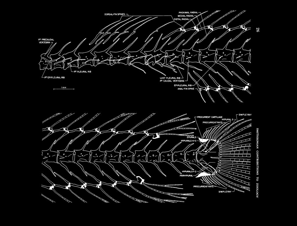
32 SIMPLE RAY Oi DORSAL-FIN SPINES «

33 FIGURE 17 (above). Xenisthmus clarus, vertebrae and unpaired fins (most segmented rays are truncated; note aberrant fusion of 8th and 9th precaudal centra). FIGURE 18 (below). Tyson belos, vertebrae and unpaired fins. -EPIPLEURAL RIB -RAYS DISTAL RADIALS - DISTAL RADIALS
.")
34 28 SMITHSONIAN CONTRIBUTIONS TO ZOOLOGY are no autogenous uroneurals, but a small piece of cartilage occurs in the area below the posterior epural, which may represent a vestige of a uroneural (not labelled in Figure 17; the cartilage is also present in the comparative specimen; autogenous, ossified uroneurals are unknown in gobioids). There is a large dorsal and a large ventral procurrent cartilage (occupying, respectively, the interneural and interhemal spaces between Pu2 and Pu3), posterior to each of which are 2 (dorsal) or 3 (ventral) small cartilages. There are 7 dorsal and 7 ventral procurrent rays (unsegmented), 1 dorsal and 1 ventral segmented, unbranched (simple) ray, and 8 dorsal and 7 ventral branched rays, for a total of 31 caudal-fin elements (32 in the comparative specimen, which has an additional ventral procurrent ray). The first dorsal fin consists of 6 spines, whose pterygiophores have the formula (Birdsong, 1975:137): 3(2,2,1,1,0), which is consistent in all of several specimens that were examined radiographically. Each of the pterygiophores has a cartilaginous ventral tip. The second dorsal fin consists of a spine and 13 segmented rays, all rays branched (12 to 14 rays, varying with the species; D.F. Hoese, in litt.). Each of the second dorsal-fin elements, except the spine and posteriormost two rays, is associated with its own tripartite pterygiophore (proximal + medial + distal radials; the distal radials are largely cartilaginous and do not appear to be differentiated into bilateral halves). The spine of the second dorsal fin is associated with a bipartite pterygiophore, the proximal portion of which possibly consists of fused proximal + medial radials. The posteriormost two rays are associated with a single pterygiophore consisting of a single proximal radial and a complex, hatchet-shaped distal radial of uncertain composition. The anal fin consists of a spine and 12 branched, segmented rays, and each of these elements except the anteriormost 2 and posteriormost 2 is associated with its own tripartite pterygiophore, similar to those in the dorsal fin. The spine lacks its own pterygiophore and is supported by the fused proximal + medial radial of the anteriormost segmented ray. The pterygiophore supporting the posteriormost 2 rays is similar to that of the dorsal fin. All the second dorsal-fin and all but the 2 anteriormost anal-fin pterygiophores insert into separate interneural or interhemal spaces. Tyson (Figure 18): The vertebral column comprises 26 vertebrae: 13 precaudal and 13 caudal. Only the posteriormost two precaudal vertebrae have complete hemal arches (but no hemal spines). The first hemal spine defines the first caudal vertebra. The atlas does not bear lateral condyles for articulation with the exoccipitals, which also lack condyles. The only epipleural ribs are attached to the second precaudal vertebra, somewhat dorsal to the ventroanterior margin of the centrum on each side. Pleural ribs occur on the third to tenth precaudal vertebrae. Pu2 has a long neural and a long hemal spine. The posteriormost vertebral centrum is similar in structure to that of Xenisthmus. An autogenous hypural 5 is absent. A large cartilaginous parhypural and comparable, single epural are present. There is no "uroneural" cartilage (see Xenisthmus). There are several accessory (procurrent) cartilages distal to the parhypural and epural, and in the interneural and interhemal spaces between Pu2 and Pu3. None of the procurrent rays and segmented caudal-fin rays are stained. There are 9 dorsal and 9 ventral procurrent rays, 3 dorsal and 3 ventral simple, segmented rays, and 5 dorsal and 4 ventral branched rays in the caudal fin, for a total of 33 caudal-fin elements. None of the dorsal- and anal-fin rays are stained. The radials of both fins are cartilaginous, except for portions of some of the proximal radials, which are unstained. There is no anterior spinous dorsalfin. The posterior dorsalfinconsists of a spinous ray and 10 simple, segmented rays (the anteriormost 1 and posteriormost 2 rays lack segmentations but consist of bilateral halves typical of segmented rays). Each element, except the 2 posteriormost rays, is associated with its own proximal and distal radials (the proximal radial probably also embodies a medial radial). The last 2 dorsal-fin rays are born on a complex proximal

35 NUMBER radial. The structure of the anal fin is similar to that of the dorsal fin except that the spine lacks both distal and proximal radials and articulates with the proximal (proximal + medial?) radial of the first segmented ray. All of the anal-fin rays are segmented. In contrast to Xenisthmus, and except for the first 2 anal-fin pterygiophores, the dorsal- and anal-fin pterygiophores are inserted 2 or 3 pterygiophores per interneural or interhemal space. Characters and Classification of the Gobioidei The gobioids have not been evaluated cladistically in the literature. It is my intent here to contribute toward such an evaluation. To establish the monophyly of the suborder Gobioidei, it is first necessary to select an outgroup as a basis for comparison. The gobioids are usually considered to belong to the order Perciformes, and although the monophyly of the Perciformes has not been established and is doubtful, indeed (however, see Rosen and Parenti, 1981:19-20, where three characters discussed would place the gobioids in their Percomorpha), I accept its monophyly for the purpose of discussion, and I accept its composition as proposed by Gosline (1968). A sister group for the Gobioidei has not been proposed, and I will not propose one formally here. 2 The main outgroup for my discussion is, therefore, the Perciformes less the Gobioidei. I also use the preperciform order Beryciformes (Zehren, 1979) as a secondary outgroup. Obviously, I have examined only a fraction of the numerous supraspecific taxa contained in the Perciformes, Beryciformes, and Gobioidei, but I have studied a large number of them over the past 30 years and have examined the literature on many others. In any event, the propositions I will introduce are subject to falsification, or corroboration, and hopefully will lead to a better understanding of the gobioids. The Gobioidei was defined in its essentials by Regan (1911), and elaborated on most substantively and recently by Gosline (1955), Miller (1973), and Birdsong (1975). Hoese (1976) recognized six families in the Gobioidei: Rhyacichthyidae, Pirskeniidae (fossil), Eleotrididae (in which he included Xenisthmus), Gobiidae, Microdesmidae, and Kraemeriidae. I propose that the Gobioidei comprises, with one exception, those taxa treated as gobioids by these authors. Miller (1973) included the Pholidichthyidae among the gobioids. The Pholidichthyidae is a monotypic family of uncertain, possibly blennioid or zoarceoid, relationships (Springer and Freihofer, 1976) that does not share, even as homoplasies, any of the four gobioid synapomorphies that I will propose. It also does not exhibit any of a number of other specializations present, apparently, in all gobioids, that are not clearly gobioid synapomorphies. The most complete attempt to characterize the gobioids is that by Birdsong (1975, table 1), who proposed approximately 30 skeletal characters to define the group. In the following section, I will first discuss the four gobioid synapomorphies (the first of which was also noted by Birdsong), and follow this by a discussion of the most important of Birdsong's characters (stated, for the most part, as he gave them). Of the four gobioid synapomorphies, number 1 was proposed by Regan (1911) and the others have not been proposed previously. Thefirstthree synapomorphies appear to be unequivocal, but it is possible that the hypothesized fourth synapomorphy is represented in a more specialized state in certain nongobioids that possibly could be related to the gobioids. 1. Parietals absent. As far as I have been able to determine, all other perciform fishes have parietals. Among nonperciforms, parietals are absent in, at least, all tetraodontiforms (Tyler, 1980:30) and some cyprinodontoids (Parenti, 1981). 2. Pelvic intercleithral cartilage present (Figure 15). I have not found a pelvic intercleithral cartilage in any other perciform or nonperciform taxon. It is probable that the pelvic intercleithral cartilage is lost in the gobioid genus Expedio Snyder, which appears to lack a pelvic girdle altogether (Birdsong, 1975). 3. Dorsal end of interhyal fails to meet dorsal end of symplectic. In many preperciforms and
.")
36 30 SMITHSONIAN CONTRIBUTIONS TO ZOOLOGY most, if not all, nongobioid perciforms, the cartilaginous dorsal ends of the symplectic and interhyal meet and articulate as a unit with the relatively slender ventral process of the hyomandibula (Figure 19 top). Additionally, the dorsal end of the interhyal, and sometimes that of the symplectic, usually impinges on the medial surface of the preopercle. All gobioids are specialized for this character complex. The most plesiomorphic configuration for this character complex among the gobioids appears to be present only in Rhyacichthys (Figure 19 middle), which differs from the generalized perciform configuration in having the interhyal displaced ventrally and articulating only with the preopercle. The remaining gobioids appear to be more specialized for the character complex (Figure 19 bottom, most generalized gobiid configuration): the ventral process of the hyomandibula has become quite broad and the dorsal ends of the interhyal and symplectic have become widely separated. The dorsal end of the interhyal maintains a close connection with the ventral process of the hyomandibula (and in this respect is less specialized than Rhyacichthys) and may or may not be attached ligamentously to the preopercle as well. 4. Basibranchial 1 cartilaginous. The primitive state for basibranchial 1 in perciforms is for this structure to be well ossified. For further discussion of this character, see character 16 and, particularly, note 6 (page 37). 5. Orbitosphenoid absent. I confirm this character, which is a synapomorphy of the Perciformes. 6. Basisphenoid absent. The basisphenoid is absent also in various other perciforms (zoarceoids, stichaeoids, and gobiesocids, at least), and may prove useful in establishing the sister group of the gobioids. 7. Intercalar reduced (lost in some species). The intercalar is well developed in many gobioids (for instance, see Figure 3), including Rhyacichthys, which appears to be the most generalized gobioid. (Miller's (1973) figure 3b, shows a very large opisthotic (= intercalar) in the ventral view of the skull of Rhyacichthys. The intercalar is inexplicably missing in the lateral view, Miller's figure 3c, of the same skull.) A well-developed intercalar is common to many perciforms and preperciforms, and will probably be of little use for establishing gobioid interrelationships. 8. Infraorbital, except lacrimal, absent in most species; one reported present in two species of eleotridids by Akihito (1969) and in Rhyacichthys by Miller (1973). I would restate this character as follows: infraorbitals represented only by lacrimal and, in Rhyacichthys only, one other. 3 (The lacrimal is most correctly termed '"infraorbital 1," but for convenience of discussion "lacrimal" is used in my study.) Birdsong's statement of this character contains some confusion, not of his own making. Akihito's (1969) "suborbital" is not a bone of the infraorbital series (sometimes termed circumorbital or suborbital series), which includes the lacrimal (termed "praeorbital" by Regan, 1911). Akihito reported the "suborbital" in two genera (Bostrichthys, = Bostrychus, and Oxyeleotris) of eleotridids in which it occurs irregularly (D.F. Hoese, in litt., states that this bone is also present in two other eleotridid genera, Micropercops Fowler and Bean and Sineleotris Herre). I have examined Akihito's suborbital and find that it is an ossification of the anteroventral portion of the membrane lining the orbital cavity. I believe that the bone is a specialization confined to certain eleotridid genera (not present in all specimens of a given species). In order to avoid confusion with other bones in the orbital region, I suggest that the term "intraorbital" be applied to it. Intraorbital has not been used previously for any bone in the orbital region. Miller (1973) used the term "prefrontal" to FIGURE 19. Medial views of suspensorial region (interopercles removed) to illustrate relationship of hyomandibula, interhyal, and symplectic. Top, Centropomus species (USNM ; Centropomidae; representative of generalized condition for perciform fishes); middle, Rhyacichthys aspro (representative of most generalized condition for gobioids; disconnection of interhyal from hyomandibula is probably an autapomorphy); bottom, Eleotris amblyopsis (representative of generalized condition for nonrhyacichthyid gobioids). The limits of bones that are obscured by others are indicated by dashed lines.


37 NUMBER OPERCLE HYOMANDIBULA MESOPTERYGOID METAPTERYGOID HYOMANDIBULA INTERHYAL PREOPERCLE MESOPTI METAPTERYGOID QUADRATE SYMPLECTIC D ERCLE SUBOPERCLE HYOMANDIBULA MESOPTERYGOID-; -METAPTERYGOID D-j / SUBOPERCLE INTERHYAL
.")
38 SMITHSONIAN CONTRIBUTIONS TO ZOOLOGY denote the lacrimal of Rhyacichthys. Prefrontal is ported in six species of eleotridids by Akihito, a term often used also for the lateral ethmoid or 1971). I prefer the term "extrascapulae" to a portion of the lateral ethmoid of dermal origin "supratemporals." In addition to Akihito's six (Harrington, 1955:287). When I first examined species, Miller (1973, fig. 2a,b) indicates one extrascapula (on each side) in Rhyacichthys. The the lacrimal in an unstained specimen of Rhyacichthys, I thought it was an antorbital, a dermal gobioid extrascapulae are lateral in position. Median extrascapulae, through which the supratem- bone that is attached to the lateral ethmoid, is dorsal and/or anterior to the lacrimal, and often poral commissural branch of the laterosensory participates in support of the nasal capsule. 4 The canal passes, are primitively present in perciforms bone in Rhyacichthys that Miller termed "suborbital" appears to be infraorbital 2 or 3, which absent in gobioids. Median extrascapulae are (in addition to lateral extrascapulae) but are is not present in other gobioids (although D.F. lacking in many perciform groups, and the value Hoese, in litt., thinks it may be represented by of this character is probably secondary to the loss the intraorbital of eleotridids). The additional of the supratemporal commissural canal in gobioids. In blenniids, for instance, the canal re- infraorbital is tightly connected by ligamentous tissue to the lateral ethmoid, from which it is only mains and the median extrascapulae appear to slightly separated (I misidentified it initially as have become fused with the surface of the skull. the lacrimal), and it bears a broad subocular shelf 10. Wide separation of symplectic from preopercle. This character (Figure 19 bottom) was first (Smith and Bailey, 1962), which in perciforms is usually confined to infraorbital 3. Inasmuch as noted by Gosline (1955). The wide separation of generalized perciforms have six infraorbitals, the the symplectic from the preopercle appears to be reduction to two in gobioids must be considered descriptive of all gobioids except the most generalized, Rhyacichthys (Figure 19 middle) in which to be a specialization. It is not possible to decide if this specialization is a gobioid synapomorphy, the two bones are scarcely more separated, if at because families such as the gobiesocids, callionymids, or batrachoidids, in which the only in- Figure 19 top). As a character, the symplectic- all, than they are in some perciforms (for instance, fraorbital present is the lacrimal, may have arisen preopercle relationship in gobioids is useful only with the gobioids from a common ancestor having as an indication that Rhyacichthys is the sister only two infraorbitals. group of all the other gobioids. Among the "orbital" bones of Rhyacichthys, 11. Preopercle attached to quadrate and hyomandibula, forming third strut of suspensorium Miller (1973, fig. 2a, b) indicated the presence of a single scleral ossification in each eye of Rhyacichthys, which presence I confirm. A single scleral Gosline (1955). The preopercle is attached to the in most species. This character was first noted by ossification occurs also in some eleotridids (D.F. quadrate and hyomandibula in many perciforms Hoese, in litt.), but other gobioids, including and preperciforms, and the arrangement of these eleotridids, I have examined have only a circular bones, as such, has little bearing on gobioid interrelationships. ribbon of scleral cartilage in each eye. Perciform fishes commonly have two scleral ossifications in 12. Symplectic dominates symplectic-metapterygoid strut of suspensorium of most species. each eye, and although I have not searched widely among the perciforms, I have seen a single The symplectic of gobioids, in contrast to most scleral ossification only in callionymids. The perciforms, is usually a larger bone than the value of a scleral ossification, particularly the metapterygoid and usually forms a stronger link presence of only one in each eye, for basing between the quadrate and hyomandibula than gobioid intra- and/or interrelationships, is, perhaps, worthy of further investigation. to be characteristic of unspecialized gobioids, but does the metapterygoid. The condition appears 9. Supratemporals absent in most species (re- its status as a synapomorphy is confused by the
, \"specialized gobiids often have a very expanded metapterygoid,\" a condition that would appear to be unspecialized. 13. Mesopterygoid reduced or absent.")
39 NUMBER absence of the metapterygoid in such groups as the gobiesocids, callionymids, and draconettids (Gosline, 1970, fig. 1; Springer and Fraser, 1976). According to D.F. Hoese (in litt.), "specialized gobiids often have a very expanded metapterygoid," a condition that would appear to be unspecialized. 13. Mesopterygoid reduced or absent. Miller (1973:408) reports, and I confirm (Figure 19 middle), that the mesopterygoid in Rhyacichthys is well developed, in contrast to its state in other gobioids that have a mesopterygoid. The reduction in size or loss of the mesopterygoid may be a basis for of Milyeringa Whitley were unavailable to me, but, D.F. Hoese informed me that all 4 of the specimens he examined had only 6 branchiostegals); and 2 fossil species (Obrhelova, 1961): 5 Pirskenius diatomaceus Obrhelova (Pirskeniidae) and Lepidocottus papyraceus (Agassiz) (Gobiidae?). Rhyacichthys, xenisthmins, and the eleotridids have 6 branchiostegals; all remaining gobioids have 4 or 5 (I have not found 4 in my material, but Akihito (1969:106), reported 4 in one of 10 specimens of the gobiid Periophthalmus cantonensis (Osbeck)). Six branchiostegals is a number that occurs frequently also in nongobioid perciforms. It establishing intragobioid, but not intersubordinal, is doubtful that the primitive number of branchiostegals relationships. Among the gobioids, the mesopterygoid is present only in Rhyacichthys and most eleotridids (D.F. Hoese, in litt.). 14. Interhyal ligamentously attached to preopercle. I have not made an extensive survey of the relationship between the interhyal and preopercle in nongobioid perciforms. In gobioids the lateral surface of the interhyal is completely or almost completely appressed against the medial surface of the preopercle, and determination of the nature of the interhyal-preopercle attachment is difficult. The eleotridids I examined did not appear to have a ligamentous attachment to the preopercle, although Birdsong (1975) reported such an attachment for Butis, which I also examined. I found a ligamentous attachment apparent in Rhyacichthys, Xenisthmus, Tyson, and the gobiid Valenciennea strigata, indicating that such an attachment is probably plesiomorphic for gobioids. Determination of whether it is a gobioid synapomorphy will require extensive study of nongobioid perciforms, but may prove important. 15. Branchiostegal rays 4 to 7. Seven branchiostegals (on each side) is a common number for perciforms and probably represents the primitive number for the group. Among gobioids, 7 branchiostegals have been reported only for: 1 of 12 (7, or 6 if, as I think probable, 7 represents a gain) in gobioids will provide much help in basing gobioid interrelationships. 16. First basibranchial absent. With possibly only one exception {Kraemeria), this character applies to no gobioid I know of. Basibranchial 1 appears to be present in all gobioids, but it is usually present only as cartilage. Hence, basibranchial 1 is probably overlooked in specimens that have not been counterstained for cartilage. 6 As an ossified basibranchial 1 is common to preperciforms and perciforms, the nonossified basibranchial 1 of gobioids is a specialization and may be a synapomorphy; however, the reductive nature of the basibranchial 1 character is confused by the fact that some forms, such as the Gobiesocidae, lack basibranchial 1 altogether (Springer and Fraser, 1976). Among the gobioids, basibranchial 1 is known to be ossified only in Tyson, possibly Kraemeria (as originally reported by Matsubara and Iwai, 1959, but questioned by Birdsong, 1975), and occasionally, perhaps, in Rhyacichthys. In my specimen of Kraemeria, basibranchial 1 appears to be a knob of bone, the ventral surface of which is attached, or weakly fused, to the anterodorsal process of the urohyal; a conspicuous joint is present between the two specimens of the eleotridid Odontobutis obscura bones. In two other specimens of Kraemeria (one (Temminck and Schlegel) (Akihito, 1969:105); of which I have not seen; information provided the monotypic Milyeringidae (McAllister, by D.E. Rosen), it is not possible to decide if the 1968:147; referred to the synonymy of the Eleotrididae by D.F. Hoese, pers. comm.; specimens knob of bone is basibranchial 1 or part of the urohyal. Perhaps of relevance here is that the

40 SMITHSONIAN CONTRIBUTIONS TO ZOOLOGY urohyal is tightly attached to basibranchial 1 in Tyson, and a fusion between the two structures might occur occasionally under such a condition. In gobioids with a cartilaginous basibranchial 1, the dorsal process of the urohyal is often closely applied to the ventral surface of the basibranchial, but the two elements can be readily separated. I consider the ossified basibranchials 1 of Tyson and Kraemeria to be homoplasies. 17. Fourth basibranchial present as cartilage. This is the generalized condition of basibranchial 4 in most, if not all, perciforms and preperciforms (Zehren, 1979:57) and is of little use for establishing gobioid interrelationships. Tyson and Xenisthmus are specialized in lacking basibranchial 4 altogether. 18. Baudelot's ligament present. This ligament is also present in a wide variety of perciforms and preperciforms (Zehren, 1979:53); hence, it is of little use for establishing gobioid interrelationships. 19. Dorsal postcleithrum absent in all groups except eleotridids. The dorsal postcleithrum is present in Rhyacichthys (Miller, 1973, fig. 6a,b), which some earlier authors placed in the Eleotrididae, and some eleotridids, but not in gobiids (sensu Miller, 1973). The presence of the dorsal and ventral postcleithra by perciforms is a primitive character found also in preperciforms (Zehren, 1979:69). What is specialized about the postcleithra in those gobioids that have retained either or both of them is that the two bones are reduced in size and are never connected when both are present, as they normally are in nongobioids. The postcleithra of perciforms have not been surveyed, and the usefulness of postcleithral characters for basing gobioid interrelationships is unknown. 20. Scapula reduced in most species. The scapula is fairly well developed in Rhyacichthys, at least, and the character as stated is of little use for basing gobioid interrelationships. Akihito (1969) noted a trend toward lack of ossification of the scapula in gobioids generally considered to be more specialized. 21. Supraneurals [= predorsal bones] absent. The presence of two predorsal bones appears to be plesiomorphic for perciforms (Smith and Bailey, 1961). However, G.D.Johnson (pers. comm.) considers three predorsal bones to be the primitive state). Therefore, the absence of predorsal bones in gobioids is a specialization. Many perciforms besides gobioids lack predorsal bones, and the character is of questionable value for establishing gobioid interrelationships. There is much confusion surrounding the interpretation of the bones referred to as supraneurals or predorsals. Birdsong (1975:163) referred to these bones as "predorsal bones" and (p. 164) "superneurals" (sic). Zehren (1979:61-62), who referred to Weitzman (1962, figs. 16 and 17), defined predorsals as bones that "resemble the proximal radials of the dorsal fin, with which they are serially homologous" (implying that these bones had lost their associated fin-ray elements). He defined supraneurals as "detached neural spines." However, it is clear from Weitzman 's discussion (1962:40-41) that Weitzman's supraneurals are the homologues of Zehren's predorsals. Gobioids lack both predorsals and supraneurals, using Zehren's terminology. An autogenous neural spine at the anterior end of the vertebral column is a condition found in relatively unspecialized perciforms and preperciforms (hence, gobioids are specialized for this character, but so are many other perciforms). 22. Penultimate vertebra [Pu2] with a short expanded neural spine and an elongated and expanded hemal spine. This is the primitive condition of Pu2 for perciforms and many preperciforms. What is specialized about the gobioid Pu2 (and Pu3) is the fusion of the hemal spine of Pu2 (and Pu3) with its respective centrum. Fusion of the hemal spines of Pu2 and Pu3 with the respective centrum of each occurs widely among perciforms, particularly benthic forms, and has probably occurred homoplasiously. 23. Procurrent caudal rays supported by cartilaginous plates in most species. This is another widely distributed character, occurring in a variety of relatively unspecialized perciforms (e.g., percichthyids, apogonids) and preperciforms. In
 pertain to numbers of pectoral-fin radials, vertebrae, spinous dorsal-fin pterygiophores, and structure of the complex urophore (last")
41 NUMBER some perciforms (for instance, serranids) only the ventral plate is present. I have not noted any perciforms in which only the dorsal plate is present. Other gobioid character states given by Birdsong (1975, table 1) pertain to numbers of pectoral-fin radials, vertebrae, spinous dorsal-fin pterygiophores, and structure of the complex urophore (last vertebra). All of the characters are descriptive of gobioids, but are either primitive for the perciforms or difficult to defend as gobioid synapomorphies, and I will not pursue them further. Two additional characters (24, 25), not treated by Birdsong, the procurrent spur (Johnson, 1975), and the ventral intercleithral cartilage (Figure 15) deserve comment. 24. The procurrent spur, which is found in preperciforms and various perciforms, is absent in gobioids, and possibly represents a specialized state (Johnson did not indicate character polarity). The absence of the spur is also characteristic of many benthic, nongobioid perciforms, such as blennioids, but may occur in free-swimming nongobioids, such as lutjanids. As such, the loss of the spur, if a specialization, may have occurred independently many times. 25. All gobioids appear to have a ventral intercleithral cartilage, which I predict will be shown to be a gobioid synapomorphy. This cartilage is also present in some pempheridids (Parapriacanthus ransonetti Steindachner, at least, but absent in Pempheris Cuvier), batrachoidids (D.E. Rosen, pers. comm.), and, perhaps (based on dissection of specimens not stained for cartilage), stichaeids, bathymasterids, nototheneids, and cheimarrichthyids. In the preceding discussion, I have alluded to the primitive nature of several of the gobioid characters as they occur in Rhyacichthys. Miller (1973) first noted Rhyacichthys' basal position among the gobioids. In the following discussion, I treat the plesiomorphic characters unique to Rhyacichthys among the gobioids. 1. Lacrimal and one other infraorbital bone present (only lacrimal present in other gobioids); see discussion above under character Three epurals (1 or 2 in other gobioids, but occasionally 3 or 1 in species normally having 2 epurals; D.F. Hoese, in litt.). 3. Lateral line present on midrow of body scales (absent in other gobioids). In addition, and not previously reported, Rhyacichthys possesses a short lateral-line scale row both just above and just below the midrow lateral line. The dorsal and ventral lateral lines commence at the caudalfin base and extend posteriorly onto the caudal fin. They appear very much as they do in Lates Cuvier and Valenciennes (Centropomidae) (Greenwood, 1976, fig. 28a). Greenwood considered the triple lateral line of Lates to be a synapomorphy of Lates, as he thought this character had not been reported in any other percoid. Rivas (1966, key), however, used the presence or absence of "accessory lateral lines on the caudal fin" to distinguish species groups in Lutjanus Bloch (Lutjanidae). The accessory lateral lines of Lutjanus are similar in appearance to those of Rhyacichthys and Lates. Besides these three genera, accessory caudal lateral lines also occur in Neoscorpis Smith (Scorpididae), Morone Mitchill (Percichthyidae) (G.D. Johnson, pers. comm.), and some Percidae (Collette and Banarescu, 1977). The presence of accessory lateral lines in these families probably indicates that the character is primitive for the perciforms, or some subgroup of the perciforms that includes, at least, percoids and gobioids. 4. Major branches of lateral-line canals present and well developed on head (Miller, 1973, fig. 10). In other gobioids, the head canals are, variously, incomplete and missing. For instance, only Rhyacichthys among the gobioids has a mandibular sensory canal, which is a common perciform and preperciform character. 5. Dorsal ends of symplectic and interhyal almost adjacent (Figure 19 middle). The two ends are well separated in other gobioids (Figure 19 bottom); see gobioid synapomorphic character 3 above. 6. Large gap between preopercle and symplectic not present; see character 10 (p. 32).
, who described it in detail (gobioid scales appear to be specialized in that only the posteriormost, peripheral ctenii may be teeth-like; the others")
42 36 SMITHSONIAN CONTRIBUTIONS TO ZOOLOGY 7. Scales with multiseriate ctenii. This character was first noted by Miller (1973), who described it in detail (gobioid scales appear to be specialized in that only the posteriormost, peripheral ctenii may be teeth-like; the others are cuboidal). Although a few other gobioids may exhibit two or three rows of ctenii, by far the majority have none or only one row, and only Rhyacichthys has more than three. Multiseriate ctenii occur commonly in other perciforms. 8. Mesopterygoid well developed; see character 13 (p. 33). 9. Caudal fin emarginate. This is the primitive condition for preperciforms and perciforms. In almost all gobioids, the caudal fin is rounded or lanceolate, but a few genera, such as Rhyacichthys, Ptereleotris Gill, Tyson, and the juveniles of various others, have emarginate caudal fins. In addition to these characters Rhyacichthys shares with other gobioids the most primitive state for numerous other characters (including, but not limited to, the tripartite structure of the dorsal- and anal-fin pterygiophores, and general configuration of the hyoid and gill arches. Miller (1973) provides a comprehensive discussion of other characters in Rhyacichthys and their status relative to other perciforms. In spite of Miller's and my contributions, I believe that a detailed osteological description of Rhyacichthys is needed (in part, to resolve conflicts between his and my findings). Such a description might best be based on specimens of mm SL, in order that advanced ossification or other ontogenetic changes not obscure significant characters. Rhyacichthys is probably the gobioid most important for determining the interrelationships of the gobioids. The only family besides the Gobiidae that Miller (1973) recognized in the Gobioidei is the Rhyacichthyidae. I believe that Miller was more impressed by the primitive characters of Rhyacichthys than by its specializations (I confess to the same bias). It is difficult to provide a list of synapomorphies for Rhyacichthys; however, the relatively tiny, ventrally directed mouth with broad patches of villiform teeth, the absence of coronomeckelian bones (associated with the anguloarticulars primitively), the greatly thickened and muscular pelvic fins and ventral pectoral-fin rays, and the disconnection of the dorsal end of the interhyal from the hyomandibula are obvious specializations not present in the generally considered primitive perch-like perciforms or generally considered primitive non-rhyacichthyid gobioids such as the eleotridids. I do not doubt that the Rhyacichthyidae will stand as a family and that Miller's Gobiidae will be found to comprise several groups worthy of family recognition, but I have little more to offer on the subject than that the Xenisthminae will probably constitute one of them. Notes 1 Steindachner (July 1906) described Kraemeria, which he placed in the family Trichonotidae. Jordan and Seale (December 1906) described Vitreola, which they placed in the Gobiidae (Eleotrinae). Regan (1908) described Psammichthys, which he placed in the Trichonotidae. Fowler (1928) First, and correctly, proposed that these three genera are synonyms and that Kraemeria is the senior name. Fowler included Kraemeria in the Trichonotidae. Regan (1911) first proposed the family Psammichthyidae (not designated as a new family, however) for Psammichthys alone. Jordan (1923) continued this allocation, at the same time placing Vitreola in the Gobiidae, and Kraemeria in the Trichonotidae. Whitley (1935) probably assumed that because Kraemeria is a senior synonym of Psammichthys that the family Psammichthyidae was invalid. Whitley (1935) erected the Kraemeriidae (not designated as a new family or replacement name, however) in which he included the Psammichthyidae as a junior synonym. Article 23 (d)(i) of the International Code of Zoological Nomenclature (1964) states: "A family-group taxon formed by the union of two or more taxa of that group takes the oldest valid family-group name among those of its components...." Psammichthyidae has priority over Kraemeriidae; however, Article 23(d)(ii) states: "If a zoologist observes that strict application of the Law of Priority to two or more synonymous family-group names would upset general usage, he is to request the Commission to decide which name is to be accepted for the Official List of Family-Group Names in Zoology." The inference here is that the Commission will decide in favor of general usage. There is little question that Kraemeriidae is the most-used of the two taxa in question, and it appears frequently in the literature. As far as I can discern, Psammichthyidae, a more or less cumbersome name, has rarely been used as a senior synonym since Whitley proposed Kraemeriidae. I do not intend to request the Commission to decide the issue of the proper family-group name for the taxon represented by the two names in ques-

43 NUMBER tion. There are two reasons for this. Foremost is the fact that I consider the infrasubordinal classification of the Gobioidei to be in a tenuous state presently. With the possible exceptions of the Rhyacichthyidae, Gobiidae, and Xenisthminae, as proposed herein, monophyletic family-groups have not been proposed in the suborder, and there is a good possibility that when they are, a family-group taxon for Kraemeria may not be needed. The second reason for not requesting a decision from the Commission is that that body more often than not fails to act or delays action inordinately, thus inhibiting nomenclatural progress. In keeping with the spirit of the Code, I recommend that those desirous of using a family-group name for the group in question, use Kraemeriidae. 2 Rosen and Parenti (1981) treated the classification and intrarelationships of the atherinomorph fishes. Among the included families is the Phallostethidae, which possibly exhibits a close relationship with the gobioids. I have no direct experience with the phallostethids and base my remarks on information in the literature. Unlike Rosen and Parenti's Division 1 atherinomorphs (in which they include the phallostethids), but like the gobioids, the phallostethids are specialized in lacking parietals and a basisphenoid, and having the pelvic-fin elements, when present, closely associated with the cleithra (Roberts, 1971). Like other Division 1 atherinomorphs but unlike the gobioids, the phallostethids are specialized in having the rostral bone (cartilage in other atherinomorphs) decoupled from the premaxillae, in having a different arrangement of ligaments in the snout region, and in lacking infrapharyngobranchial 4 (which is lacking in Tyson, however). Other characters given by Rosen and Parenti to define the atherinomorphs variously apply to, or have not been investigated in, gobioids. Determining whether the phallostethids are more closely related to the gobioids than to Division 1, or other, atherinomorphs, will require more study, but the possibility of such a relationship should be considered. 3 Obrhelova (1961) reported that the lacrimal was absent in the fossil gobioid family Perskeniidae. As the lacrimal is a small, thin, loosely attached, superficial bone in most gobioids, its loss during fossilization might be expected. 4 Although I have not seen the "lacrimal" defined as such, the term, as applied in the literature on teleosts, refers to a bone that articulates with the usually cartilaginously tipped, transverse process of the lateral ethmoid. In most fishes, but not gobioids, the lacrimal is a canal-bearing bone. See Gosline (1961:25-31) and Weitzman (1962:28-31) for discussion of the antorbital, and Weitzman (1962:28, footnote 7) for discussion of names of various series of bones in the orbital region. 5 Obrhelova (1961) gives contradictory information on the number of branchiostegals in her new, monotypic family Pirskeniidae. On page 108 she states, in the diagnosis of the family, that the Pirskeniidae have 6 or 7 branchiostegals, gives the generic composition of the family as, "Gattungen: Pirskenius n. g.," as if to indicate more than one genus was to be included (but none were), and states in the diagnosis of the genus that Pirskenius has 6 or 7 branchiostegals. On page 110, in a comparison of Pirskenius with Lepidocottus aries (Agassiz), which has 5 branchiostegals, she reports only that Pirskenius has 7 branchiostegals; and on pages she describes the positions of these branchiostegals on the epihyal and ceratohyal (= anterior and posterior ceratohyals). Finally, on pages (table 21) she records only 7 branchiostegals for each of the 10 specimens of Pirskenius for which branchiostegal counts are given (she had an additional 20 specimens, but gives no data on these). The confusion about the number of branchiostegals in Pirskenius may have resulted from Obrhelova's belief (1961:111) that the closest [related] species to the monotypic Pirskenius is the fossil Lepidocottus papyraceus (Agassiz), "bei welcher L. Agassiz die Anwesenheit von 6-7 radii branchiostegi anfuhrt, wie es bei der von uns beschriebenen Gattung der Fall ist." She, additionally, leaves open the possibility that L. papyraceus might be the same species as her Pirskenius diatomaceus. In short, I believe Obrhelova diagnosed the Pirskeniidae and Pirskenius as having 6 or 7 branchiostegals because during preparation of her study she considered including L. papyraceus in that family and genus, but having changed her mind, neglected to correct the diagnoses. 6 Lack of cartilage stain is probably the reason Miller (1973) and Birdsong (1975) missed basibranchial 1 in their material. Miller's illustration (fig. 5g) of the basibranchials of Rhyacichthys has basibranchial 2 labelled as basibranchial 1, basibranchial 3 labelled as basibranchial 2, and does not illustrate basibranchial 4 (which is cartilaginous and cuboid in my specimen). Instead he shows a separate, elongate bone, which he labels as basibranchial 3, in the position of basibranchial 4. This last-named structure appears to be the posterior end of basibranchial 3, which in gobioids extends posteriorly ventral to basibranchial 4, which is invariably a cuboidal structure (Figure 12) filling most of the area outlined by the anterior ends of the ceratobranchials 4 and the medial ends of the hypobranchials 3. Miller indicates that this piece of basibranchial 3 is ossified and disconnected from the main body of basibranchial 3 (his 2). I cannot explain this discrepancy between my specimen and his, which is about 40 mm SL shorter than the larger of the two specimens of Rhyacichthys I examined, and which would be expected to be less well ossified. Miller shows a gap between the anterior end of basibranchial 2 (his 1) and the basihyal. In my specimen, this gap is filled dorsally by connective tissue and ventrally by the small, cartilaginous basibranchial 1, which extends almost directly ventral from the ventrally directed anterior end of basibranchial 2. The anteriormost tip of basibranchial 1 in my specimen is stained faintly pinkish, indicating that a slight amount of ossification has occurred. The dorsal surface of the urohyal lies directly beneath the ventral surface of basibranchial 1 and is at-
44 38 SMITHSONIAN CONTRIBUTIONS TO ZOOLOGY tached to it by tough connective tissue. D.F. Hoese informed me that basibranchial 1 is completely ossified in his 110 mm SL specimen of Rhyacichthys. I would like to observe this for myself, but in view of the partial ossification of basibranchial 1 in my specimen, it is possible that variation in the degree of ossification of this element occurs. If this is so, the fourth synapomorphy I gave for the Gobioidei would have to be amended to: tendency for basibranchial 1 to fail to ossify.
:93-114, figures 1-8. 1971. On the Supratemporals of Gobiid Fishes. Japanese Journal of Ichthyology, 18(2):57-64. Birdsong, R.S. 1975.")
45 Literature Cited Akihito (Prince) The Systematic Examination of the Gobiid Fishes Based on the Mesopterygoid, Postcleithra, Branchiostegals, Pelvic Fins, Scapula, and Suborbital. Japanese Journal of Ichthyology, 16(3):93-114, figures On the Supratemporals of Gobiid Fishes. Japanese Journal of Ichthyology, 18(2): Birdsong, R.S The Osteology of Microgobius signatus Poey (Pisces: Gobiidae), with Comments on Other Gobiid Fishes. Bulletin of the Florida State Museum, Biological Sciences, 19(3): , figures Collette, B.B., and P. Banarescu Systematics and Zoogeography of the Fishes of the Family Percidae. Journal of the Fisheries Research Board of Canada, 34(10): Dawson, C.E A Review of the Microdesmidae (Pisces: Gobioidea), 1: Cerdale and Clarkichthys with Descriptions of Three New Species. Copeia, 1974(2) : Fowler, H.W The Fishes of Oceania. Memoirs of the Bernice P. Bishop Museum, 10:1-540, plates Gosline, W.A The Osteology and Relationships of Certain Gobioid Fishes, with Particular Reference to the Genera Kraemeria and Microdesmus. Pacific Science, 9: Some Osteological Features of Modern Lower Teleostean Fishes. Smithsonian Miscellaneous Collections, 142(3): The Suborders of Perciform Fishes. Proceedings of the United States National Museum, 124(3647): 1-78, figures A Reinterpretation of the Teleostean Fish Order Gobiesociformes. Proceedings of the California Academy of Sciences, series 4, 28(19) : , figures 1-6. Greenwood, P.H A Review of the Family Centropomidae (Pisces, Perci formes). Bulletin of the British Museum (Natural History), Zoology, 29(1): 1-81, figures Harrington, R.W., Jr The Osteocranium of the American Cyprinid Fish, Notropis bifrenatus, with an Annotated Synonymy of Teleost Skull Bones. Copeia, 1955(4): Hoese, D. F [1978]. Higher Classification of Gobioid Fishes [abstract]. Revue des Travaux de I'lnstitul dt Peches Maritimes, 40(3-4) :605. Johnson, G.D The Procurrent Spur, an Undescribed Perciform Character and Its Phylogenetic Implications. California Academy of Sciences Occasional Papers, 121:1-23. Jordan, D.S A Classification of Fishes Including Families and Genera as Far as Known. Stanford University Publications, University Series, Biological Sciences, 3(2): i-x. Jordan, D.S., and A. Seale The Fishes of Samoa. Bulletin of the Bureau of Fisheries, 25: , plates Larson, H.K., and D.F. Hoese The Species of the Indo-West Pacific Genus Calumia (Pisces: Eleotridae). Proceedings of the Linnean Society of New South Wales, 104(1-2): Matsubara, K., and T. Iwai Description of a New Sandfish, Kraemeria sexradiata, from Japan, with Special Reference to Its Osteology. Journal of the Washington Academy of Sciences, 49(l): McAllister, D.E Evolution of Branchiostegals and Classification of Teleostome Fishes. National Museum of Canada Bulletin, 221:xiv pages, foldout, endpapers. Miller, PJ The Osteology and Adaptive Features of Rhyacichthys aspro (Teleostei: Gobioidei) and the Classification of Gobioid Fishes. Journal of Zoology (London), 171: , figures Obrhelova, N Vergleichende Osteologie der tertiaren Siisswasserfische Bohmens (Gobioidei). Sbornik Ustredniho Ustavu Geologickeho, 1959, 26(Paleontologicky): , foldout, 15 plates. Parenti, L.R A Phylogenetic and Biogeographic Analysis of Cyprinodontiform Fishes (Teleostei, Atherinomorpha). Bulletin of the American Museum of Natural History, 168(4): Regan, C.T Report on the Marine Fishes Collected by Mr. J. Stanley Gardiner in the Indian Ocean. The Transactions of the Linnean Society of London, series 2 (Zoology), 12(3): , plates
: 117-136. Roberts, T.R. 1971.")
46 40 SMITHSONIAN CONTRIBUTIONS TO ZOOLOGY The Osteology and Classification of the Gobioid Fishes. Annals and Magazine of Natural History, series 8,8: Rivas, L.R Review of the Lutjanus campechanus Complex of Red Snappers. Quarterly Journal of the Florida Academy of Sciences, 29(2): Roberts, T.R Osteology of the Malaysian Phallostethoid Fish Ceratostethus bicomis, with a Discussion of the Evolution of Remarkable Structural Novelties in its Jaws and External Genitalia. Bulletin of the Museum of Comparative Zoology, 142 (4): Rosen, D.E., and L.R. Parenti Relationships of Oryzias, and the Groups of Atherinomorph Fishes. American Museum Novitates, 2719:1-25. Schultz, L.P Order Percomorphida, Suborder Gobiina, Superfamily Gobioidea. In L.P. Schultz, L.P. Woods, and E.A. Lachner, Fishes of the Marshall and Marianas Islands. United States National Museum Bulletin, 202(3): 1-13, figures Smith, C.L., and R.M. Bailey Evolution of the Dorsal-Fin Supports of Percoid Fishes. Papers of the Michigan Academy of Science, Arts, and Letters, 46: The Subocular Shelf of Fishes. Journal of Morphology, 110(l):l-17. Snyder, J.O The Fishes of Okinawa, One of the Riu Kiu Islands. Proceedings of the United States National Museum, 42(1913): , plates Springer, V.G., and T.H. Fraser Synonymy of the Fish Families Cheilobranchidae (=Alabetidae) and Gobiesocidae, with Descriptions of Two New Species of Alabes. Smithsonian Contributions to Zoology, 234:23 pages, 14 figures. Springer, V.G., and W.C. Freihofer Study of the Monotypic Fish Family Pholidichthyidae (Perciformes). Smithsonian Contributions to Zoology, 216: 43 pages, frontispiece, 23 figures. Steindachner, F Zur Fischfauna der Samoa-Inseln. Sitzungsberichten der Kaiserlichen Akademie da Wissenschaflen in Wien, Mathematisch-Naturwissenschaflliche Klasse, 115(1): Travers, R.A The Interarcual Cartilage: A Review of Its Development, Distribution and Value as an Indicator of Phyletic Relationships in Euteleostean Fishes. Journal of Natural History, 15(5) : Tyler, J.C Osteology, Phytogeny, and Higher Classification of the Fishes of the Order Plectognathi (Tetraodonti formes). United States Department of Commerce, NOAA Technical Report NMFS Circular, 434: Weitzman, S.H The Osteology of Brycon meeki, a Generalized Characid Fish, with an Osteological Description of the Family. Stanford Ichthyological Bulletin, 8:1-77, figures Whitley, G.P Studies in Ichthyology, No. 9. Records of the Australian Museum, 19(4): , plate 18. Zehren, S.J The Comparative Osteology and Phylogeny of the Beryciformes (Pisces: Teleostei). Evolutionary Monographs, 1: 389 pages.


, with 1'A\" margins, submitted as ribbon copy (not carbon or xerox), in")
47 REQUIREMENTS FOR SMITHSONIAN SERIES PUBLICATION Manuscripts intended for series publication receive substantive review within their originating Smithsonian museums or offices and are submitted to the Smithsonian Institution Press with Form SI-36, which must show the approval of the appropriate authority designated by the sponsoring organizational unit. Requests for special treatment use of color, foldouts, casebound covers, etc. require, on the same form, the added approval of the sponsoring authority. Review of manuscripts and art by the Press for requirements of series format and style, completeness and clarity of copy, and arrangement of all material, as outlined below, will govern, within the judgment of the Press, acceptance or rejection of manuscripts and art. Copy must be prepared on typewriter or word processor, double-spaced, on one side of standard white bond paper (not erasable), with 1'A" margins, submitted as ribbon copy (not carbon or xerox), in loose sheets (not stapled or bound), and accompanied by original art. Minimum acceptable length is 30 pages. Front matter (preceding the text) should include: title page with only title and author and no other information; abstract page with author, title, series, etc., following the established format; table of contents with indents reflecting the hierarchy of heads in the paper; also, foreword and/or preface, if appropriate. First page of text should carry the title and author at the top of the page; second page should have only the author's name and professional mailing address, to be used as an unnumbered footnote on the first page of printed text. Center heads of whatever level should be typed with initial caps of major words, with extra space above and below the head, but with no other preparation (such as all caps or underline, except for the underline necessary for generic and specific epithets). Run-in paragraph heads should use period/dashes or colons as necessary. Tabulations within text (lists of data, often in parallel columns) can be typed on the text page where they occur, but they should not contain rules or numbered table captions. Formal tables (numbered, with captions, boxheads, stubs, rules) should be submitted as carefully typed, double-spaced copy separate from the text; they will be typeset unless otherwise requested. If camera-copy use is anticipated, do not draw rules on manuscript copy. Taxonomic keys in natural history papers should use the aligned-couplet form for zoology and may use the multi-level indent form for botany. If cross referencing is required between key and text, do not include page references within the key, but number the keyed-out taxa, using the same numbers with their corresponding heads in the text. Synonymy in zoology must use the short form (taxon, author, yeanpage), with full reference at the end of the paper under Literature Cited. For botany, the long form (taxon, author, abbreviated journal or book title, volume, page, year, with no reference in Literature Cited") is optional. Text-reference system (author, yeanpage used within the text, with full citation in "Literature Cited" at the end of the text) must be used in place of bibliographic footnotes in all Contributions Series and is strongly recommended in the Studies Series: (Jones, 1910:122)" or "...Jones (1910:122)." If bibliographic footnotes are required, use the short form (author, brief title, page) with the full citation in the bibliography. Footnotes, when few in number, whether annotative or bibliographic, should be typed on separate sheets and inserted immediately after the text pages on which the references occur. Extensive notes must be gathered together and placed at the end of the text in a notes section. Bibliography, depending upon use, is termed "Literature Cited," References," or "Bibliography." Spell out titles of books, articles, journals, and monographic series. For book and article titles use sentence-style capitalization according to the rules of the language employed (exception: capitalize all major words in English). For journal and series titles, capitalize the initial word and all subsequent words except articles, conjunctions, and prepositions. Transliterate languages that use a non- Roman alphabet according to the Library of Congress system. Underline (for italics) titles of journals and series and titles of books that are not part of a series. Use the parentheses/colon system for volume(number):pagination: ' 10(2):5-9." For alignment and arrangement of elements, follow the format of recent publications in the series for which the manuscript is intended. Guidelines for preparing bibliography may be secured from Series Section, SI Press. Legends for illustrations must be submitted at the end of the manuscript, with as many legends typed, double-spaced, to a page as convenient. Illustrations must be submitted as original art (not copies) accompanying, but separate from, the manuscript. Guidelines for preparing art may be secured from Series Section, SI Press. All types of illustrations (photographs, line drawings, maps, etc.) may be intermixed throughout the printed text. They should be termed Figures and should be numbered consecutively as they will appear in the monograph. If several illustrations are treated as components of a single composite figure, they should be designated by lowercase italic letters on the illustration; also, in the legend and in text references the italic letters (underlined in copy) should be used: Figure 9b. Illustrations that are intended to follow the printed text may be termed Plates, and any components should be similarly lettered and referenced: Plate 9b." Keys to any symbols within an illustration should appear on the art rather than in the legend. Some points of style: Do not use periods after such abbreviations as mm, ft, USNM, NNE. Spell out numbers "one" through "nine" in expository text, but use digits in all other cases if possible. Use of the metric system of measurement is preferable; where use of the English system is unavoidable, supply metric equivalents in parentheses. Use the decimal system for precise measurements and relationships, common fractions for approximations. Use day/month/year sequence for dates: 9 April 1976." For months in tabular listings or data sections, use three-letter abbreviations with no periods: "Jan, Mar, Jun, etc. Omit space between initials of a personal name: J.B. Jones. Arrange and paginate sequentially every sheet of manuscript in the following order: (1) title page, (2) abstract, (3) contents, (4) foreword and/or preface, (5) text, (6) appendixes, (7) notes section, (8) glossary, (9) bibliography, (10) legends, (11) tables. Index copy may be submitted at page proof stage, but plans for an index should be indicated when manuscript is submitted.

48 i
-8- spinous. nape caudal fin. body depth. pectoral fin. anus. total length Fig. 4
 click for previous page -8-1.3 Illustrated Glossary of Technical Terms and Measurements External Morphology and Measurements spinous dorsal fin soft nape caudal fin interorbital body depth snout lateral
- 7 - DESCRIPTION OF SPECIES
 I - 7 - DESCRIPTION OF SPECIES./' Anguilla bicolor McClelland ' Level-finned eel (Figs.i & 2) Length of head 6-8 times in length of body; Diameter of eye 8-10 times, Inter-orbital length 2-2.5 times, Gape
2. SYSTEMATIC CATALOGUE
 click for previous page 15 2. SYSTEMATIC CATALOGUE 2.1 General Aids to Identification 2.1.1 Diagnostic Features of the Family Caesionidae Oblong to fusiform, moderately compressed, medium-sized to small
* A New Species of Cichlid Fish From Lake Malawi. Pseudotropheus tursiops, \(I75 Tropical Fish Hobbyist a'l (3) : 8 L-? 0. ,$ IOU.
 : 8 L-? 0. ,$ IOU.") ,$ IOU. \(I75 Tropical Fish Hobbyist a'l (3) : 8 L-? 0. * 2.37 Pseudotropheus tursiops, A New Species of Cichlid Fish From Lake Malawi by Warren E. Burgess and Dr. Herbert R. Axelrod Among the cichlid
,$ IOU. \(I75 Tropical Fish Hobbyist a'l (3) : 8 L-? 0. * 2.37 Pseudotropheus tursiops, A New Species of Cichlid Fish From Lake Malawi by Warren E. Burgess and Dr. Herbert R. Axelrod Among the cichlid
Lubbockichthys myersi, a new species of dottyback fish from Guam (Pseudochromidae: Pseudoplesiopinae)
") Zootaxa : 43 48 (2006) www.mapress.com/zootaxa/ Copyright 2006 Magnolia Press ISSN 1175-5326 (print edition) ISSN 1175-5334 (online edition) Lubbockichthys myersi, a new species of dottyback fish from
Zootaxa : 43 48 (2006) www.mapress.com/zootaxa/ Copyright 2006 Magnolia Press ISSN 1175-5326 (print edition) ISSN 1175-5334 (online edition) Lubbockichthys myersi, a new species of dottyback fish from
FAO SPECIES IDENTIFICATION SHEETS MUGILOIDIDAE. (Parapercidae of some authors) Sandsmelts, sandperches, grubfishes
 Sandsmelts, sandperches, grubfishes") click for previous page MUGILO 1983 FAO SPECIES IDENTIFICATION SHEETS FISHING AREA 51 (W. Indian Ocean) MUGILOIDIDAE (Parapercidae of some authors) Sandsmelts, sandperches, grubfishes Body elongate, subcylindrical,
click for previous page MUGILO 1983 FAO SPECIES IDENTIFICATION SHEETS FISHING AREA 51 (W. Indian Ocean) MUGILOIDIDAE (Parapercidae of some authors) Sandsmelts, sandperches, grubfishes Body elongate, subcylindrical,
FAO SPECIES IDENTIFICATION SHEETS CAESIONIDAE. Fusiliers
 click for previous page CAES FAO SPECIES IDENTIFICATION SHEETS FISHING AREA 51 (W. Indian Ocean) CAESIONIDAE Fusiliers Lutjanoid fishes, moderately deep-bodied to slender and fusiform, laterally compressed.
FAO SPECIES IDENTIFICATION SHEETS CONGIOPODIDAE* Horsefishes
 click for previous page CONGIO 1983 FAO SPECIES IDENTIFICATION SHEETS FISHING AREA 51 (W. Indian Ocean) CONGIOPODIDAE* Horsefishes Bottom fishes, with large heads and strongly compressed bodies. Snout
Microbrotula randalli Cohen and Wourms, Samoa and Vanuatu at 30 to 38 m near reef-sand interface. Rare.
 click for previous page Ophidiiform Fishes of the World 107 Diagnosis and description: Body completely covered with small imbricate scales; head partly naked; snout depressed; eyes small, more than 6 times
LIBRARY. Class\ V"^ A *Ii:T_
 LIBRARY Class\ V"^ A *Ii:T_ ^ Publications OP FIELD MUSEUM OF NATURAL HISTORY ZOOLOGICAL SERIES Volume X Chicago, U. S. A. 1909-1923 7/,3 ^Issued September 18, 19 12. 69 NEW SPECIES OF FISHES FROM
Review of Stathmonotus, with Redefinition and Phylogenetic Analysis of the Chaenopsidae (Teleostei: Blennioidei)
") Review of Stathmonotus, with Redefinition and Phylogenetic Analysis of the Chaenopsidae (Teleostei: Blennioidei) PHILIP A. HASTINGS and VICTOR G. SPRINGER I W 9\ SMITHSONIAN CONTRIBUTIONS TO ZOOLOGY NUMBER
Review of Stathmonotus, with Redefinition and Phylogenetic Analysis of the Chaenopsidae (Teleostei: Blennioidei) PHILIP A. HASTINGS and VICTOR G. SPRINGER I W 9\ SMITHSONIAN CONTRIBUTIONS TO ZOOLOGY NUMBER
oxfitates AMiiiui?can JMllselIm Threadfin from New Guinea BY J. T. NICHOLS A New Blenny from Bali and a New
 AMiiiui?can JMllselIm oxfitates PUBLISHED BY THE AMERICAN MUSEUM OF NATURAL HISTORY CENTRAL PARK WEST AT 79TH STREET, NEW YORK 24, N.Y. NUMBER i68o JUNE 30, 1954 A New Blenny from Bali and a New Threadfin
AUSTRALIAN MUSEUM SCIENTIFIC PUBLICATIONS
 AUSTRALIAN MUSEUM SCIENTIFIC PUBLICATIONS Rennis, D. S., and Douglass F. Hoese, 1987. Aioliops, a new genus of Ptereleotrine fish (Pisces: Gobioidei) from the tropical Indo-Pacific with descriptions of
Vanderhorstia bella, a New Goby from Fiji (Teleostei: Gobiidae)
") PROCEEDINGS OF THE CALIFORNIA ACADEMY OF SCIENCES Fourth Series Volume 56, No. 32, pp. 619 623, 4 figs. December 30, 2005 Vanderhorstia bella, a New Goby from Fiji (Teleostei: Gobiidae) David W. Greenfield
PROCEEDINGS OF THE CALIFORNIA ACADEMY OF SCIENCES Fourth Series Volume 56, No. 32, pp. 619 623, 4 figs. December 30, 2005 Vanderhorstia bella, a New Goby from Fiji (Teleostei: Gobiidae) David W. Greenfield
Osteology and relationships of Pseudotrichonotus altivelis (Teleostei: Aulopifornies: Pseudotrichonotidae)
") Osteology and relationships of Pseudotrichonotus altivelis (Teleostei: Aulopifornies: Pseudotrichonotidae) G. David Johnson, 1 Carole C. Baldwin, 1 Muneo Okiyama 2 & Yoshiaki Tominaga 3 1 Division of Fishes,
Osteology and relationships of Pseudotrichonotus altivelis (Teleostei: Aulopifornies: Pseudotrichonotidae) G. David Johnson, 1 Carole C. Baldwin, 1 Muneo Okiyama 2 & Yoshiaki Tominaga 3 1 Division of Fishes,
Paraxenisthmus springeri, New Genus and Species of Gobioid Fish from the West Pacific, and Its Phylogenetic Position within the Xenisthmidae
 Paraxenisthmus springeri, New Genus and Species of Gobioid Fish from the West Pacific, and Its Phylogenetic Position within the Xenisthmidae Anthony C. Gill; Douglass F. Hoese Copeia, Vol. 1993, No. 4.
Larvae of two Indo-West Pacific anthiine fishes, Giganthias immaculatus and Serranocirrhitus latus (Perciformes: Serranidae)
") Larvae of two Indo-West Pacific anthiine fishes, Giganthias immaculatus and Serranocirrhitus latus (Perciformes: Serranidae) Yoshinobu Konishi, Chiyuki Sassa and Makoto Okamoto Seikai National Fisheries
Larvae of two Indo-West Pacific anthiine fishes, Giganthias immaculatus and Serranocirrhitus latus (Perciformes: Serranidae) Yoshinobu Konishi, Chiyuki Sassa and Makoto Okamoto Seikai National Fisheries
Three New Australian Species of the Fish Genus Xenisthmus (Gobioidei: Xenisthmidae)
") Copyright Australian Museum, 2004 Records of the Australian Museum (2004) Vol. 56: 241 246. ISSN 0067-1975 Three New Australian Species of the Fish Genus Xenisthmus (Gobioidei: Xenisthmidae) ANTHONY C.
Copyright Australian Museum, 2004 Records of the Australian Museum (2004) Vol. 56: 241 246. ISSN 0067-1975 Three New Australian Species of the Fish Genus Xenisthmus (Gobioidei: Xenisthmidae) ANTHONY C.
FAO SPECIES IDENTIFICATION SHEETS ACROPOMATIDAE. (= "Percichthyidae") Glow-bellies and splitfins
 Glow-bellies and splitfins") click for previous page ACRO 1983 FAO SPECIES IDENTIFICATION SHEETS FISHING AREA 51 (W. Indian Ocean) ACROPOMATIDAE (= "Percichthyidae") Glow-bellies and splitfins Body oblong, more or less compressed.
click for previous page ACRO 1983 FAO SPECIES IDENTIFICATION SHEETS FISHING AREA 51 (W. Indian Ocean) ACROPOMATIDAE (= "Percichthyidae") Glow-bellies and splitfins Body oblong, more or less compressed.
64 FAO Species Catalogue Vol. 18
 click for previous page 64 FAO Species Catalogue Vol. 18 Epetriodus Cohen and Nielsen, 1978 Type species: Epetriodus freddyi Cohen and Nielsen, 1978 by original designation. Fig. 61 Epetriodus freddyi
OCCASIONAL PAPERS OF THE MUSEUM OF ZOOLOGY UNIVERSITY OF MICHIGAN
 OCCASIONAL PAPERS OF THE MUSEUM OF ZOOLOGY UNIVERSITY OF MICHIGAN NOTES ON THE GOBIOID FISHES OF CALIFORNIA, WITH DESCRIPTIONS OF TWO NEW GENERA These notes result from our collecting along the Californian
The osteology of Eucyclogobius newberryi and Quietula guaymasiae (Teleostei: Gobiidae), two closely related Gobionellines from the East Pacific
, two closely related Gobionellines from the East Pacific") Ann. Naturhist. Mus. Wien 108 B 13-56 Wien, Mai 2007 The osteology of Eucyclogobius newberryi and Quietula guaymasiae (Teleostei: Gobiidae), two closely related Gobionellines from the East Pacific G. Kindermann*,
Ann. Naturhist. Mus. Wien 108 B 13-56 Wien, Mai 2007 The osteology of Eucyclogobius newberryi and Quietula guaymasiae (Teleostei: Gobiidae), two closely related Gobionellines from the East Pacific G. Kindermann*,
Jurassic Fish and their Stratigraphic significance from the Hengyang Region of Lingling Co., Hunan Province
 Jurassic Fish and their Stratigraphic significance from the Hengyang Region of Lingling Co., Hunan Province Nianzhong Wang Institute of Vertebrate Paleontology and Paleoanthropology, Academia Sinica Vertebrata
Study of the Monotypic Fish Family Pholidichthyidae (Perciformes)
") Study of the Monotypic Fish Family Pholidichthyidae (Perciformes) VICTOR G. SPRINGER and WARREN C. FREIHOFER SMITHSONIAN CONTRIBUTIONS TO ZOOLOGY NUMBER 216 SERIAL PUBLICATIONS OF THE SMITHSONIAN INSTITUTION
Study of the Monotypic Fish Family Pholidichthyidae (Perciformes) VICTOR G. SPRINGER and WARREN C. FREIHOFER SMITHSONIAN CONTRIBUTIONS TO ZOOLOGY NUMBER 216 SERIAL PUBLICATIONS OF THE SMITHSONIAN INSTITUTION
THE CERATIOID FISHES OF THE GENUS THAUMATICHTHYS
 THE CERATIOID FISHES OF THE GENUS THAUMATICHTHYS OSTEOLOGY, RELATIONSHIPS, DISTRIBUTION AND BIOLOGY BY E. BERTELSEN and PAUL J. STRUHSAKER Zoological Museum University of Copenhagen National Marine Fisheries
Coryphaenoides longifilis (Günther, 1877) Fig MACROUR Cory 9
 Fig MACROUR Cory 9") click for previous page 211 First dorsal fin with 2 spines and 9 to 11 rays; teeth along leading edge of spinous first dorsal ray slender, sharp, and prominent; pectoral fin rays i18 to i21; pelvic fin
click for previous page 211 First dorsal fin with 2 spines and 9 to 11 rays; teeth along leading edge of spinous first dorsal ray slender, sharp, and prominent; pectoral fin rays i18 to i21; pelvic fin
1.5. Systematics of Butidae and Eleotridae
 CHAPTER 1.5 Systematics of Butidae and Eleotridae Christine Thacker Eleotridae and Butidae are two clades of gobioid fishes, both differing from relatives Gobiidae and Gobionellidae in that they are generally
AACL BIOFLUX Aquaculture, Aquarium, Conservation & Legislation International Journal of the Bioflux Society
 AACL BIOFLUX Aquaculture, Aquarium, Conservation & Legislation International Journal of the Bioflux Society Descriptive osteology of the endemic spined loach Cobitis linea from Iran 1 Pariya Jalili, 1
THE percomorph family Caristiidae includes seven
 Copeia 2011, No. 3, 385 399 Revision of the Manefish Genus Paracaristius (Teleostei: Percomorpha: Caristiidae), with Descriptions of a New Genus and Three New Species Duane E. Stevenson 1 and Christopher
Original Article Description of skeletal structure and cranial myology of Cobitis keyvani (Cypriniformes: Cobitidae)
") International Journal of Aquatic Biology (2014) 2(6): 337-345 ISSN: 2322-5270; P-ISSN: 2383-0956 Journal homepage: www.npajournals.com 2014 NPAJournals. All rights reserved Original Article Description
International Journal of Aquatic Biology (2014) 2(6): 337-345 ISSN: 2322-5270; P-ISSN: 2383-0956 Journal homepage: www.npajournals.com 2014 NPAJournals. All rights reserved Original Article Description
Sphagemacrurus Fowler, MACROUR Spha. S. decimalis Fig Genus with Reference : Sphagemacrurus Collett, 1896, by original designation).
.") click for previous page 287 Sphagemacrurus Fowler, 1925 MACROUR Spha Genus with Reference : Sphagemacrurus Collett, 1896, by original designation). Fowler, 1925, Am.Mus. Novit. 162:3 (type species Macrurus
click for previous page 287 Sphagemacrurus Fowler, 1925 MACROUR Spha Genus with Reference : Sphagemacrurus Collett, 1896, by original designation). Fowler, 1925, Am.Mus. Novit. 162:3 (type species Macrurus
The significance of osteology in identification of Schizothorax species
 2016; 4(1): 442-447 ISSN: 2347-5129 (ICV-Poland) Impact Value: 5.62 (GIF) Impact Factor: 0.352 IJFAS 2016; 4(1): 442-447 2016 IJFAS www.fisheriesjournal.com Received: 12-12-2015 Accepted: 15-01-2016 Shabanum
30 a. Allothunnus fallai Fig b.
 click for previous page - 18-30 a. Jaw teeth tiny, 40 to 55 on each side of upper and lower jaws; gillrakers fine and numerous, total of 70 to 80 on first arch; body elongate; distance from snout to second
Systematics & Diversity of Amblyopine gobies. Edward O. Murdy & Koichi Shibukawa IPFC9
 Systematics & Diversity of Amblyopine gobies Edward O. Murdy & Koichi Shibukawa IPFC9 Amblyopinae Composition: Trypauchen Group: Amblyotrypauchen, Caragobius, Ctenotrypauchen, Karsten, Paratrypauchen,Trypauchen,Trypauchenichthys
Osteology of Bluegill Sunfish,
 J. Fac. Fish. Anim. Husb. Hiroshima Univ. (1971), 10:73-101 Osteology of Bluegill Sunfish, Lepomis macrochirus RAFINESQUE Nirmal K. THAKUR*, Masao TAKAHASHI, and Shiro MuRACHI Department of Fisheries,
soft dorsal-fin and caudal-fin margin pale small embedded scales on maxilla
 click for previous page 82 FAO Species Catalogue Vol. 16 13a. Juveniles brown, with 7 dark brown bars on body, the last covering most of caudal peduncle, its upper half black; distance between fourth and
Materials: Field notebook and pencil INTRODUCTION:
 Field Methods of Fish Biology 2014 Exercise 1: Basic Anatomy and Finding and Measuring Characters *Labs modified from Caillet et al. 1986 and Eric Schultz s Biology of Fishes lab Materials: Field notebook
Natural History of Vertebrates Characters Used in Fish Identification (modified )
") Natural History of Vertebrates Characters Used in Fish Identification 1-9-03 (modified 20050118) This lab is designed to familiarize the student with characters used in the identification of fishes. Only
Natural History of Vertebrates Characters Used in Fish Identification 1-9-03 (modified 20050118) This lab is designed to familiarize the student with characters used in the identification of fishes. Only
Field Identification of Tunas from Indian Waters
 3 Field from Indian Waters Subal Kumar Roul and Retheesh T. B. Pelagic Fisheries Division The Family Scombridae is one of the largest and most economically important fish family which comprises of most
Article. Opistognathus albicaudatus, a new species of jawfish (Teleostei: Opistognathidae) from the Andaman Islands
 from the Andaman Islands") Zootaxa 3085: 34 40 (2011) www.mapress.com/zootaxa/ Copyright 2011 Magnolia Press Article ISSN 1175-5326 (print edition) ZOOTAXA ISSN 1175-5334 (online edition) Opistognathus albicaudatus, a new species
Zootaxa 3085: 34 40 (2011) www.mapress.com/zootaxa/ Copyright 2011 Magnolia Press Article ISSN 1175-5326 (print edition) ZOOTAXA ISSN 1175-5334 (online edition) Opistognathus albicaudatus, a new species
Symphurus ocellaris, a new shallow-water symphurine tonguefish collected off Pacific Panama (Pleuronectiformes: Cynoglossidae)
") PROCEEDINGS OF THE BIOLOGICAL SOCIETY OF WASHINGTON 118(3):576 581. 2005. Symphurus ocellaris, a new shallow-water symphurine tonguefish collected off Pacific Panama (Pleuronectiformes: Cynoglossidae)
PROCEEDINGS OF THE BIOLOGICAL SOCIETY OF WASHINGTON 118(3):576 581. 2005. Symphurus ocellaris, a new shallow-water symphurine tonguefish collected off Pacific Panama (Pleuronectiformes: Cynoglossidae)
Remarks: Allen & Swainston (1988) have erroneously recorded this species from northwestern Australia.
 have erroneously recorded this species from northwestern Australia.") click for previous page 71 Literature: Masuda et al. (1975, 1984); Kyushin et al. (1977); Smith (1977); Randall et al. (1978, as P. townsendi); Rau & Rau (1980); Kyushin et al. (1982); Fischer & Bianchi
click for previous page 71 Literature: Masuda et al. (1975, 1984); Kyushin et al. (1977); Smith (1977); Randall et al. (1978, as P. townsendi); Rau & Rau (1980); Kyushin et al. (1982); Fischer & Bianchi
Chapter 30 Nonvertebrate Chordates, Fishes, and Amphibians Name
 Chapter 30 Nonvertebrate Chordates, Fishes, and Amphibians Name Lab Dissecting a Perch Background Information Fish are the largest group of vertebrates found in fresh and salt water. In fact, over 25,000
Description of a new species of Istigobius (Teleostei: Gobiidae) from Australia and Indonesia
 from Australia and Indonesia") Description of a new species of Istigobius (Teleostei: Gobiidae) from Australia and Indonesia DOUGLASS F. HOESE Senior Fellow, Australian Museum, 1 William Street, Sydney 2010, NSW Australia Email: dough@austmus.gov.au
Description of a new species of Istigobius (Teleostei: Gobiidae) from Australia and Indonesia DOUGLASS F. HOESE Senior Fellow, Australian Museum, 1 William Street, Sydney 2010, NSW Australia Email: dough@austmus.gov.au
FAO SPECIES IDENTIFICATION SHEETS NEMIPTERIDAE. (including Scolopsidae of authors) Threadfin breams, monocle breams and dwarf monocle breams
 Threadfin breams, monocle breams and dwarf monocle breams") click for previous page NEMIP 1983 FAO SPECIES IDENTIFICATION SHEETS FISHING AREA 51 (W. Indian Ocean) NEMIPTERIDAE (including Scolopsidae of authors) Threadfin breams, monocle breams and dwarf monocle
click for previous page NEMIP 1983 FAO SPECIES IDENTIFICATION SHEETS FISHING AREA 51 (W. Indian Ocean) NEMIPTERIDAE (including Scolopsidae of authors) Threadfin breams, monocle breams and dwarf monocle
FAO SPECIES IDENTIFICATION SHEETS FISTULARIIDAE. Cornetfishes, flutemouths
 click for previous page FIST 1982 FAO SPECIES IDENTIFICATION SHEETS FISHING AREA 51 (W. Indian Ocean) FISTULARIIDAE Cornetfishes, flutemouths Body elongate and depressed. Mouth small, at end of a long
FAO SPECIES IDENTIFICATION SHEETS NOMEIDAE. Man-of-war fishes, also driftfishes
 click for previous page NOM 1983 FAO SPECIES IDENTIFICATION SHEETS FISHING AREA 51 (W. Indian Ocean) NOMEIDAE Man-of-war fishes, also driftfishes Slender to deep, laterally compressed fishes (in Psenes
Description of five new species of marine gobies (Teleostei: Gobiidae) of the genus Grallenia from the tropical western Pacific Ocean
 of the genus Grallenia from the tropical western Pacific Ocean") Description of five new species of marine gobies (Teleostei: Gobiidae) of the genus Grallenia from the tropical western Pacific Ocean Abstract GERALD R. ALLEN Department of Aquatic Zoology, Western Australian
Description of five new species of marine gobies (Teleostei: Gobiidae) of the genus Grallenia from the tropical western Pacific Ocean Abstract GERALD R. ALLEN Department of Aquatic Zoology, Western Australian
Acentrogobius limarius, a new species of goby (Pisces: Gobiidae) from West Papua Province, Indonesia
 from West Papua Province, Indonesia") Acentrogobius limarius, a new species of goby (Pisces: Gobiidae) from West Papua Province, Indonesia GERALD R. ALLEN Department of Aquatic Zoology, Western Australian Museum, Locked Bag 49, Welshpool DC,
Acentrogobius limarius, a new species of goby (Pisces: Gobiidae) from West Papua Province, Indonesia GERALD R. ALLEN Department of Aquatic Zoology, Western Australian Museum, Locked Bag 49, Welshpool DC,
MATSUBARICHTHYSINUSITATUS, A NEW GENUS AND SPECIES OF VELVETFISH (SCORPAENIFORMES: APLOACTINID AE) FROM THE GREAT BARRIER REEF
 FROM THE GREAT BARRIER REEF") 25 September 1991 PROC. BEX. SOC. WASH. 104(3), 1991, pp. 468-473 MATSUBARICHTHYSINUSITATUS, A NEW GENUS AND SPECIES OF VELVETFISH (SCORPAENIFORMES: APLOACTINID AE) FROM THE GREAT BARRIER REEF Stuart G.
25 September 1991 PROC. BEX. SOC. WASH. 104(3), 1991, pp. 468-473 MATSUBARICHTHYSINUSITATUS, A NEW GENUS AND SPECIES OF VELVETFISH (SCORPAENIFORMES: APLOACTINID AE) FROM THE GREAT BARRIER REEF Stuart G.
NOVITATES PUBLISHED BY THE AMERICAN MUSEUM OF NATURAL HISTORY CITY OF NEW YORK APRIL 27, 1954 NUMBER 1655
 AtMERIICAN MUSEUM NOVITATES PUBLISHED BY THE AMERICAN MUSEUM OF NATURAL HISTORY CITY OF NEW YORK APRIL 27, 1954 NUMBER 1655 Review of the Deep-Sea Fishes of the Genus Asquamiceps Zugmayer, With Descriptions
Xyrichtys trivittatus, a New Species of Razorfish (Perciformes: Labridae) from Hong Kong and Taiwan
 from Hong Kong and Taiwan") Zoological Studies 39(1): 18-22 (2000) Xyrichtys trivittatus, a New Species of Razorfish (Perciformes: Labridae) from Hong Kong and Taiwan John E. Randall 1, * and Andrew S. Cornish 2 1 Bishop Museum,
Zoological Studies 39(1): 18-22 (2000) Xyrichtys trivittatus, a New Species of Razorfish (Perciformes: Labridae) from Hong Kong and Taiwan John E. Randall 1, * and Andrew S. Cornish 2 1 Bishop Museum,
FAO SPECIES IDENTIFICATION SHEETS KUHLIIDAE * Flagtails, daras
 click for previous page KUH 1983 FAO SPECIES IDENTIFICATION SHEETS FISHING AREA 51 (W. Indian Ocean) KUHLIIDAE * Flagtails, daras Body oblong, compressed. Maxilla mostly exposed, without supramaxilla;
Eviota lentiginosa, a new dwarfgoby from Norfolk Island, Australia (Teleostei: Gobiidae)
") Eviota lentiginosa, a new dwarfgoby from Norfolk Island, Australia (Teleostei: Gobiidae) DAVID W. GREENFIELD Research Associate, Department of Ichthyology, California Academy of Sciences, 55 Music Concourse
Eviota lentiginosa, a new dwarfgoby from Norfolk Island, Australia (Teleostei: Gobiidae) DAVID W. GREENFIELD Research Associate, Department of Ichthyology, California Academy of Sciences, 55 Music Concourse
GROUP II : 2a. Dorsal fin high, spinous second ray about as long as, or longer than, head length
 click for previous page 123 GROUP II : 1a. A prominent blackish blotch, sometimes ocellated, above and behind pectoral fin base; anus slightly removed from anal fin; underside of snout ; anterolateral
Haemulon chrysargyreum Günther, 1859
 click for previous page 1538 Bony Fishes Haemulon chrysargyreum Günther, 1859 Frequent synonyms / misidentifications: Brachygenys chrysargyreus (Günther, 1859 ) / None. FAO names: En - Smallmouth grunt;
INDO-PACIFIC DISTRIBUTION OF MICRODESMID FISHES (GOBIOIDEA)*
*") /. mar. biol. Ass. India, 1973, 15 (1) : 318-322 INDO-PACIFIC DISTRIBUTION OF MICRODESMID FISHES (GOBIOIDEA)* C. E. DAWSON Gulf Coast Research Laboratory, Ocean Springs, Mississippi 39564, USA ABSTRACT
/. mar. biol. Ass. India, 1973, 15 (1) : 318-322 INDO-PACIFIC DISTRIBUTION OF MICRODESMID FISHES (GOBIOIDEA)* C. E. DAWSON Gulf Coast Research Laboratory, Ocean Springs, Mississippi 39564, USA ABSTRACT
BANDTAIL FROGFISH, ANTENNATUS STRlGATUS (GILL 1 863) (PISCES: ANTENNARIIDAE)
 (PISCES: ANTENNARIIDAE)") EARLY STAGES OF THE BLOODY FROGFISH, ANTENNARlUS SANGUlNWS GILL 1863, AND THE BANDTAIL FROGFISH, ANTENNATUS STRlGATUS (GILL 1 863) (PISCES: ANTENNARIIDAE) WILLIAM WATSON National Marine Fisheries Service
EARLY STAGES OF THE BLOODY FROGFISH, ANTENNARlUS SANGUlNWS GILL 1863, AND THE BANDTAIL FROGFISH, ANTENNATUS STRlGATUS (GILL 1 863) (PISCES: ANTENNARIIDAE) WILLIAM WATSON National Marine Fisheries Service
from Guam, Marianas lsl~nds
 Taenioides limicola, A New Goby from Guam, Marianas lslnds by C. Lavett SMITH Assistant Curator, Department of Ichthyology, The American Museum of Natural Historv. The gobioid fishes allied to Ta enioid
NOTES ON EGGS, LARV^ AND JUVENILES OF FISHES FROM INDIAN WATERS
 NOTES ON EGGS, LARV^ AND JUVENILES OF FISHES FROM INDIAN WATERS V. Euthynnus affinis (Cantor)* BY S. JONES {Central Marine Fisheries Research Station) LARVAL stages of Katsuwonus pelamis (Linnaeus) and
click for previous page BONY FISHES
 click for previous page BONY FISHES 1540 Bony Fishes General Remarks GENERAL REMARKS by K.E. Carpenter Bony fishes constitute the bulk, by far, of both the diversity and total landings of marine organisms
122 FAO Species Catalogue Vol. 18
 click for previous page 122 FAO Species Catalogue Vol. 18 Diagnosis and description: Skin over head and fin bases thick, less so over body, body covered with tiny scales arranged in regular rows; small
The Osteology and Relationships of Certain Gobioid Fishes, with Particular Reference to the Genera Kraemeria and Microdesmus 1
 The Osteology and Relationships of Certain Gobioid Fishes, with Particular Reference to the Genera Kraemeria and Microdesmus 1 WLLAM A. GOSLNE 2 NTRODUCTON AMONG TROPCAL MARNE PERCFORM FSHES there is a
A New Indo-Pacific Fish of the Genus Cirripectes (Blenniidae, Salariini)l
l") Pacific Science (1980), vol. 34, no. 4 1981 by The University Press of Hawaii. All rights reserved A New Indo-Pacific Fish of the Genus Cirripectes (Blenniidae, Salariini)l BRUCE A. CARLSON 2 ABSTRACT:
Pacific Science (1980), vol. 34, no. 4 1981 by The University Press of Hawaii. All rights reserved A New Indo-Pacific Fish of the Genus Cirripectes (Blenniidae, Salariini)l BRUCE A. CARLSON 2 ABSTRACT:
Sueviota pyrios, a new species of coral-reef dwarfgoby from the Red Sea (Teleostei: Gobiidae)
") Sueviota pyrios, a new species of coral-reef dwarfgoby from the Red Sea (Teleostei: Gobiidae) DAVID W. GREENFIELD Research Associate, Department of Ichthyology, California Academy of Sciences, 55 Music
Sueviota pyrios, a new species of coral-reef dwarfgoby from the Red Sea (Teleostei: Gobiidae) DAVID W. GREENFIELD Research Associate, Department of Ichthyology, California Academy of Sciences, 55 Music
DESCRIPTION OF A BATHYPELAGIC FISH, LESTIDIUM BLANCI SP. NOV. (FAMILY PARALEPIDIDAE) FROM THE ARABIAN SEA*
 FROM THE ARABIAN SEA*") J.: mar. biol. Ass. India, 10, 12 (1 & 2): 146-150 DESCRIPTION OF A BATHYPELAGIC FISH, LESTIDIUM BLANCI SP. NOV. (FAMILY PARALEPIDIDAE) FROM THE ARABIAN SEA* K. N. RASACHANDRA KARTHA Central Marine Fisheries
J.: mar. biol. Ass. India, 10, 12 (1 & 2): 146-150 DESCRIPTION OF A BATHYPELAGIC FISH, LESTIDIUM BLANCI SP. NOV. (FAMILY PARALEPIDIDAE) FROM THE ARABIAN SEA* K. N. RASACHANDRA KARTHA Central Marine Fisheries
THE GENUS PHOTIS (CRUSTACEA: AMPHIPODA) FROM THE TEXAS COAST WITH THE DESCRIPTION OF A NEW SPECIES, PHOTIS MELANICUS
 FROM THE TEXAS COAST WITH THE DESCRIPTION OF A NEW SPECIES, PHOTIS MELANICUS") THE GENUS PHOTIS (CRUSTACEA: AMPHIPODA) FROM THE TEXAS COAST WITH THE DESCRIPTION OF A NEW SPECIES, PHOTIS MELANICUS L. D. McKinney Texas A&M University at Galveston, Moody College of Marine Technology,
THE GENUS PHOTIS (CRUSTACEA: AMPHIPODA) FROM THE TEXAS COAST WITH THE DESCRIPTION OF A NEW SPECIES, PHOTIS MELANICUS L. D. McKinney Texas A&M University at Galveston, Moody College of Marine Technology,
FAO SPECIES IDENTIFICATION SHEETS SYNODONTIDAE. Lizardfishes
 click for previous page SYNOD 1474 FAO SPECIES IDENTIFICATION SHEETS FISHING AREAS 57,71 (E Ind. Ocean) (W Cent. Pacific) SYNODONTIDAE Lizardfishes Body elongate, usually cylindrical and with adipose fin.
BONY FISHES TECHNICAL TERMS
 previous page 1 TECHNICAL TERMS Principal Measurements Used (shortest distance between the points marked) preorbital length head length eye standard length postorbital length 1st dorsal fin base total
Perciformes: Stromateoidei: Nomeidae 3771 NOMEIDAE. Driftfishes (cigarfishes)
") click for previous page Perciformes: Stromateoidei: Nomeidae 3771 NOMEIDAE Driftfishes (cigarfishes) by P.R. Last Diagnostic characters: Small to medium-sized (to 1 m) fishes with a compressed, slender
click for previous page Perciformes: Stromateoidei: Nomeidae 3771 NOMEIDAE Driftfishes (cigarfishes) by P.R. Last Diagnostic characters: Small to medium-sized (to 1 m) fishes with a compressed, slender
Slide 1. Slide 1. Next. 5:30:08 AM
 Slide 1 Slide 1 http://www3.utep.edu/leb/mosquito/larvslide1.htm10/27/2004 5:30:08 AM Slide 1 Slide 2 Recognition that the specimens are mosquito larvae is a prerequisite to identification of the genera.
Internal Anatomy of Fish
 Internal Anatomy of Fish The Systems of a Fish Skeletal System Muscular System Respiratory System Digestive System Circulatory System Nervous System Reproductive System Special Organs Skeletal System
FAO SPECIES IDENTIFICATION SHEETS LETHRINIDAE. Emperors, scavengers
 click for previous page LETH 1974 FAO SPECIES IDENTIFICATION SHEETS FISHING AREAS 57,71 (E Ind. Ocean) (W Cent. Pacific) LETHRINIDAE Emperors, scavengers Moderate-sized perch-like fishes with a large head;
click for previous page D E
 click for previous page D E DREP FAO SPECIES IDENTIFICATION SHEETS 1974 FISHING AREAS 57, 71 (E Ind. Ocean) (W Cent. Pacific) DREPANIDAE Sicklefishes (placed by some authors, together with the Platacidae,
-G\Su---c9 ~ ~ THE KANSA$ $CHOOL NATURALIST THE CARP A MANUAL STRESSING OBSERVATION. Kansas State Teachers College Emporia, Kansas. No.3. Vo l.
 THE CARP A MANUAL STRESSNG OBSERVATON -G\Su---c9 ~ ~ THE KANSA$ $CHOOL NATURALST Kansas State Teachers College Emporia, Kansas Vo l. 22 No.3 February 1976 The Ka nsa s School N aturalist Published by Emporia
Perch Dissection Lab
 Name: Block: Due Date: Perch Dissection Lab Background The fish in the class Osteichthyes have bony skeletons. There are three groups of the bony fish: ray-finned, lobe-finned, and the lungfish. The perch
FAO SPECIES IDENTIFICATION SHEETS. FAMILY: LETHRINIDAE FISHING AREA 51 (W. Indian Ocean) Lethrinus conchyliatus (Smith, 1959)
 Lethrinus conchyliatus (Smith, 1959)") click for previous page LETH Leth 9 1983 FAO SPECIES IDENTIFICATION SHEETS FAMILY: LETHRINIDAE FISHING AREA 51 (W. Indian Ocean) Lethrinus conchyliatus (Smith, 1959) OTHER SCIENTIFIC NAMES STILE IN USE:
click for previous page LETH Leth 9 1983 FAO SPECIES IDENTIFICATION SHEETS FAMILY: LETHRINIDAE FISHING AREA 51 (W. Indian Ocean) Lethrinus conchyliatus (Smith, 1959) OTHER SCIENTIFIC NAMES STILE IN USE:
Four new species of Trimma (Pisces: Gobiidae) from Papua New Guinea and Indonesia
 from Papua New Guinea and Indonesia") Four new species of Trimma (Pisces: Gobiidae) from Papua New Guinea and Indonesia GERALD R. ALLEN Department of Aquatic Zoology, Western Australian Museum, Locked Bag 49, Welshpool DC, Perth, Western Australia
Four new species of Trimma (Pisces: Gobiidae) from Papua New Guinea and Indonesia GERALD R. ALLEN Department of Aquatic Zoology, Western Australian Museum, Locked Bag 49, Welshpool DC, Perth, Western Australia
Eric J. Hilton Geology Department, Field Museum, 1400 South Lake Shore Drive, Chicago IL,
 ISSN 0097-3157 PROCEEDINGS OF THE ACADEMY OF NATURAL SCIENCES OF PHILADELPHIA REDESCRIPTION OF ORTHOSTERNARCHUS TAMANDUA (BOULENGER) 156: 1-25 1 JUNE 2007 Redescription of Orthosternarchus tamandua (Boulenger,
AUSTRALIAN MUSEUM SCIENTIFIC PUBLICATIONS
 AUSTRALIAN MUSEUM SCIENTIFIC PUBLICATIONS Anderson, M. Eric, 1994. Studies on the Zoarcidae (Teleostei: Perciformes) of the southern hemisphere. VII. A new species of Dieidolycus Anderson, 1988 from the
PROCEEDINGS i j OF THE BIOLOGICAL SOCIETY OF WASHINGTON
 e3 if Vol. 86, No. 27, pp. 333-338 28 September 1973 PROCEEDINGS i j OF THE ' BIOLOGICAL SOCIETY OF WASHINGTON A NEW SPECIES OF SESARMA, S. (HOLOMETOPUS) RUBINOFFORUM, FROM THE PACIFIC COAST OF PANAMA
Article.
 Zootaxa 3701 (1): 093 100 www.mapress.com/zootaxa/ Copyright 2013 Magnolia Press Article http://dx.doi.org/10.11646/zootaxa.3701.1.8 http://zoobank.org/urn:lsid:zoobank.org:pub:59c67bfe-fd32-45f8-a15e-095b6216dc07
4. Two New Species of Mbuna (Rock-Dwel ling Cichl ids) from Lake Malawi. by Warren E. Burgess
 from Lake Malawi. by Warren E. Burgess") STUDIES ON THE FAMILY CICHLIDAE: 4. Two New Species of Mbuna (Rock-Dwel ling Cichl ids) from Lake Malawi. by Warren E. Burgess and Dr. Herbert R. Axelrod As the mbuna become better known scientifically
STUDIES ON THE FAMILY CICHLIDAE: 4. Two New Species of Mbuna (Rock-Dwel ling Cichl ids) from Lake Malawi. by Warren E. Burgess and Dr. Herbert R. Axelrod As the mbuna become better known scientifically
Oreoglanis infulatus, a new species of glyptosternine catfish (Siluriformes: Sisoridae) from central Vietnam
 from central Vietnam") Journal of Fish Biology (2001) 59, 1164 1169 doi:10.1006/jfbi.2001.1730, available online at http://www.idealibrary.com on Oreoglanis infulatus, a new species of glyptosternine catfish (Siluriformes: Sisoridae)
Journal of Fish Biology (2001) 59, 1164 1169 doi:10.1006/jfbi.2001.1730, available online at http://www.idealibrary.com on Oreoglanis infulatus, a new species of glyptosternine catfish (Siluriformes: Sisoridae)
First record of the engraulid fish Encrasicholina macrocephala (Clupeiformes) from Somalia
 from Somalia") NOTES ON GEOGRAPHIC DISTRIBUTION Check List 13 (4): 47 51 https://doi.org/10.15560/13.4.47 First record of the engraulid fish Encrasicholina macrocephala (Clupeiformes) from Somalia Harutaka Hata, 1 Hiroyuki
NOTES ON GEOGRAPHIC DISTRIBUTION Check List 13 (4): 47 51 https://doi.org/10.15560/13.4.47 First record of the engraulid fish Encrasicholina macrocephala (Clupeiformes) from Somalia Harutaka Hata, 1 Hiroyuki
Jam BOB IBS. kuuhnh. H HhBbHEs. HNfliHBK
 mm Jam BOB IBS kuuhnh H HhBbHEs HNfliHBK L I U N I OF B R.ARY OF THE VERS ITY ILLINOIS 5TO-5 I LL v. Z5-30 ceo. 2, r The person charging this material is responsible for its return to the library from
Thresher Shark Alopias vulpinus
 Thresher Shark Alopias vulpinus 30 Upper tooth Lower tooth Diagnosis The only Alaska species of shark with a distinctly elongate upper caudal-fin lobe. Dentition Teeth small, somewhat triangular, without
Dogfish Shark Dissection
 Dogfish Shark Dissection Name Date Period Fun Facts: Materials: The teeth of sharks are modified scales embedded in the skin of its mouth Sharks have pits on their face used to detect electric fields Sharks
Landmarking protocol
 Landmarking protocol Jonathan Chang Introduction You will be marking key points on images of fish, which will help determine the shape of different fishes and how that affects their performance in the
TWO NEW SPECIES OF COPEPODS JAPANESE FISHES. Author(s) Yamaguti, Satyu; Yamasu, Terufumi.
 Yamaguti, Satyu; Yamasu, Terufumi.") Title TWO NEW SPECIES OF COPEPODS JAPANESE FISHES PARASIT Author(s) Yamaguti, Satyu; Yamasu, Terufumi Citation PUBLICATIONS OF THE SETO MARINE BIO LABORATORY (1960), 8(1): 137-140 Issue Date 1960-05-30
Title TWO NEW SPECIES OF COPEPODS JAPANESE FISHES PARASIT Author(s) Yamaguti, Satyu; Yamasu, Terufumi Citation PUBLICATIONS OF THE SETO MARINE BIO LABORATORY (1960), 8(1): 137-140 Issue Date 1960-05-30
Hemigymnus fasciatus (Bloch, 1792)
") click for previous page 3438 Bony Fishes Hemigymnus fasciatus (Bloch, 1792) Frequent synonyms / misidentifications: None / None. FAO names: En - Barred thicklip; Fr - Tamarin à bandes noires; Sp - Tamarín
click for previous page 3438 Bony Fishes Hemigymnus fasciatus (Bloch, 1792) Frequent synonyms / misidentifications: None / None. FAO names: En - Barred thicklip; Fr - Tamarin à bandes noires; Sp - Tamarín
Revision of the Manefish Genera Caristius and Platyberyx (Teleostei: Percomorpha: Caristiidae), with Descriptions of Five New Species
, with Descriptions of Five New Species") Revision of the Manefish Genera Caristius and Platyberyx (Teleostei: Percomorpha: Caristiidae), with Descriptions of Five New Species Author(s): Duane E. Stevenson and Christopher P. Kenaley Source: Copeia,
Revision of the Manefish Genera Caristius and Platyberyx (Teleostei: Percomorpha: Caristiidae), with Descriptions of Five New Species Author(s): Duane E. Stevenson and Christopher P. Kenaley Source: Copeia,
FAO SPECIES IDENTIFICATION SHEETS ALBULIDAE. Bonefishes
 click for previous page ALBU 1983 FAO SPECIES IDENTIFICATION SHEETS FISHING AREA 51 (W. Indian Ocean) ALBULIDAE Bonefishes Elongate, fusiform fishes possessing a distinctive conical snout projecting beyond
Longnose Skate Raja rhina
 Longnose Skate Raja rhina 42 dorsal ventral egg case Nuchal Orbital Dorsal diagram showing thorns on disc. Diagnosis The only Alaska species of Raja with an elongate snout and deeply notched pelvic fins.
Exercise 18B Class Chondrichthyes Cartilaginous Fishes
 AP Biology Chapter 24 Exercise #18: Chordates: Fish Cartilaginous Fishes Lab Guide Exercise 18B Class Chondrichthyes Cartilaginous Fishes This group contains about 970 species that are characterized by
2. SYSTEMATIC CATALOGUE
 click for previous page 14 2. SYSTEMATIC CATALOGUE 2.1 Diagnostic Features of the Family Lethrinidae Perchlike sparoid marine coastal fishes of the tropical Indo-West Pacific and West Africa. Dorsal fin
Myripristis adusta Bleeker, 1853
 Myripristis adusta Bleeker, 1853 English Name: Shadowfin soldierfish Family: HOLOCENTRIDAE Local Name: Hiyani dhanbodu Order: Beryciformes Size: Common to 25 cm; max. 32 cm Specimen: MRS/0322/88 Distinctive
Lubricogobius nanus, a new species of goby (Pisces: Gobiidae) from eastern Papua New Guinea
 from eastern Papua New Guinea") Lubricogobius nanus, a new species of goby (Pisces: Gobiidae) from eastern Papua New Guinea GERALD R. ALLEN Department of Aquatic Zoology, Western Australian Museum, Locked Bag 49, Welshpool DC, Perth,
Lubricogobius nanus, a new species of goby (Pisces: Gobiidae) from eastern Papua New Guinea GERALD R. ALLEN Department of Aquatic Zoology, Western Australian Museum, Locked Bag 49, Welshpool DC, Perth,
Umbrina broussonnetii (Cuvier, 1830)
") click for previous page Perciformes: Percoidei: Sciaenidae 1647 Umbrina broussonnetii (Cuvier, 1830) Frequent synonyms / misidentifications: None Umbrina coroides (Cuvier, 1830) FAO names: En - Striped
click for previous page Perciformes: Percoidei: Sciaenidae 1647 Umbrina broussonnetii (Cuvier, 1830) Frequent synonyms / misidentifications: None Umbrina coroides (Cuvier, 1830) FAO names: En - Striped
Perch Dissection Lab
 Perch Dissection Lab Introduction: The fish in the class Osteichthyes have bony skeletons. There are three groups of the bony fish - -- ray-finned fish, lobe-finned fish, and the lung fish. The perch is