SBORNIK NARODNIHO MUZEA V PRAZE
|
|
|
- Clementine Briggs
- 5 years ago
- Views:
Transcription
1 SBORNK NARODNHO MUZEA V PRAZE ACTA MU SE NATONALS PRAGAE B XLV (1991), No. 1-4 REDAKTOR: JR CEJKA STANSLAV STAMBERG Regional Museum of Eastern Bohemia at Hradec Kralove ACTNOPTERYGANS OF THE CENTRAL BOHEMAN CARBONFEROUS BASNS NTR ODUCTON The following work r eviews the actinopterygian fishes found in the Central Bohemian Carboniferous Basins. The revision is based on the types and referred specimens of Prof. Antonin Fric on previously unstudied material from the collections of the P aleontological Department 0 the National Museum at Prague and the Regional Museum of West Bohemia at Plzeii, and on new material from boreholes in the collection of the Geological Survey in Prague. A revision of t he Carboniferous fishes of t he Central Bohemian Basins is necessary for several reasons. Firstly, it is now clear that the original descriptions by A. F ric no longer correspond t o our knowledge of the anatomy of t hese Carboniferous fishes and that some structures were misint erpreted. Secondly, a precise classification of t he int act fish specimens originally described by A. Fric is a prerequisite for the determination of t he many osteological fragments and scales of fishes found during more recent geological exploration of Permocarboniferous coalfields. The completion of this revis ion will form t he basis of objective comparisons with other material from similar basins and facies. t gives us the possibility of underst anding aspects of t he evolution of some of these groups of fishes, and, at t he same time, it is a contribution to the resolution of the stratigraphy of t he Perm ocarboniferous basins. am pleased to thank everyone who has helpe d me in t his work. would particularly like to thank Prof. Dr Zde nek Spinar for his int erest and his frequent advice and valuable comments. This work woul d not have been possible without t he amicable help of t he entire staff of the P aleontological Department of the National Museum at Prague, in particular Dr M. Maiio urova, Dr V. Turek, CSc., Dr R. P rokop, CSc., and others. also wish t o thank Dr J. Zajic (Geo logical Survey, Prague) for t he loan of specimens from t he boreholes. am obliged to Dr A. A. Selezneva (Paleont ological nstitute of Academy of Science of t he USSR, Moscow), t o Dr C. Poplin (Musee National d'h istoire Naturelle, P aris) for frequent discussion and much advice and t o Dr A. Milner (B irkbeck College, University of London) for correct ion of t he english translation of t he first half of the text and for many valuable suggestions. 3 25
, as a designation for all covered and exposed areas of the Central Bohemian upper Palaeozoic.")
2 TERMNOLO GY This paper summarizes results of the revision of actinopterygian fishes from the Carboniferous sediments of the Central Bohemian basins. The term "Central Bohemian basins" is used here in the sense of HAVLENA and PESEK (1975), as a designation for all covered and exposed areas of the Central Bohemian upper Palaeozoic. The Central Bohemian Carboniferous basins are geographically divided into following: the Plzeii Basin, the Manetin Basin, the Radnice Basin, the Zihle Basin, the Rakovnik Basin, the Kladno Basin, the Roudnice Basin and the Msec Basin. The stratigraphical correlation of the Central Bohemian Basins is given in text. table 1. The system used for the description of this fish material is essentially that used by GARDNER (1967). The terminology used by LEHMAN (1966) is applied in the descriptions together with detailed anatomical terms used by NELSEN (1942, 1949) and GARDNER (1963, 1967, 1984). New terms used in this work are explained in the following paragraphs. The aim has been to describe the species to a uniform standard and the descriptions are divided into following parts: 1. Neurocranium a) Endoskeleton of the neurocranium b) Exoskeleton of the neurocranium - skull roof, rostrum, dermal bones of the ventral part of the neurocranium, cheek bones 2. Vis c e roc ran i u m - endoskeleton and exoskeleton of the palatomaxillary apparatus, lower jaw, hyoid arch and gill arches 3. S h 0 u de r g i r die 4. T run k - vertebral column, fins and squamation n the interests of precision, the dimensions of the fishes and of individual bones are given in mm, exact scale counts are given and the size of the fins is indicated by the number of lepidotrichiao The descriptions of bones are based on measurable values, namely length, width and height. The length of a bone is its orocaudal dimension. The term "width" is used for the mediolateral dimension of the bones of the dorsal and ventral part of the head, namely the bones of the skull roof, the rostral region, the posttemporal, the gular bones and also the clavicle. The "height" of a bone is its dorsoventral dimension, and this term is used for bones on the lateral surface of the head. Similar terms are used for the squamation. "The inclination of the suspensorium" is a term widely used in the description of fishes. Terms such as "suspensorium oblique", "suspenosrium moderately oblique" or "suspensorium almost vertical" are often used but are imprecise. The changes in the inclination of the suspensorium (os hyomandibulare) have an immediate effect on the shape and orientation of the preopercular which covers the hyomandibular. Because of this relationship, the terms "angle of obliqueness of the preoprecular" and "angle of the bend of the preopercular" are used in this work. The angle of obliqueness ofthe preopercular is the angle between the dorsocaudal margin of the preopercular and the horizontal plane. The angle of the bend of the preopercular is the angle between the caudal margin of the narrow ventral part of the preopercular and the dorsocaudal margin of the broader dorsal part of the preopercular. The method of measurement of these angles is illustrated in fig. 1. The angle of obliqueness of the opercular is also used for the same reasons. t is the angle subtended by the axis of the opercular (situated in the direction of dorsoventral elongation of the bone) against the horizontal plane (fig. 1). t should be noted that the definition of the angle of obliqueness of the opercular used in this work differs from that used by KAZANCEVA SELEZNEVA (1981). The text. tables in the following text contain the dimensions of the bodies and fins of the fishes and the scale counts. The methods of measurement are demonstrated in fig. 2 n the text. tables, the numbered columns 1-9 refer to the following data. 1. Scale count The number in the numerator is the number of scale rows dividing the supracleithrum from the anterior base of the dorsal fin. The first number in the denominator is the number of scale rows between the supracleithrum and the anterior part of the pelvic fin base; the second one is the number of scale rows between the supracleithrum and the anal fin base; the third one is the number of scale rows between the supracleithrum and the caudal fin base. The final number represents the total number of scale rows from the supracleithrum to the point at which the lobe of the caudal fin ascends (i.e. where the scales change their direction). The number of scale rows was determinated, as far as possible, by counting the number of scales along the lateral sensory canal. 26
3 to Text. tab. 1. Lithostratigraphical division of the Central Bohemian Basins (after V. HAVLENA, J. PESEK 1980) with occurence of actinopterygians. Age Litho stratigraphical unit Main Horizons Occurrence of fishes Lower ~. ~ Q.. E Autun ian Line c. ~ c ro B.r; en 0- :J (lj 0 -f-' CJ1 (lj ""' 'H..-< c 0 Formation C - Hiatu s 51any Formation - Klobuky Horizon <i: Sphaerolepi~ kounoviensis - Zdet n Horizon Spinarichthys dispersu5 Kamenny Most Member { Progyrolepis speciosus Zaborichthys fragmentalis Kounov - Kounov Coal Seams Acrolepis gigas Member Sphaerolepis kounoviensis Spinarichthys dispersus Ledce Member. Hfedle Member Msec ~ Acrolepis gigas.d Member Watsonichthys sphaerosideritarum ro ""' Watsonichthys krejcii u Jele ni ce - Melnik Coal Seams Member t:-:l -::J CD l 0 Tynec Formatio n - Kladno Nyrany Member r-< Hiatus. f;. Formation C Radnice - Lubna Coa l Seams - Radnice Coal Seams B - Pl zen Coal Seams! ~ 1"-" Member - Tatina Horizon -Plachtin Hor izo n - Nevren Coal Seams ~ Sceletophorus biserialis - Nyrany Coal Seams Scele t ophorus ver rucosus - Mi rosov Horizon Pyritocephalus sculptus
4 Fig. 1. Schematic demonstration methods of measurement. a - the angle of obliqueness of the preopercular. fj - the angle of the bend of the preopercular, y - the angle of obliqueness of the opercular. hm - height of the maxillary plate; hp - horizontal plane; m -length of the maxillary plate; 10-length of the opercular. K >t Fig. 2. The methods of measurements, the results of which are recorded on text. tables in text. Explanations of measurements are given in pages
5 2. Tot a len g tho f the bod y, This was measured in the body axis from the rostral tip to the caudal end of the dorsal lobe of the tail. 3. Len g t h of the t run k. This was measured in the body axis from the caudal margin of the supracleithrum to the angle included by the dorsal and ventral lobes of the caudal fin. 4. The dis tan c e fro m the c a u d a mar gin 0 f the sup rae lei t h rum tot h e 0 r a mar gin 0 f the d 0 r s a fin bas e The length of the dorsal fin base measured at the level of the basal segments The number of dorsal fin lepidotrichia The length of the longest lepidotrichia of the dorsal fin. 5. The dis tan c e fro m the c a u d a mar gin 0 f the sup rae lei t h rum tot h e 0 r a mar gin 0 f the a n a fin bas e The length of the anal fin base The number of anal fin lepidotrichia The length of the longest lepidotrichium of the anal fin. 6. The dis tan c e fro m the c a u d a mar gin 0 f the sup rae lei t h rum tot h e 0 r a mar gin 0 f the pelvic fin bas e The length of the pelvic fin base The number of pelvic fin lepidotrichia The length of the longest lepidotrichium of the pelvic fin The len g tho f the d 0 r s a lob e 0 f the c a u d a fin The len g tho f the v e n t r a lob e 0 f the c a u d a fin. 8. The g rea t est h e i g h t 0 f the bod y. 9. The h e i g h t 0 f the c a u d a p e dun c e. All measurements are given in mm. The presence of a question mark with the number of scales or lepidotrichia indicates that that number is an approximate estimate, because of the imperfect state of preservation of some specimens. Systematic section Subclass: Actinopterygii Order: Palaeonisciformes HAY, 1929 Family: Cosmoptychiidae GARDNER, 1963 D i a g nos is (after GARDNER 1963, emended): Body fusiform. Dorsal fin shifted orally. Dorsal and anal fins triangular. Caudal fin deeply cleft with unequal lobes. Pectoral fin with bases of principal rays unjointed. Anal fin with long base. All fins with numerous small fulcra. Lepidotrichia distally bifurcated. Skull rounded anteriorly and without well-developed rostrum. Suspensorium oblique and orbit large. Antorbital bearing teeth. Opercular much larger than subopercular. Dermohyal and epipreopercular present. Branchiostegal rays numerous, suborbital bones present. Dentition consisting of a series of a few large laniary teeth flanked laterally by series of smaller, more numerous teeth. Scales rhomboidal, with pronounced striae. T y peg e nus: Cosmoptychius TRAQUAR, 1877 Rem ark s : The family Cosmoptychiidae was erected by GARDNER (1963) to include the Carboniferous genera Cosmoptychius TRAQUAR, 1877 and Watsonichthys ALDNGER, These two genera had previously been included in the family Acrolepididae by ALDNGER (19~7). The family Cosmo- 29

6 ptychiidae was diagnosed by GARDNER (1963) particularly by the presence of accesory bones situated anterior to the opercular and by the shape of the long-based pelvic fin. nc l u d e d g e n e ' a: Cosmoptychius TRAQUAR, 1877; Watsonichthys ALDNGER, 1937; Grassator KAZANCEVA, S t rat i g ' a phi c a ' a n g e: Lower Carboniferous - Upper Carboniferous. G e 0 g 'a phi c a dis t 'i bution: Great Britain, Bohemia, USSR, South Africa. Watsonichthys ALDNGER, Watsonichthys, nov. gen.; H. ALDNGER, Permische Ganoidfische, p Watsonichthys ALDNGER, 1937; B. G. GARDNER, Certain Palaeoniscoid Fishes, p Watsonichthys ALDNGER, 1937; L. S. BERG, A. A. KAZANCEVA, D. V. OBRU TSCHEV, Osnovy paleontologii, p Watsonichthys ALDNGER; J. P. LEHMAN, Traite de Paleontologic, p Watsonichthys ALDNGER, 1937; D. HEYLER, Sur e genre Amblypterus, p T y p e 8 pee i e s: Watsonichthys pectinatus (TRAQUAR, 1877). Lo c u stypic us: Gilmerton, Scotland. S t rat u m t y pic u m: Lower Carboniferous. D i a g nos i s (after GARDNER 1963, emended): Body fusiform, some species reaching almost one metre in length. Pectoral fin long with principal epidotrichia unjointed in proximal third of their length. Dorsal fin arising opposite anal fin or shifted forward oflevel of anal fin. Caudal fin deeply cleft and unequally lobed. All fins with numerous small fulcra anteriorly. Lepidotrichia bifurcated distally. Rostral region of head not conspicuously convex orally. Two pairs of extrascapular bones present. Maxilla with orocaudally elongated maxillary plate. Lower jaw strong. Dentition comprising two types of teeth. Suborbital hones present. Suspensorium very oblique. Opercular of oval shape, 2-3 times higher than the subopercular and narrowing ventrally. Branchiostegal rays numerous. Accesory bones, namely the dermohyal, epipraeopercular and prospectively the antopercular, present orally from the opercular. Rhomboidal scales with peg and socket articulation and with sculpture formed by mounds of enamel. Caudal margin of scales either serrate or straight. Rem a ' k s: ALDNGER (1937) described the genus Watsonichthys on the basis of the type species Elonichthys pectinatus (TRAQUAR, 1877). GAR DNER (1962) added a further species, W. lotzi (GUERCH, 1923) from the Dwyka Series of South Africa. HEYLER (1976) considered W. pectinatus to be a synonym of W. eupterygius (AGASSZ, ) originally described by AGASSZ as Amblypterus eupterygius. A detailed discussion of the interrelationships of the species of the genus Watsonichthys follows the concluding paragraph of the description of W. sphaerosideritarum. nc ud e d s p e c i e s: Watsonichthys pectinatus (TRAQUAR, 1877); Watsonichthys eupterygius (AGASSZ, ); Watsonichthys lotzi (GV RCH, 1923); Watsonichthys krejcii (FRTSCH, 1895); Watsonichthys sphaerosideritarum (FRTSCH, 1895). o c cur e n c e and dis t ' i but ion: Lower Carboniferous - Upper Carboniferous; Great Britain, Bohemia, South Africa. 30
7 Watsonichthys krejcii (FRTSCH, 1895) (figs. 3-5, pls. -) 1895 Acrolepis KrejCii, Fr.; A. FRTSCH, Fauna der Gaskohle, Band 3, p , pl. 128, figs Acrolepis KrejCii Fr.; F. BAYER, Kataog, p Acrolepis krejcii FRTSCH, 1895; J. ZAJC, S. STAMBERG, Summary of the Permocarboniferous, p. 71. Ho 0 t Y P e (by original designation): Specimen figured by FRTSCH (1895) on P. 128, figs 1-8, deposited in the National Museum, Prague as M 1208 (both counterparts) and M 890 (galvanic cast). The holotype is refigured in this work (fig. 5, and pls, -). L 0 c u stypic u s: Malesice, distr, Plzeii - north, Czech Rep. S t r at u m t y pic u m: Msec member, Stephanian B, Upper Carboniferous. Mat e l' i a : Holotype specimen only. D i a g nos is: Body fusiform reaching a total length of 130 mm. Lepidotrichia of pectoral fin long, reaching to oral margin of pelvic fin base. Anal fin with long base, about 1.5 times the length of the oral margin of anal fin. Anal fin composed of approximately 50 lepidotrichia. Base of dorsal fin partly opposite the space between pelvic and anal fins and partly opposite to the oral region of anal fin. Oral margin of dorsal fin shorter or same length as oral margin of anal fin. First branchiostegal ray ventral to the subopercular is high, the rest being narrow. Ventral margin of supracleithrum reaching up to half the height of subopercular. Sccles stout, sculpture formed by conspicuous ridges. Scales with 25 straight caudal margin. Scale count? ? Des c r i P t ion: The holotype is relatively well preserved, missing only the caudal fin and the oral part of the head. HEAD Skull roof The bones of the skull roof are indistinctly preserved. The sculpture on the frontal is formed by tubercles and short pits, mostly orocaudally arranged. The rostral region of the head is not conspicuously convex orally. Palatomaxillary apparatus o sma x i a l' e (fig. 5, p., fig. 2). The maxilla is 22 mm long with a well developed oblong maxillary plate, 12 mm long. The maxillary plate bears a process in its caudoventral region. The height of the plate, including this process, is 8 mm, The maxilla is conspicuously sculptured on its lateral surface by 01'0 caudally arranged tubercles and short ridges. There are conspicuous ridges on the ventral part of the lateral face of the maxilla. The maxillary dentition is formed by two distinct series of teeth. The outer marginal series comprises numerous minute pointed teeth of mm length, while the inner series is formed by a few larger stout pointed teeth. There is no sculpture on the tooth surfaces. Of the dermal bones on the medial side of the palatoquadrate only the impression of the ectopterygoid is preserved. The ectopterygoid is oro caudally elongated but its oral margin does not reach the level of the oral margin of the maxillary plate. 31

8 ~ t-:l Fig. 3. Watsonichthys krejcii (FRTSCH, 1895). Reconstruction of whole fish. Drawn after holotype M 1208, X 2.0
9 tnx. Pop..., PSc Sop rbr... rng Fig ca Watsonihcthys krejcii (FRTSCH, 1895). Reconstruction of head lateral view. Drawn after holotype M 1208, x 2.7 Fig. 5. Watsonichthys krejcii (FRTSCH, 1895). Maxilla and lower jaw in lateral view. M 890, X
.")
10 Lower jaw (fig. 5, pl., fig. 2). The lower jaw is 20 mm long, well-developed and apparently strong. ts maximum height is 5.5 mm at the caudal end, and it is narrower orally. The sculpture on the lateral side is formed by conspicuous ridges arranged in orocaudal rows. The dentition mirrors the dentition of the maxillary. o shy 0 man d i b u are (p., fig. 2, pl., fig. 1). The hyomandibular is only partly preserved. The bone is very oblique orally. The hyomandibular is bent orally at the level of the dividing line between the opercular and the subopercular. ts ventral region is narrow and the dorsal region is broader. o s p rae 0 per c u l are (p., fig. 2, p., fig. 1). The preopercular is only partly preserved. t consists of a narrow ventral part and a broader dorsal region which is orally inclined. The angle of the inclination of the preopercular is 25 degrees, and the angle of the bend of the preopercular is 145 degrees. The oral margin of the dorsal part of the bone is at the same level as the oral margin of the maxillary plate. The preopercular is bent orally in the same manner as the hyomandibular, at the level of the dividing line between the dorsal part of the opercular and subopercular. Accesory bones are present between the dorsal part of the preopercular and the opercular. They are the dermohyal, the antopercular and the epipreopercular. Poor preservation has precluded the recognition of dermal sculpture or sensory canals. o s d e r m 0 h Y a e (pl., fig. 2). The dermohyal is a relatively small triangular bone, considerably dorsoventrally elongated. t is 10.5 mm high and, dorsally, 2 mm long. t is situated. between the oral part of the opercular and the dorsocaudal part of the preopercular. The dorsal part of the dermohyal borders the small antopercular. Gill arch Only some parts of the gill arch endoskeleton are present. n the dorsal region of the gill arch are several small stick-like fragments of bone. They are dorsoventrally elongated and 7 mm high, and consider them to be the remains of the epibranchials (pl., fig. 2, p., fig. 1). Remains of the ventral region of the endoskeleton are also preserved and appear to represent the ceratobranchial bones. The dermal cover of the gill arches comprises the following dermal bones: os operculare, os suboperculare, radii branchiostegales, os gulare laterale and a single os gulare mediale. As well as these bones, there are also some accessory bones present namely: os epipraeoperculare and os antoperculare. o sop e r c u l are (p., fig. 2, p., fig. 1). The opercular is of oval shape and dorsoventrally elongated. t is 12 mm high and 5.5 mm long at the dorsal end. t is narrower ventrally, and because of this there is a space oroventral to it which is occupied by an anamnestic ossification - the os epipraeoperculare. The antopercular and the dermohyal are situated beside the epipreopercular orally from the opercular. The caudal margin of the opercular overlaps the presupracleithrum and the supracleithrum. The opercular is very oblique orally and its angle of inclination is 30 degrees. The sculpture on the lateral face of the bone is formed only by fine, concentrically arranged striae. 34
11 o s sub 0 per cui are (pl., fig. 1). The subopercular is poorly preserved. t is square in. shape and is 6 mm in height and length. Orally from it is the ventral part of the preopercular, while its caudal margin overlaps the oral region of the supracleithrum. Radii branchiostegales There were probably a large number of branchiostegal rays present but in the preserved material only those that are ventral to the subopercular can be seen. The first ray is overlapped by the subopercular and is relatively large in comparison with the others. t is 2.5 mm high and 4.5 mm long. The second ray is only 1.5 mm high and the height of the others does not exceed 1 mm. They are very narrow. Five branchiostegal rays are preserved ventral to the subopercular. They overlap one another in dorsoventral direction. The branchiostegal rays in the oroventral region of the gill arch are preserved fragmentarily. t appears from the preserved fragments that there were approximately twenty branchiostegal rays in all. Os gulare laterale The lateral gular is 4 mm long and 2 mm wide. t is situated caudally to the median gular. The sculpture is formed by ridges situated parallel to the lateral margin of the bone. Os gulare mediale This bone is triangular in shape. ts sculpture is formed by ridges situated orocaudally. Two short pit lines are present in the medial region of the ventral face of the bone and these connect with one another. o santop ere u are (p., fig. 1). The antopercular is a small oval bone, 2 mm high and 2.5 mm long. The sculpture resembles that of the opercular. o s e pip rae 0 per cui are (p., fig. 1). The epipreopercular is a small supplemental bone of triangular shape, 2.5 mm in length and 2 mm high. t borders the opercular, the subopercular and the preopercular. Shoulder girdle The shoulder girdle consists of the following paired dermal bones: os posttemporale, os supracleithrum, os praesupracleithrum, os cleithrum and os claviculare. Os posttemporale This bone is indistinctly preserved on the dorsal part of the head. o s sup rae lei t h rum (p., fig. 2, p., fig. 1) The supracleithrum is oval in shape and is dorsoventrally elongated, being 8 mm high and 5 mm long. The ventral margin of the supracleithrum reaches up to a level halfway up the height of the subopercular. The sculpture on the lateral face of the bone is formed by conspicuous ridges. n the dorsal part of the bone these are arranged in orocaudal rows, but in the ventral part, the rows are parallel with the oral and caudal margins of the bone. The lateral line sensory canal passes through the dorsal third of this bone. 35
![o s p rae sup rae lei t h rum (p., fig. 1) This is a smal] oval bone 3 mm long and 3 mm high. t is situated caudally from the opercular and partly overlaps the orodorsal region of the supracleithrum.](/docs-images/88/115207117/images/12-0.jpg "ts dermal sculpture is formed by conspicuous short ridges situated in orocaudal rows. Os cleithrum Only a fragment of this bone is preserved. o sci a vic u are (p., fig.")
12 o s p rae sup rae lei t h rum (p., fig. 1) This is a smal] oval bone 3 mm long and 3 mm high. t is situated caudally from the opercular and partly overlaps the orodorsal region of the supracleithrum. ts dermal sculpture is formed by conspicuous short ridges situated in orocaudal rows. Os cleithrum Only a fragment of this bone is preserved. o sci a vic u are (p., fig. 2) The clavicle is situated orally from the ventral region of the cleithrum. Only the long ventral part of the clavicle is preserved and is 13 mm in length. The caudal part of the clavicle is at the level of the caudal margin of the maxillary. TRUNK Paired fins Pee tor a fin (p., fig. 1, p., fig. 1) The pectoral fin is conspicuously long and contains approximately 17 lepidotrichia. The longest lepidotrichia are 26 mm long and are articulated and distally dichotomously branching. Only 6-7 orally placed lepidotrichia are not articulated in the proximal third of their length. Numerous small fulcra are present on the oral margin of the fin. Pel vic fi n (p., fig. 1, pl., fig. 1) The pelvic fin is approximately equidistant between the pectoral and anal fins. t is composed of articulate lepidotrichia. The oral margin of the fin base is situated behind the tenth scale row. Unpaired fins A n a fi n (p., fig. 1, pl., fig. 2) The anal fin is very well preserved and characterized by its long base and numerous lepidotrichia. t is composed of 50 articulated and dichotomously branching lepidotrichia. The oral margin of the fin is protected by fringing fulcral scales. D 0 r s a fin (p., fig. 1) The base of the dorsal fin is situated partly above the space between the pelvic and anal fins and partly above the base of the anal fin. The oral margin of the fin base begins behind the twenty-fifth scale row. The dorsal fin base is half the length of the anal fin base. Alliepidotrichia are articulated. There are four ridge scales orally from the dorsal fin. Squamation The trunk is covered by stout, rhomboidal scales. The scales on the flank of the fish are of rhomboidal shape but near the ventral edge of the trunk, they are orocaudally elongated. The dimensions of representative scales along the lateral sensol'y canal is: in the tenth row, 2 mm high and 1.5 mm long (pl., fig. 2); in the twentieth row (near the oral margin of the anal fin), 0.7 mm high and 1.5 mm long. The scale count is _ 25? ? The scales are of medium size and overlap each other only slightly. All scales are ornamented with the ridges arranged parallel with the ventral and dorsal margins of the scales. Over the centre ofthe scale, ridges are arranged diagonally. 36

13 The ridges are connected in the caudal part of each scale. The caudal margin of the scale is not denticulate. The ridge scales situated orally from the dorsal fin are ornamented with orocaudally arranged ridges. The lateral sensory canal is clearly visible from the supracleithrum to the level of the anal fin. No. 1 2 M1208 To 20 40'1 Text. tab. 2. Watsonichthys krejcii (FRTSCH, 1895). Dimensions of the body. Explanations see on p Rem arksan d re a t ion s hip s: The interrelationships of the species of the genus Watsonichthys are discussed following the description of W. sphaerosideritarum, Watsonichthys sphaerosideritarum (FRTSCH, 1895). (figs. 6-11, p. 4) 1895 Acrolepis sphaerosideritarum, Fr.; A. FRTSCH, Fauna der GaskoWe, Band 3, p p. 127, figs Acrolepis sphaerosideritarum Fr.; F. BAYER, Katalog, p Acrolepis sphaerosideritarum FRTSCH, 1895; J. ZAJC, S. STAMBERG, Summary of the Permocarboniferous, p. 71. Ho 0 t y p e (by original designation): Specimen figured by FRTSCH (1895) on p. 127, figs. 1-7, deposited in the National Museum, Prague as M 888 (positive) and M 889 (negative). The holotype is refigured in this work (figs. 6, 7 and P. V). L 0 c u stypic u s: Zilov, distr. Plzeii - north, Czech Rep. S t rat u m t y pic u m: Msec member, Stephanian B, Upper Carboniferous. Mat e ria : Holotype and another well-preserved specimen YA 135 b, together with five specimens showing fragments of heads and scales. D i a g nos i s: Body fusiform reaching a total length of 100 mm. Lepidotrichia of pectoral fin long, reaching to caudal margin of pelvic fin base. Pelvic fin composed of approximately 30 lepidotrichia. Anal fin base about the same length as oral margin of anal fin. Dorsal fin base entirely opposite anal fin base. Oral margin of dorsal fin is 25% longer than oral ma:rgin of anal fin. First branchiostegal ray ventral to the subopercular is high, the rest being narrow. Ventral margin of supracleithrum not reaching subopercular. Scales stout, sculpture formed by conspicuous ridges. Scales with denticulated caudal margin. Large ridge scales extending from head to anterior base of dorsal fin. Scale count THE HEAD DESCRPTON Sku roo f (figs. 7, 6) The skull roof is composed of the following paired bones: os parietale, os frontale, os dermopteroticum, os dermosphenoticum. Caudally it is separated from the dermal bones of the shoulder girdle by the row of extrascapular bones. 37
14 ft dsph J>t,,, c.hy - epi Fig. 6. Watsonichthys sphaerosideritarum (FRTSCH, 1895). Dermal bones of the skull roof, gill arches and pectoral girdle in dorsoventral view. Ma88, X 3.5,,,,,,,,,, dsph po ext r chy? 38
15 Extrascapular bones These bones are situated caudally from the parietal and dermopterotic. Two or three pairs of extrascapular bones are present. Because of their poor preservation, no trace of the sensory canal could be found. Os parietale The parietal is s quare in shape and is two-fifths of the length of the frontal. The dermopterotic borders it laterally. The dorsal surface of the parietal is ornamented with tubercles and at the caudolateral end of the bone, short grooves represent traces of the sensory lines. Os frontale The frontal is considerably orocaudally elongated, its length being three times its width. The frontal is orally in contact with the postrostral and nasal, and laterally with the dermopterotic and dermosphenotic. The sculpture on the dorsal face of the bone is formed by numerous tubercles, partly arranged in orocaudal rows. The remains of the supraorbital sensory canal are preserved in the oral region of the bone. Os dermopteroticum This bone is of oblong shape and also orocaudally elongated. ts caudal margin extends to the extrascapular bones and its oral margin contacts the dermosphenotic. The infraorbital sensory canal passes along the lateral margin of the bone and continues on the dermosphenotic. Os dermosphenoticum This bone is triangular in shape with the caudal margin contacting the dermopterotic, the oral margin contacting the nasal, and the lateral edge forming the orbit margin. The sculpture on the dorsal face ofthe bone is formed by irregularly arranged tubercles (fig. 7). Parts ofthe sensory canal are present. The infraorbital canal continues from the dermopterotic on to this bone, turning ventrally in the oral region and passing along the dorsocaudal margin of the orbit. ndistinct traces of the infraorbital canal suggest that it continues orally to the nasal (fig. 7). R 0 s t r a (fig. 7) The rostral region of the head is poorly preserved as in other material. judge from the visible remains that the rostral region is made up of a single postrostral, paired nasals and paired rostropremaxillaries. The oral region of the head is blunt and the rostrum is not conspicuously convex orally. Only a fragment of the rostropremaxillary is present and is sculptured with irregularly arranged tubercles.. Palatomaxillary apparatus and dermal bones of the cheek The palatomaxillary apparatus includes the following visible ossifications: os maxillare, os praeoperculare and os dermohyale. Other bones are not preserved. Two of the circumorbital series of dermal cheek bones have also been identified namely: os jugale and os suborbitale. Os maxillare The upper jaw is very indistinct but its fragments do show very well, the broad oblong maxillary plate, orocaudally elongated. The maxillary plate bears a lobular process in its ventrocaudal region. The dentition is not preserved. 39

16 o s p rae 0 per cui are (fig. 6) The preopercular is composed of broad and orally inclined dorsal region and a narrow ventral region. The preopercular is inclined orally at an angle of 20 to 25 degrees. The angle of the bend of the preopercular is 140 degrees. The oral margin of the dorsal part of the preopercular is at the level of the oral margin of the maxillary plate. The dermohyal and antopercular lie caudal to the dorsal broadened part of the preopercular. The next supplemental bone, the os epipraeoperculare lies caudally from the bend of the preopercular. Consequently the supplemental dermohyal and epipreopercular ossifications entirely separate the preopercular and opercular. Os dermohyale The dermohyal is triangular and dorsoventrally elongate. t is 6 mm high and ventrally is in contact with the preopercular. o s jug a e (fig. 7) The jugal (also known as the os infraorbitale inferior) is crescent-shaped. t is situated orally from the maxillary plate and the preopercular. The oral margin of the bone is arched and forms the ventrocaudal margin of the orbit. Neither sculpture nor traces of the sensory canals are preserved. o s sub 0 r bit a e (fig. 6, 7) n the holotype, there is an indistinct fragment of this oval bone situated between the caudal margin of the orbit and the oral margin of the preopercular. The orbit is of medium size and bordered orally by the nasal. The nasal extends up to half of the dorsal margin of the orbit. Dorsally the orbit is bordered by the dermosphenotic and ventrocaudally by the jugal. The remains of the ring of oblong sclerotic ossicles is present (fig. 7). The gill arch The dermal cover of the gill arches comprises the os operculare, os suboperculare, radii branchiostegales, ossa gularia, os antoperculare and os epipraeoperculare. o sop e r c u l are (figs. 6, 7) The opercular is oval and is twice as high as it is long. tis onger dorsally and narrower ventrally, the later feature providing space for the supplemental bone - the epipreopercular. The opercular is substantially inclined orally. The angle of inclination of the opercular is 35 degrees. The opercular partly covers the oral margin of the presupracleithrum and the supracleithrum. Os suboperculare This bone is approximately square in shape, and is half the height of the opercular. The ventral margin of the supracleithrum reaches the level of the boundary between the opercular and subopercular. Os antoperculare The antopercular is very obscure. t is a small bone situated between the dermohyal and the oral margin of the opercular. o s e pip rae 0 per c u are (fig. 6) The epipreopercular is a triangular bone bordering the opercular, suhopercular and preopercular. The dorsal margin of this bone reaches the dermohyal. 40
17 Radii branchiostegales The branchiostegal rays are indistinctly preserved. The fragments suggest that there was a great number of narrow branchiostegal rays. The shoulder girdle The following dermal bones are present in the shoulder girdle: os posttemporale, os supracleithrum, os praesupracleithrum, os cleithrum and os claviculare. s.r. 110 "_ ptr - -- ju - dpt. dsph sbo re po soc -, ext fr" ' -, ', -,, ",,. }. -,... " <,.. '-"... ~ ~-~...-~"""-' /' /'./ /' /' / /' /' /' e ;' dhy ;' /' ;' pop / ;' Psc/ epi sop rprnx rbr de.spl mg c/o- - cl Fig. 8. Watsonichthys sphaerosideritarum (FRTSCH, 1895). Reconstruction of head in lateral view, X 3.2 o s p 0 s t t e m p 0 r a e (fig. 6) The posttemporal is situated caudallyfrom the extrascapular bones. ts lateral margin covers the presupracleithrum and the supracleithrum. The dorsal face of the bone is sculptured by ridges arranged in an orocaudal direction. o s sup r a c lei t h rum (fig. 6) The supracleithrum is a dorsoventrally elongated oval bone. Orally from its dorsal region lies the presupracleithrum. The ventral part of the supracleithrum. does not reach the level of the dividing line between the opercular and subopercular, The sculpture on the lateral face of the bone is formes by conspicuous ridges arranged in a dorsoventral direction. The sculpture dorsal to the lateral 4 41
18 sensory canal is formed by irregularly arranged ridges and tubercles. The lateral sensory canal passes through the dorsal region from the caudal margin of the bone obliquely to the dorsal margin. Os cleithrum The cleithrum is dorsoventrally elongate with an arch-like orally directed bend in the ventral region. The oral margin of the cleithrum is covered by the subopercular and the branchiostegal rays. The clavicle lies orally from the cleithrum. o sc a vic u are (fig. 10) The clavicle forms the oroventral part of the dermal skeleton of the shoulder girdle. t connects to the ventral part of the cleithrum, The oral edge of the clavicle reaches up to the level of the caudal margin of the orbit. o s p rae sup r a c lei t h rum (fig. 6) The presupracleithrum is a small bone situated orally from the dorsal part of the supraeleithrum. ts sculpture is a ridge pattern similar to that on the supracleithrum. TRUNK The shape of the trunk is illustrated in fig. 9 and its dimensions are given in text. tab. 3. No. M 889 Text. tab. 3. Watsonichthys sphaerosideritarum (FRTSCH, Explanations see on p Dimensions of the body. Paired fins P e c tor a fin (p. V) The pectoral fin is conspicuously long, the longest lepidotrichia in the holotype being 25 mm long. The lepidotrichia are articulated and distally dichotomously branching. Only the proximal third of the oral lepidotrichia is not articulate. f the pectoral fin could be stretched caudally, it would extend beyond the caudal margin of the base of the pelvic fin. Numerous small fringing fulcra are present. Pelvic fin The pelvic fin is approximately equidistant between the pectoral and anal fins, or may be nearer the pectoral fin. The oral margin of the pelvic fin base begins at the eight scale row. The fin is composed of about 15 articulated epidotrichia. Median fins Anal fin The anal fin is smaller than the dorsal fin and lies opposite to it. The oral 42
19 >!' * t Fig. 9. Watsonichthys sphaerosideritarum (FRTSCH, 1895). Reconstruction of whole fish, X 1.6
20 margin of the anal fin base begins at scale rows The fin consists of articulated lepidotrichia. Dorsal fin The dorsal fin is triangular and consists of articulated lepidotrichia. t begins above scale rows There are fringing fulcra on the leading edge and orally from the fin lie a row of large ridge scales. :/ / -~( ~~~~~'" -,... /' 0<7 ~==::, «~n <, 2mm ~- ~c1 ~ u~ ~ ~:== c:? U) '-~_.~~~~ l~~~/' ====~ Fig. 10. Watsonichthys sphaerosideritarum (FRTSCH, 1895). Clavicle in ventrolateral view. YA Fig.. Watsonichthys sphaerosideritarum (FRTSCH, 1895). Large ridge scales protect the dorsal side of the caudal peduncle. Arrow indicates the directio cranialis. M 889, X 8.0 Caudal fin The caudal fin is deeply cleft with well developed dorsal and ventral lobes. The lepidotrichia are branched and jointed. There are ridge scales along the dorsal margin of the fin and caudal peduncle, and fulcral scales along the ventral margin of the fin. Squamation The trunk is covered by rhomboidal scales which are ornamented with orocaudally arranged ridges. On the lateral face of each scale are 5-7 ridges arranged in parallel and rarely linked. The ridges terminate caudally in small 44

21 teeth. n the holotype, representative scales along the lateral sensory canal have the following dimensions: in row 10: height 2 mm, length 1.3 mm in row 25: height 0.8 mm, length 1.3 mm. The scale count is ? The dorsal margin of the trunk between the posttemporal and the dorsal fin is protected by ridge scales. Similar ridge scales are present caudal to the dorsal fin on the peduncle of the caudal fin. o c cur e nee inc z e c h 0 s 0 v a k i a: Zilov (Kladno and Rakovnik Basins), Netovice (Borehole Nt-). G e 0 g rap hi c a dis t rib uti 0 n: Czech Rep. S t rat i g rap hie a ran g e: Msec member, Stephanian B, Upper Carboniferous. Rem arksan d r e l a t ion s hip s: Two species of the genus Watsonichthys are now known from the Carboniferous basins of Central Bohemia, namely W. krejcii and W. sphaerosideritarum. They are very similar but have been clearly differentiated in this study. The basic differences are as follows: 1. n W. krejcii, the longest lepidotrichia of the pectoral fin reach the level of the oral margin of the pelvic fin base. n W. sphaerosideritarum, the same lepidotrichia reach the caudal margin of the pelvic fin base. 2. n W. krejcii, the anal fin is composed of 50 epidotrichia and its base is 1.5 times longer than the length of its oral margin. n W. sphaerosideritarum, the anal fin is composed of approximately 30 lepidotrichia and the base is approximately the same lenght as the oral margin. 3. n W. krejcii, the dorsal fin base lies partly above the space between the pelvic and anal fin, and partly above the anal fin. n W. sphaerosideritarum, the base of the dorsal fin lies entirely above the base of the anal fin. 4. n W. krejcii, the scales are not denticulated on their caudal margin. n W. sphaerosideritarum, the scales are denticulated on their caudal margin. 5. n W. krejcii, the ventral margin of the supracleithrum extends to the level of half of the height of the subopercular. n W. sphaerosideritarum, the ventral margin of the supracleithrum does not reach the subopercular. Comparison of W. krejcii and W. sphaerosideritarum with the description of the type species W. pectinatus given by TRAQUAR ( ), ALDNGER (1937) and GARDNER (1963) reveals that the Bohemian basin species share some distinct features not found in the type species: a) W. krejcii and W. sphaerosideritarum share the possession of a conspicuously long pectoral fin, and a long anal fin base. b) W. krejcii and W. sphaerosideritarum do not possess the long base to the pelvic fin, as described in W. pectinatus. c) n W. krejcii and W. sphaerosideritarum, the branchiostegal ray ventral to the subopercular is twice as high as the other branchiostegal rays. This size discrepancy does not occur in W. pectinatus. d) W. krejcii and W. sphaerosideritarum posses 3 a presupracleithrum which is absent in W. pectinatus. 45
 and W. eupterygius (AGASSZ, 1833-1843). n contradistinction to HEYLER (1976), consider W. eupterygius to be distinct from W. pectinatus and closer to the two Bohemian species.")
22 As a result of this work, the genus Watsonichthys is considered to include the species W. pectinatus (TRAQUAR, 1877), W. krejcii (FRTSCH, 1895), W. sphaerosideritarum (FRTSCH, 1895), W. lotzi (GUERCH, 1923) and W. eupterygius (AGASSZ, ). n contradistinction to HEYLER (1976), consider W. eupterygius to be distinct from W. pectinatus and closer to the two Bohemian species. Several features of the body and fins support this argument. The pelvic fin does not have a long base, but the anal fin does have a long base and the pectoral fin is elongate. Unfortunately, it is not possible to make comparison of the bones of the head as they are poorly preserved in W. eupterygius. Family: Pygopteridae ALDNGER, 1937 D i a g nos i s: (after ALDNGER 1937, emended): Body fusiform. Pectoral fin with bases of principal rays unjointed. Pelvic fin very small. Anal fin with long base. Caudal fin deeply cleft and unequally lobated. Lepidotrichia distally bifurcated. Endocranium only partly ossified. Parietal bones short. One pair of extrascapular bones present. Parasphenoid short with processus ascendens posterior and processus ascendens anterior. Skull with well-developed rostrum. Postrostral large and wide. Maxilla with well-developed oro caudally elongated maxillary plate. Dentition consisting of a series of a few large laniary teeth flanked laterally by a series of smaller, more numerous teeth. Suspensorium very oblique. Subopercular longer than opercular. Branchiostegal rays numerous. Accessory bones present between dorsal parts of opercular and preopercular. Sculpture on dermal bones of the head well-developed. Scales small, in oral part of the trunk oblong, dorsoventrally elongated, usually sculptured with ridges. The scales in caudal part ofthe trunk rhomboidal, smooth. Ridge scales developed orally from the dorsal fin and on the dorsal side of the caudal fin. Typ e g en us : Pygopterus AGASSZ, ncluded genera: Pygopterus AGASSZ, ; Nematoptychius TRAQUAR, 1875; Progyrolepis FRTSCH, 1895; tararichthys BELTAN, 1977; Zaborichthys n, g. S t rat i g l' a phi c a l' a n g e: Lower Carboniferous - Triassic. G e 0 g l' a phi c a dis t l' i but ion: Europe, South America. Progyrolepis FRTSCH, Progyrolepis, Fr.; A. FRTSCH, Fauna der Gaskohle, Band 3, p T y pes pee i e s: Progyrolepis speciosus (FRC, 1875). L 0 c u stypic us: Kounov, distr. Rakovnlk, Czech Rep. S t l' at u m t y pic u m: Kounov member, Stephanian B, Upper Carboniferous, Kladno and Rakovnik Basins. D i a g nos is: Fish approximately 60 cm in length. Lepidotrichia of pectoral fin unjointed in their proximal part, distally articulated and dichotomously branched. Endocranium ossified. Frontal 2.5 times longer than the wide, sculpture formed by conspicuous tubercles. Rostral region of head conspicuously convex orally, comprising large single postrostral, paired nasal and rostro- 46

23 premaxillar. Rostro-premaxillar with teeth. Medial side of palatoquadratum formed hy entopterygoid, ectopterygoid, dermometapterygoid and small dermopalatines. Maxilla with long and low maxillary plate, the length-height ratio of maxillary plate is 1.6; the ratio of the length of the maxillary plate to the length of the oral narrow maxillary part is 0.9. Sculpture on maxilla formed hy tubercles and ridges. Lower jaw very stout. Dentition comprising two types of teeth in two rows. Preopercular conspicuously hent and inclined orally. The angle of ohliqueness of the preopercular is 27 degrees, angle of hend of preopercular is 137 degrees. Orbit small, placed orally. Opercular of oval shape, three times as high as long. The angle of ohliqueness of the opercular is 30 degrees. Suhopercular of square shape. One anamnestic hone, the epipreopercular, present. Numerous hranchiostegal rays. Rhomboidal scales small and stout, with peg and socket articulation. Sculpture on scales formed hy several ridges. Caudal margin of scales not serrated. Rem ark san d r e l at ion s: According to the results of this work, the genus Progyrolepis includes the type species Progyrolepis speciosus only. DUN CLE (1946) descrihed Progyrolepis tricessimalaris from the Lower Permian of Texas. consider this species to he distinct from the genus Progyrolepis, namely the maxilla and preopercular differ in shape. Remaining hones are not comparable. Progyrolepis speciosus (FRC, 1875) (figs , ps. V-X) 1875 Gyrolepis speciosus Fr.; A. FR~, Uber die Fauna, p Gyrolepis speciosus Fr.; A. FRH], Zur Fauna der Gaskohle, p Gyrolepis speciosus Fr.; A. FRC, Neue Ubersicht, p Gyrolepis speciosus Fr.; A. FRTSCH, Fauna der Gaskohle, Band 1, p Elonichthys speciosus; A. S. WOODWARD, Catalogue of the Fossil Fishes, p Progyrolepis speciosus, Fr.; A. FRTSCH, Fauna der Gaskohle, Band 3, p , fig. 308, p. 131, figs. 1-15, p. 132, figs Progyrolepis speciosus Fr.; F. BAYER, Kataog, p Progyrolepis speciosus, A. S. ROMER, Late Carboniferous, p Progyrolepis speciosus FRTSCH; A. DUNCLE, A new palaeoniscoid fish, p Progyrolepis speciosus Fr.; BERG, KAZANCEVA, OBRUTSCHEV, Osnovy paeontoiogii, p Progyrolepis speciosus (FRC, 1875); J. ZAJC, S. STAMBERG, Summary of the Permocarboniferous, p. 72. Lee tot Y p e (designated herein): Specimen figured hy FRTSCH (1895) on p. 131, fig. 12, deposited in the National Museum, Prague as M 1217 (positive) and M 881 (negative). The lectotype is refigured in this work on figs. 12, 14 and pls. V, V - fig. 1, V. Par a lee tot Y pes: Specimens figured hy FRTSCH (1895) on fig. 308, pls, 131, 132, deposited in the National Museum, Prague as M 1204 (cast only - fig. 308), M 1216 (p. 131, figs. 1-9), M 886 (cast only - p. 131, fig. 10), M 823 (p. 131, fig. 14), M 2070 and M 2071 (positive and negative, p. 132, fig. 1), M 884 (p. 132, figs. 2-7). L 0 c u stypic u s: Kounov, distr, Rakovnik, Czech R ep. S t rat u m t y pic u m: Kounov member, Stephanian B, Upper Carboniferous, Kladno - Rakovnik hasin. 47
24 Mat e ria : Together with lect ot ype and paralectotypes additional other five fragments of specimens were used. Material is deposited in the National Museum, Prague. D i a g nos i s : Same as for genus. DESCRPTON Samples provide the most complete information about some dermal bones of t he head, part of endocranium and scales. Pectoral fin is poorly preserved. Shape of the body and position of fins are not known. -- r <, (". -, r: '-:-:' ". r '--'.. ',":''.'. J ~ "j :.:.:'~ ~"'''~,.." _ - font m (b --- ~7.-~. _)..~ - c.p.ot Fig. 12. Progyrolepis speciosus (FRC, 1875). Otical part of the neurocranium in dorsal view M 1217, X 3.5 c.p.ot - caudal processus of otical part; fh - fossa Bridgei; font - fontanella, m mould in place of semicircular canal. HEAD End 0 s k e l e t 0 n of the n e u roc ran i urn (fig. 12). Endoskeleton of the nerocranium is preserved on lect ot ype M t is probably preserved whole, but we can study only it s caudal part because the remaining part is covered with dermal bones of the skull roof. The dorsal part of regio oticalis is distinguished. The round foramen lying medially consider to be fontanelle anterior. This fontanelle is not prolonged in oral direction, as it is on Kansasiella eatoni (see POPLN 1974). n our sample there is only a deep furrow orally from the fontanelle anterior. Laterocaudally from t he font anelle anterior lies mould in the place of semicircular canal which is lat erally bordered by deep narrow cut with two foramens or deep pits in its caudal part. Two small foramens also lie in oral part of this cut. This cut probably corresponds with the formation described as a Fossa Bridgei anterior and posterior. Other small foramens on dorsal side of the endocranium are difficult to determine. Sku 11 roo fan d r 0 s t r a par t. Dermal bones of the skull roof and rostral part of the head are conserved on lect ot ype M 1217 and isolate bones are on M 2070 and M
25 o s fro n t a e. Only the frontal of the bones of the skull roof is present. t is orocaudally elongated, its length being two-fifth times its width. The postrostral and nasal border the frontal orally. Sculpture on the dorsal face of bone is formed by tubercles. Rostral The rostral part is conspicuously convex orally. t is made up of a single postrostral, paired nasals and rostropremaxillaries. o s p 0 s t r 0 s t r a e (fig. 14). The postrostral is aproximately of square shape, broadened orally. The width of the bone in its oral part on M 1217 is 18 mm and caudally is narrower. The length of the same bone is 16 mm. The sculpture is formed by conspicuous tubercles. Fig. 13. Progyrolepis speciosus (FRC, 1875). Nasal in orodorsal view. M 2070, X 2.0 o s n a sal e (fig. 13). The nasal is partly preserved on M 1217, M 1204 and M t is orocaudally elongated, 15 mm long; in its oral part lateral margin formes large shallow notch for anterior nostril. The oral part of the nasal is narrow, caudally broadening. Caudal margin is concave. The sculpture is made by short ridges parallel with the caudal and lateral margins of the bone, which pass into tubercles anteriorly. o s r 0 s t r 0 - P l' a e m a x i a r e. The rostropremaxillary lies orally to the postrostral. The right and left rostropremaxillaries are in connection medially. This bone on M 2070 measures 16 mm in lateromedial direction and 7 mm in dorsoventral direction. The ventral margin is provided by teeth arranged in two rows. The outer marginal series comprises numerous minute sharp pointed teeth, while the inner series is formed of a few larger stout pointed teeth. The remains of the infraorbital sensory canal are pre erved along the ventral margin of the bone. Dermal bones surrounding the orbit The bones surrounding the orbit between the orbit and the preopercular are poorly preserved. As samples M 1217 and M 881 show, the orbit is small, bounded ventrally by infraorbital bones, which partly cover orodorsal margin of the palatomaxillary apparatus. solate infraorbital bone on M 2070 is partly curved and sculptures by short ridges and tubercles. Oroventrally orbit is bordered by the antorbital which is sculptured with tubercles; the teeth are not developed. 49
26 Ot o - pt pop sbo rbr fr hm----- ih ptr - -na -r.pmx ant - - de.sp/ Fig. 14. Progyrolepis speciosus (FRC, 1875). Bones of palatomaxillary apparatus, lower jaw and rostral region. M 1217, X 1.0
27 oup 1 mx enpt dmptg ecpt Fig. 15. Progyrolepis speciosus (FRC, 1875). Dermal bones of the palatomaxillary apparatus. M 2070, X 2.0 Fig. 16. Progyrolepis speciosus (FRC, 1875). Reconstruction of head in lat eral view, X

28 Two oval suborbital bones lie anteriorly to the preopercular and they are preserved on M 1217 and M The sculpture on their lateral surface is formed by conspicuous ridges running parallel with caudal margin of the bone, appart from the oral part of the bone, which was covered in natural position with the jugal. Palatomaxillary apparatus Endoskeleton Os p a a t 0 qua d rat urn (fig. 14). Orocaudally elongated bone with narrow oral pars autopalatina, without any processes. Pars autopalatina form with the oral part of maxilla the compact unit as on M 823 (fig. 19). On the boundary line of coalescent there are rests of sensory canal. Exoskeleton Os palatoquadratum is medially and laterally covered with dermal bones. Medially lies os entopterygoideum, os ectopterygoideum, os dermometapterygoideum and several small dermopalatines. The lateral cover of the palatoquadratum consists of maxilla and partly of preopercular. Os en top t e r y g 0 ide u m (figs. 15, 19). The entopterygoid is the largest dermal bone on medial side of palatoquadratum. On M 2070 it is 38 mm long and 11 mm high. t lies orally, its oral margin is frayed and it does not extend to the oral margin of the palatoquadratum. Caudal part of the entopterygoid extends to the level of half length of the maxillary plate. The centre of ossification lies near the ventral margin of the bone. Fig. 17. Progyrolepis speciosus (FRC, 1875). Lower jaw in lateral view. M 884, X 1.4 o sec top t e r y go ide u m (figs. 15, 19). The ectopterygoid is on M mm long and 10 mm high. Oral margin of the bone extends to the level of the oral margin of the maxillary plate. o s d e r mom eta pte r y g 0 ide u m (figs. 15, 19). The dermometapterygoid is on M mm long. Orally is bounded by entopterygoid and ventrally by ectopterygoid. o s de r mop a a tin u m (figs. 15, 19). Four dermopalatines 5-7 mm long are preserved on sample M 823 ventrally from the entopterygoid. o sma x i l are (figs. 14, 15, 19, 20). Maxilla is well preserved on several specimens. t consists of narrow oral part and broadened caudal maxillary plate. The shape of maxillary plate is very characteristic; it is low and bears a process in its caudoventral region. Ratio of the length of maxillary plate to its height is 1.6; ratio of the length of the maxillary plate to the narrow oral part of the maxilla is 0.9. Length of the upper jaws on specimens oscillates from 62 md to 64 mm and height of the maxillary plate from 19 to 20 mm. 52
29 Sculpture on the lateral side of the maxilla is formed by tubercles and ridges. The ventral margin of the maxilla is sculptured with tubercles which pass into short and lastly to long ridges (fig. 14). Oral narrow part of maxilla is sculptured only along its ventral margin, while the dorsal part is without sculpture and has been covered with infraorbitals and antorbital. Fine remainders of the sensory lines are on the boundary between the narrow oral part of the maxilla and maxillary plate. Two rows of teeth cover stronger ventral border of the jaw. The outer marginal series comprises numerous pointed teeth, while the inner series consists of a few larger stout pointed teeth. The detailed description of the teeth will be presented further. < Fig. 18. Progyrolepis speciosus (FRC, 1875). Sculpture on lower jaw in lateral view. Arrow indicates the directio cranialis. M 714, X 3.5 Lower jaw (figs. 14, 17, 18, 20). The lower jaw is preserved on several samples. t is well developed, strong, lateral side is formed by dentalosplenial and angular. Length of the lower jaws on samples ranges from 56 mm to 72 mm and height in their caudal parts from 12 to 14 mm. o s den t a los pe n i a e, by far the largest dermal bone on the lateral side of the lower jaw. Caudally it bears a processus which is presumed to joint with the palatoquadrate. The sculpture along the ventral margin of the dentalosplenial is formed by long ridges parallel with its ventral margin, which are shorten dorsally. Near to the dorsal margin of the bone the sculpture consists of tubercles (fig. 18). o san g u a ' e. The angular covering the lateral surface of the lower jaw is restricted to caudal part of the lower jaw and is poorly preserved. Ventromedial side of the lower jaw is covered with a narrow strip of dentalosplenial which is sculptured by ridges. Dorsally, on medial side of the lower jaw is poorly preserved os praearticulare. The teeth on the lower jaw are similar as on the maxilla. t is possible to describe the teeth in detail on the basis of samples M 1217 and M 884. Medial row bears large teeeth 3-5 mm long. Orally lying teeth are 3 mm long, in caudal direction are teeth longer (5 mm), and two last are again shorter. The teeth have a wide base (2 mm) and they are sharp pointed distally. Lower jaw bears from 11 to 12 large teeth. Lateral row consists of small teeth about 1.2 mm lon g. There are more numerous than the first ones. There are 4 or 5 small teeth 53
30 pop hm dmptg enpt. 1 1 mx ~ 0 ' 04t~: ", ~,.#, ~ool '.... ".:.-. 41_:::-':: ::0 ;.:.'l ecpt / / / / V dpf Fig. 19. Progyrolepis sp eciosus (FRC, 1875). Dermal hones of the palatomaxillary apparatus. M 823, X 2.0. in the space hetween the two large teeth in inner row. n the lateral row there are also teeth not exceeding 0.5 mm in length. Hyoid arch Of the endoskeletal hones building the hyoid arch there are partly preserved the hyomandibular, interhyal, ceratohyal and the preopercular covers the endoskeletal bones laterally. Os h yom and i b u are (figs. 14, 19). The hyomandibular is how-like dent bone making the dorsal part of the endoskeleton of the hyoid arch. On sample M 1217 its dorsal part is orally inclined at the same angle as the preopercular, Processu s opercularis is not developed. Ventral margin is bordered with a short strong bone, which we can consider as the interhyal. Os in t e r h y ale (fig. 14). The interhyal on M 1217 is short, very strong, not well preserved. t mediates connection between the hyomandibula, ceratohyal and jaws. Os c era t 0 h Yale (fig. 21). The ceratohyal forms ventral part of the endoskeleton of the hyoid arch. This bone is partly preserved, orocaudally 54 t aup
31 mg de.spl g ~"O... ','. 1 '{ -: clav ang mm, Fig. 20. Progyrolepis speciosus (FRC, 1875). Dermal bones of the gill arches and clavicle in vetral view. M elongated. Approximately at mid-length it is narrow, broadening distally and orally. Oral margin of the bone is convex. Over the whole bone runs a grove, which regard as the groove with the hyoid artery (NELSEN 1942) on Pteronisculus aldingeri. o s p rae 0 per c u are (figs. 14, 19). The preopercular is partly preserved on M t is acutely bent anteriorly, orally expanded and ventrocaudally narrow. The angle of the obliqueness of the preopercular is 27 degrees, the angle of the bend of the preopercular is 137 degrees. Gill arch The endoskeleton of the gill arch of Progyrolepis speciosus is entirely obscure. The dermal cover of the gill arch consists of paired opercular, subopercular, epipreopercular, branchiostegal rays, gular lateral and singl gular medial. 55

32 Os 0 per c u l are (fig. 16). The opercular is known on samples M 881, M 2070, M t is dorsoventrally elongated, three times as high as long, ventrally narrow. The ventral margin of the bone partly overlaps the subopercular and epipreopercular. The angle of obliqueness of the opercular is 30 degrees. The sculpture consists of ridges and tubercles arranged parallel with the elongation of the bone. o.hy > Fig. 21. Progyrolepis speciosus (FRC, 1875). Ceratohyalin lateral view. Arrow indicates the directio cranialis. M 2070, X 2.0 Fig. 22. Progyrolepis speciosus (FRC, 1875). Clavicle in dorsal view. Arrowindicates the directio cranialis, M 2070, X 1.4 o s e pip rae 0 per c u l are. The epipreopercular is preserved on M 881; it is triangular, dorsoventral dimension is 6 mm and orocaudal dimension 3.5 mm. Bone is surrounded by the opercular, subopercular and preopercular. Other anamnestic bones have not been found. o s sub 0 per cu are. The subopercular on samples M 881 and M 2070 has square shape with concave dorsal and oral margins. Orally it is in contact with the ventrocaudal part of the preopercular, Caudal margin of the subopercular is convex. The sculpture consists of ridges and tubercles. R a d i i bra n chi 0 s t ega e s (fig. 16). t is possible to presume from several very narrow branchiostegal rays that they have been very numerous. o s g u are ate r a e (fig. 20). The paired gular is narrow in its oral part, caudally expanded, lying orally from the branchiostegal rays. The gular lateral is known on sample M Ventral side of the bone is provided with fine ridges and tubercles. The rests of sensory canals are preserved in the central part of the bone and they also form groove on oral part of the bone. Os g u a ' e me d i a e (fig. 20). The gular medial is poorly preserved on M Pectoral girdle The following dermal bones form the pectoral girdle: os posttemporale, os supracleithrum, os cleithrum and os claviculare. o s p 0 s t t e m p 0 r a e. The posttemporal is of oval shape, mediolaterally elongated, sculptured with the conspicuous ridges running parallel with the caudal and oral margins of the bone. o sc e i t h rum. The cleithrum is dorsoventrally elongated, sculptured by conspicuous r idges placed dorsoventrally. 56
33 o sci a vic u are (figs. 20, 22). The clavicle is preserved on M 1204 and M t consists of the short and broad oroventral part and narrow dorsal part. On sample M 2070 clavicle is preserved in dorsal view. The oroventral part is 22 mm long and 15 mm wide. From the form of clavicle the wide shape of head may be inferred. Pectoral fin The pectoral fin is partly preserved on M 1216 and M t is composed of numerous lepidotrichia not articulated in proximal third; distally they are articulated and dichotomously branching. A Fig. 23. Progyrolepis speciosus (FRC, 1875). M 1216, x 6.0 A - scale from between the pectoral and pelvic fins in lateral view. B - scale from the part caudally from the pelvic fin in lateral view. Squamation Scales on the trunk are stout, very small, conspicuously sculptured. The preserved scales have a rhombic shape, orocaudally elongated, twice as long as high. The peg and socket articulation is developed on the scales from oral part of the trunk. The sculpture consists of ridges arranged diagonally across the lateral side of scales. Caudal margin of scales is not denticulated. Zaborichthys n. g. D e r i vation 0 min is: After locality Zahof (distr. Plzeii-North}, Czech Rep., with the occurrence of typical species Zaborichthys fragmentalis. T y pes p e c i e s: Zaborichthys fragmentalis n, sp. L 0 c u stypic us: Zahof, distr. Plzeii-North, Czech Rep. S t rat u m t y pic u m: Kounov member, Stephanian B, Upper Carboniferous, Kladno and Rakovnik Basin. D i a g nos i s: Head with rostral part conspicuously convex orally. Frontal in its oral part broader than in caudal, four times long as wide. Postrostral lar ge, wide. Maxilla with very low and long maxillary plate, its length/width ratio 2.3. Dentition on jaws comprising two types of teeth in two rows. Preopercular very ohlique, the angle of ohliqueness is 30 degrees, the angle of its hend is 145 degrees. Orodorsal part of the preopercular short, not reaching up to oral margin of maxillary plate. Lower jaw strong. Opercular of oval shape, three times as high as long, ventrally narrow. Epipreopercular probahly present. Subopercular of square shape, lower orally hy one third than caudal part. Subopercular one and half times lower hut one and half times longer than the opercular. Clavicle short and wide. Dermal hones sculptured mostly with tuber- 5 57
34 des and rare ridges. Rhombic scales small, stout, sculptured with diagonally arranged ridges. Caudal margin of scales denticulated. Rem ark sand re a t ion s: Only type species Z. fragmentalis is known. The interrelationships with other closely related genera is discussed in the concluding paragraph of the description of Z. fragmentalis. Zaborichthys fragmentalis n. sp. (Figs , pls. X-X) De r i vation 0 min i s: After poorly conserved holotype; fragmentum, = fragment. Ho 0 t Y P e: Specimen figured in this work on figs. 24, 25, 27 and pls, X -X, deposited in the National Museum, Prague as M 2065 and M 2066 (positive and negative). L 0 c u sty pic u s: Zabof, distr. Plzeii-North, Czech Rep. S t rat u ill t Y pic u m: Kounov member, Stephanian B, Upper Carboniferous, Kladno and Rakovnik Basin. Mat e ria : Holotype and another isolate lower jaw as YA D i a g nos is: Same as for genus. DESCRPTON Only holotype and isolate lower jaw have been used for the study. The bones and scales on holotype are dissociated and fragmentarily preserved. Skull roof o s fro n t a e (fig. 24). Right and left frontals in dorsal view are preserved. The frontal is orocaudally elongated, nearly four times as long as wide (19 mm long and 5 mm wide), in oral part wider than in caudal. Oral margin is medially slanting and it has been in contact with the postrostral. The sculpture is formed exclusively by tubercles, several short ridges are on the caudal part of the bone only. The ossification centre lies in the narrowest part of the bone. Sensory lines have not been observed. Os parietale, os dermopteroticum and os dermosphenoticum are conserved very obscurely and their detailed description is not possible. Rostral o s po s t r 0 s t r a e (fig. 24). This bone is partly preserved, is 10 mm wide and 5.5 mm long; caudal margin is concave (part is broken off), oral margin is convex. The sculpture consists exclusive of tubercles irregularly placed on the whole dorsal side of the bone. o s n a s a e. This bone is poorly preserved. Palatomaxillary apparatus o spa at 0 q a d rat u m (fig. 24). Pars metapterygoidea is relatively well preserved and pars quadrata indistinctly. Pars metapterygoidea makes on its orodorsal margin a distinct notch for the articulation with the basipterygoid process. The processes which are described from Pteronisculus magnus (NELSEN 1942) are missing. The presence of a notch 58
35 c sci op po pop de.spl Fig. 24. Zaborichthys fragmentalis n. sp. Holotype, M 2066, X 1.2 for articulation of pars metapterygoidea with the basipterygoid process only, demonstrates progressive changes in articulation between the palatoquadratum and endocranium. This articulation is mediated in Devonian paleoniscoids Myothomasia durgaringa, Mimia toombsi and in Carboniferous genus Kentuckia by a hole in the dorsal part of pars metapterygoidea (GARDNER 1973, 1984; GARDNER, BARTRAM 1977; RAYNER 1951). On Zaborichthys this hole is reduced to notch, which loosened the articulation and allowed freer motion of the palatoquadrate. o sma x i l l are (fig. 24). The upper jaw is well preserved on the holotype. Orally the maxilla is slender broadening caudally into long and low maxillary plate. The maxilla is 37 mm long, maxillary plate is 21 mm long, ventrocaudally bears a process. The height of the plate, including the process is 9 mm, without the process 7 mm. The shape of the maxillary plate is considered to be very characteristic, especially the length/height ratio of the plate (2.3). Maxilla is medially smooth, along the ventral margin stretches horizontal longitudinal lamin a (lamina horizontalis). At the boundary between oral slender part and maxillary plate four foramina occur, one of theme is larger than the others; 59

36 they probably contained nerves leading to the sensory line on the lateral side of the maxilla. The sculpture on lateral side of the bone is preserved in dorsocaudal part. t consists of tubercles and short ridges. On sample YA 1355 the sculpture on lateral side of the bone is formed by short ridges and the dentition consists of two types of teeth in two rows. The outer marginal row comprises numerous minute teeth, while the inner row is formed of a few larger stout teeth. Lower jaw (fig. 24). The lower jaw on holotype is 44 mm long, in caudal part it is 10 mm high and apparently strong. Laterally it is formed by the dentalosplenial which is sculptured by irregularly arranged tubercles on the whole lateral side except the oral part of the dentalosplenial, where it is sculptured with ridges. n medial view of lower jaw orocaudally elongated prearticular is seen in its caudal part. Hyoid arch Of the bones of the hyoid arch only the preopercular is preserved. o s p rae 0 per cui are (fig. 24). The preopercular shows the medial face and part impression of lateral surface is observable. Bone is bent orally and consists of broadened oral part and narrow ventral part. The angle of the bend of the preopercular is 145 degrees and it may be presumed that the angle of obliqueness of the preopercular was 30 degrees. Oral margin of broadened oral part is moderately concave. The preopercular rims the maxillary plate dorsally and caudally. t is rather small relative to the maxillary plate, so that it has not been in contact with the whole dorsal and caudal margins of the maxillary plate. Bone is medially smooth. Well preserved sensory canal lies in oral part of the bone and pass ventrocaudally to the ventral margin of the preopercular. The sculpture on the lateral face consists of irregularly placed tubercles. The ventral narrow part of the preopercular is caudally probably bordered with the subopercular and dorsocaudally with the opercular. With regard to the narrow ventral part of the opercular it is possible to presume the presence of triangular epipreopercular between the opercular, subopercular and preopercular. Gill arch o sop ere u are (fig. 24). The opercular is considered to be the dorsoventrally elongated bone on the holotype close to the subopercular. The height of the bone is 21 mm, maximum length in dorsal part is 7 mm, ventrally it is narrow (3.5 mm). ts ventral margin covers the subopercular. The presence of the epipreopercular is presupposed. With regard to the shape of the maxillary plate and preopercular, the inclination of the opercular about degrees may be presumed. The sculpture on the bone lateral face is formed by ridges passing dorsoventrally. o s sub 0 per cu are (figs. 24, 25). The right and left suboperculars of approximately square shape are preserved. Height of the bone is 14 mm and length 11 mm, Dorsal margin is concave, orodorsally the bone protrudes into a small process. Caudal margin is convex. The bone is orally one third lower than in its caudal part. The whole lateral face of the bone is sculptured with numerous tubercles except the smooth processus and a strip along the dorsal side of the bone which 60
37 was covered with opercular and probably epipreopercular. Along the oral margin of the subopercular several short ridges are developed (fig. 25). Pectoral girdle Of the bones of pectoral girdle the supracleithrum, cleithrum and clavicle are preserved. Fig. 25. Zaborichthys fragmentalis n, sp. Sub opercular in lateral view. Arrow indicates the directio cranialis. M 2065, X 4.1 o s sup rae lei t h rum (fig. 24). t is a dorsoventrally elongated bone, with the sensory canal on medial side in dorsal third of the bone. o sci e it h rum (fig. 24). The bone is sculptured with conspicuous ridges arranged dorsoventrally. o sci a vic u are (fig. 24). The clavicle lies orally from the cleithrum. Oroventrally it is triangular, short and wide. Length/wide ratio of oroventral part of the bone is 1.6. S qua mat ion (fig. 27). The scales covering the trunk are of rhombic shape, small and stout. They are not well preserved. Peg and socket articulation developed, the sculpture on the lateral face consists of ridges which posteriorly terminate in sharp points. Fins The fins were orally probably protected with fulcral scales. The oral part of the pectoral fin consists of lepidotrichia, which are not articulated in their proximal part. Rem ark sand rei a t ion s: The species Zaborichthys fragmentalis is similar to Progyrolepis, but it differs in several features, which consider to be significant: 1. The shape of the maxilla. The maxillary plate of Zaborichthys fragmentalis apparently low, with length/ height ratio 2.3; ventrocaudally well developed process on maxillary plate. 61
38 set i op / / / / ext / / / pa / pop dpt / / / / / / / / / / / / / fr dsph / / / / ""ptr ;' /' /' "" _ -- na ~~~~~~~~ de.spl c sop ciav mx Fig. 26. Zaborichthys fragmentalis n. sp, Reconstruction of head in lateral view, X 1.8 ~~~ C Fig. 27. Zaborichthys fragmentalis n. sp, M 2065, X 6.0 A - scale in lateral view; B - scale in medial view; C - scale from ventral part of tie trunk in lateral view. 62 8

39 The maxillary plate of Progyrolepis speciosus well developed, but not so low as at Z. fragmentalis. Length/height ratio of the maxillary plate is Shape of the preopercular. The preopercuar of Z. fragmentalis is orally bent at an angle of 145 degrees, oral part of the preopercular is short relative to the maxillary plate and it does not extend to the level of its oral margin. The angle of bend of the preopercuar of P. speciosus is 137 degrees, oral part of it is broadened and long so that it extends to the level of the oral margin of the maxillary plate. 3. Shape of the subopercular. The shape of the subopercular of Z. fragmentalis very characteristic, orally one third lower than in the caudal part. 4. The sculpture of the bones of Z. fragmentalis (frontal, dentalosplenial, subopercular, preopercular, partly maxilla) formed predominantly by conspicuous tubercles. The ridges are developed in a low degree and some form the sculpture on the opercular, cleithrum and supracleithrum. The sculpture on the bones of P. speciosus consists predominantly of ridges. 5. Scales. Caudal margin of scales of Z. fragmentalis pectinated. Caudal margin of scales of P. speciosus straight, not pectinated. Family: Acrolepididae ALDNGER, 1937 D i a g nos i s (after ALDNGER 1937, emended): Body fusiform, some species reaching almost two metres in length. Proximal lepidotrichia of pectoral fin not articulated. Pelvic fin small. Caudal fin deeply cleft, unequally lobed, advanced species with equal lobes. Endocranium of ancient species partly or totally ossified. Number of extrascapular bones oscillates. Parietal of square shape, small, frontal either narrow, long, or in oral part wider than in its caudal part. Parasphenoid short with teeth on its ventral side. Dentition on jaws consists of stout teeth in two rows, in advanced species the number of teeth is reduced. Ancient species with suspensorium very oblique, later vertical. Besides the dermohyal also several antopercular bones may be present. Opercular narrower than subopercular. Sculpture well developed on the dermal bones of the skull roof. Sculpture on the dermal bones of gill arch formed by tubercles and short ridges. The trunk provided with stout scales, usually with sculpture formed by conspicuous ridges. Caudal margin of scales either straight or denticulated. T y peg e nus: Acrolepis AGASSZ, Rem ark s: The family Acrolepididae was erected by ALDNGER (1937) to include initially numerous genera, which ALDNGER (1937) divided into groups A, B, C. With regard to the fact that it was a really heterogeneous complex, some of these genera were later classified in others families. Two of them, Acrolepididae and Cosmoptychiidae, are mentioned in this paper. nc l u d e d g en era: Acrolepis AGASSZ, ; Acropholis AL DNGER, 1937; Acrorhabdus STENSO, 1921; Hyllingea ALDNGER, 1935; Plegmolepis ALDNGER, 1937; Reticulolepis ALDNGER, 1937; Mesonichthys GARDNER, 1963; Gondwanichthys BELTAN, 1977; Carbonilepis BELTAN, 1977; Avamia KAZANCEVA-SELEZNEVA,

40 S t rat i g rap hi c a ran g e: Lower Carboniferous - Upper Triassic. G e 0 g rap h i c a dis t rib uti 0 n: Europe, USSR, North and South America. Acrolepis AGASSZ, Acrolepis Agas.; L. AGASSZ, Recherches sur es Poissons, p Acrolepis, Agassiz; partim, A. S. WOODWARD, Catalogue, p. 50l Acrolepis, Agassiz; H. ALDNGER, Permische Ganoidfische, p Acrolepis, Agassiz; MOY THOMAS, B. DYNE, On the Actinopterygian, p T y pes p e c i e s: Acrolepis sedgwicki AGASSZ, L 0 c u stypic u s: Great Britain. S t rat u m t y pic u m: Upper Permian. D i a g nos is: Body fusiform reaching almost one metre in length. Pelvic fin small, dorsal and anal fins of triangular shape and approximately of the same dimensions. Caudal fin deeply cleft and unequally lobed. All fins protected with small fulcral scales. Lepidotrichia articulated except those on proximal part of the pectoral fin. Endocranium probably only partly ossified. One pair of extrascapular bones, small postparietal bones present. Parietal of square shape, frontal of oblong shape, three times longer than the parietal, orally broader than in its caudal part. Dermopterotic orocaudally elongated with caudal process. Dermosphenotic orally from the dermopterotic. Conspicuous sculpture on all dermal bones of the skull roof. Rostral part of the head blunt, postrostral wide. Suborbital bones present. Maxilla with well developed maxillary plate. Ratio of the length of the maxillary plate to the length of narrow oral part of maxilla is 1. Length/height ratio of the maxillary plate is 1.1. Lower jaw strong. Dentition consists of two types of teeth in two series. The inner series is formed of a few larger teeth, while the outer comprises numerous minute teeth. The angle of the obliqueness of preopercular is about 35 degrees, bone is apparently bent orally. Anamnestic bones between opercular and preopercular. Opercular relatively small, narrower than subopercular. Angle of the obliqueness of the opercular about 40 degrees. Branchiostegal rays numerous. Scales stout, minute, widely overlapping. Sculpture on scales and dermal bones conspicuous, forming by anastomosing ridges. Ridge scales on dorsal margin of caudal fin and caudal peduncle, sometimes also orally from dorsal fin. Lateral sensory line on trunk 48 scales not well distinct. Scale count of type species Rem ark sand re a t ion s: The deficient description of the type species Acrolepis sedgwicki is the main obstruction to the classification of the species of genus Acrolepis. Type species A. sedgwicki was described by L. AGASSZ (1833 to 1843, p ) only on the basis of the part of trunk and caudal fin. Reconstruction ofthe head of type species is known from the manuscript of WESTOLL (1934), which was taken over by ALDNGER (1937). ndistinct determination of the genus has been the cause of classification of 23 species with the genus Acrolepis. They are discussed in detail by ALDNGER (1937). n determining the diagnosis of genus Acrolepis in this paper the works of AGASSZ (1833 to 1843), TRAQUR ( and ALDNGER (1937) are used. However, 64
41 consider a new revrsion of the type species A. sedgwicki and whole family Acrolepididae to be necessary. S t rat i g rap hie a ran g e: Lower Carboniferous - Upper Permian. G e 0 g rap hie a dis t rib uti 0 n: Europe, USSR, USA. Acrolepis gigas (FRC, 1877) (figs , pls. X-XV) 1877 Amblypterus gigas Fr.; A. FRC, Zur Fauna der Gaskohle, p Acrolepis gigas; R. H. TRAQUAR, The Ganoid Fishes, p Amblypterus gigas Fr.; A. FRTSCH, Fauna der Gaskohle, Band, p Elonichthys (?) gigas (FRTSCH); A. S. WOODWARD, Catalogue of the Fossil Fishes, p Acrolepis gigas, Fr.; A. FRTSCH, Fauna der Gaskohle, Band 3, p , pl Acrolepis gigas Fr.; F. BAYER, Katalog, p Acrolepis gigas FRTSCH; H. ALDNGER, Permische Ganoidfische, p Acrolepis gigas (FRTSCH, 1877); J. ZAJC, S. STAMBERG, Summary of the Permocarboniferous, p. 71. Lee tot Y p e (here designated): Specimen figured by A. FRTSCH (1895, pls. 129, 130, fig. 1) deposited in the National Museum, Prague as M 125 and refigured here on pls. X, X. L 0 c us t Ypic us: Zilov, distr. Plzeii-North, Czech Rep. S t rat u m t y pic u m: Msec Member, Stephanian B, Upper Carboniferous, Plzeii basin. Mat e ria : Holotype and isolated scales only. D i a g nos is: Body fusiform, reaching a total length of 1250 mm. Base of dorsal fin opposite to the base of pelvic fin. Caudal peduncle long. Branchiostegal rays about 30, considerably overlapping one another. Height of subopercular half the height of opercular. Angle of obliqueness of the preopercular is 25 degrees, angle of obliqueness of opercular is 30 degrees. Scales with sculpture formed by diagonally arranged ridges. Scales are considerably overlapping one 55 another, caudal margin not denticulated. Scale count ? DESCRPTON The description is based on the lectotype, which shows well preserved trunk with the pelvic and dorsal fins; partly preserved are anal fin and ventral lobe of the caudal. The pectoral fin is missing. On the head it is possible to study dermal bones of the gill arch and dermal bones of the pectoral girdle. HEAD Skull roof Only obscure fragments of the dermal bones with the sculpture formed by tubercles are preserved. Rostral The rostral part of the head is not conspicuously convex orally. A fragment of the square-shaped postrostral with the sculpture formed by conspicuous tubercles is present. ndistinct fragment of the bone lying oroventrally is considered to be the rostro-premaxilla. 65
42 ptr mx - pop <, <, r.pmx- - _.- op <, <, <, <, <, <, <, pt ",, ", "- "- <, / /./,/./ /./ pscl,/ / / / - sd sop rbr de.spl - / mg././ / / rbr J / ang / / / / cia / / c Fig. 28. Acrolepis gigas (FRC, 1877). Reconstruction of head in lat eral view. Drawn after lectotype M 125, X 0.4. Palatomaxillary apparatus Part of the palatoquadratum is preserved with the broad caudal part. The maxilla is missing but from the shape of palatoquadratum a maxilla with broad, orocaudally elongated maxillary plate may be presumed. Lower jaw The lower jaw is strong, caudally formed by the angular. Dentition on jaws is not preserved. o s p ' a e 0 per cui a ' e. The preopercular is partly preserved. t is orally bent, the angle of the bend is 140 degrees, the angle of obliqueness is 25 degrees, therefore the suspensorium is very oblique. The preopercular is dorsocaudally bordered by the opercular and it is not clear if the dermohyal is 66
43 present. Caudally the preopercular is also bordered by the subopercular and by the first branchiostegal ray. Gill arch The dermal bones of the gill arch are relatively well preserved and give an idea of the construction of this part of head. The dermal part of the gill arch consists of the opercular, subopercular, branchiostegal rays, gular lateral and also unpaired medial gular was probably present. o sop e ' c u l are (p. X). The opercular is of oblong shape, dorsoventrallyelongated. t is 30 mm long and 65 mm high. The angle of obliqueness of the opercular is 30 degrees. Orally lies the preopercular, anamnestic bones are not developed. The presence of the dermohyal is uncertain. The sculpture on opercular is formed by conspicuous tubercles and short ridges, mostly parallel with the dorsoventral prolongation of the bone. o s sub 0 per c u l a ' e (p. X). The height of subopercular is one half of the opercular height but on the contrary it is longer. The height of the subopercular is 30 mm and its length 35 mm. Subopercular is dorsally concave, orally is bordered by the ventral part of preopercular. The sculpture on the subopercular is formed by conspicuous tubercles. R a d i i b ' abc h i 0 s t ega l c s (p. X). The branchostegal rays are narrow and numerous. The branchiostegal ray lying ventrally from the subopercular is 10 mm high and 45 mm long, the nineth (take account from the subopercular) is 4 mm high and 40 mm long. The one lying caudally from the gular lateral is 9 mm high and 40 mm long. They overlap one another by one half. They are overlapping both in dorsoventral direction from the subopercular and in oro caudal direction from the gular lateral. Uncovered part is sculptured by tubercles, overlapped part of the lateral face is smooth. According to preserved remains, about thirty branchiostegal rays could be developed. o s g u a ' e ate ' a e (p. X). Part of the bone is preserved with the sculpture formed by tubercles. Pectoral girdle o s po s t t e m po' a e (p. X). The posttemporal is of oval shape, lateromedially elongated (63 mm). Sculpture consists of ridges. o s sup ' a c lei t h r u m (p. X). The supracleithrum is oval, dorsoventrally elongated. The height is approximately 130 md, length 40 mm. ts ventral margin reaches up to the level of the boundary between subopercular and first branchiostegal ray. The sculpture is formed by conspicuous and numerous ridges. Os p ' a e sup r a c lei t h r u m (pl. X). Presupracleithrum is indistinctly preserved orally from the dorsal part of the supracleithrum. Height of the bone is 17 mm and length 10 mm, The sculpture is formed by tubercles. o sc e i t h ' U m. t is indistinctly preserved. o sc a vic u a ' e (p. X). The clavicle is relatively short, length of its ventral part is 70 mm. Connection of t l.e cleithrum with clavicle is approximately at the level of bend of the preopercular. Orally, clavicle reaches up to the level of the oral margin of the preopercular. 67
44 68
45 TRUNK The trunk is fusiform, elongated, dorsally not conspicuously convex. Th e dimensions are on text. table 4. No / 63 ~~ J ?! / ? M ? 95? 320 no 330!170? Text. tab. 4. Acrolepis gigas (FRC, 1877). Dimensions of the body, Explanations see one p Pai re d f i n s P e c to r a f in is no t preserved. P el v ic fi n (pl. X). The pelvic fin consists approximately of 38 articula t ed epidot richia and is placed at the mid-length of t he fish. Fin is orally protected by fu lcral scales. Dimensions of the fin are in text. tab. 4. U n p a i r e d f ins D o r s a f in (pl. X ). The dorsal fin is moved orally. The dorsal fin is unusu ally locat ed because its oral margin begins nearly opposite t he beginning A c B o Fig. 30. Acrolepis gigas (FRC, 1877). M 125, X 2.4 Arrow indicates directio cranialis. Lat eral view. A - scales from rows 12 and 13; B - scales from rows 34 and 35; C - scales from rows (from caudal peduncle); D - scales from rows on ventral part of the trunk. 69

46 of the pelvic fin. t has a triangular shape and consists of numerous articulated lepidotrichia. The fulcral scales are indistinct. C au d a fin (p. X). Only the ventral lobe of anal fin is preserved and is formed by articulated lepidotrichia. The caudal peduncle is relatively long. Squamation The trunk is covered with stout scales sculptured by diagonally arranged ridges. The peg and socket articulation is developed. Caudal margin of scales is straight. The shape of scales is changing in accord with their position on trunk (fig. 30). Scales are relatively small and overlapping one another by one third. Scales of the twelfth row on the flank are oblong (fig. 30A), mm high and 6 mm long; in the thirty-fifth row there are rhombic scales 8 mm high and 7 mm long (fig. 30B). n the eighty row, on the peduncle caudal are rhombic scales 5 mm long and 4 mm.high. On the ventral part of the trunk, orally from the pelvic fin are oblong scales, 9 mm long and 2 mm high (fig. 30D). The scale count ? 95? S t rat i g rap hie a ran g e: Stephanian B, Upper Carboniferous. G e 0 g rap hie a dis t rib uti 0 n: Czech Rep. o c cur e nee inc z e c h 0 s 0 va k i a: Zilov, Kounov (Kladno and Rakovnik Basins, PlzeiiBasin). Rem arksan d r e l at ion s: Three species of the genus Acrolepis were initially described by A. FRTSCH (1895) from the Central Bohemian Carboniferous basins: A. gigas, A. krejcii and A. sphaerosideritarum. After the revision of these three species it is evident that only the species A. gigas belongs to the genus Acrolepis, whereas the remaining two are classified as Watsonichthys krejcii and Watsonichthys sphoerosuleritarum, The species Acrolepis gigas is exceptional not only by its dimensions, it is the largest Permocarboniferous fish in Czechoslovakia, but it has also characteristic anatomical features. t is distinguished by the following features: numerous branchiostegal rays, the scale count, movement of the dorsal fin orally, long peduncle caudal, oblique suspensorium, conspicuous sculpture on dermal bones and scales. Family: Trissolepididae FRTSCH, 1895 D i a g nos i s: Fusiform body of moderate size, reaching a total length of 15 em. Paired and unpaired fins formed by a few articulated lepidotrichia, uncommonly dichotomically bifurcated. Lepidotrichia of pectoral fin articulated also in their proximal part. Head short, without conspicuously convex oral part. Maxilla with maxillary plate short and high. Dentition consists of teeth of one type. Suspensorium nearly vertical. Opercular large, a few branchiostegal rays. Presupracleithrum present. Scales of medium size, thin, rhombic or cycloida. T y peg e nus: Sphaerolepis FRC, 1877 Rem ark s: The family Trissolepididae was erected by FRTSCH (1895) on the basis of genus Trissolepis, for which its original name Sphaerolepis is now used. The family includes now two genera: Sphaerolepis and Sceletophorus. Both are placed in this family with regard to the relatively universal structure 70

47 of the dermal cover of palatomaxillary apparatus, pectoral gidle and to the tendency towards the decrease of the number of branchiostegal rays. ncud e d g en era: Sphaerolepis FRC, 1877; Sceletophorus FRTSCH, S t rat i g rap hie a ran g e : Westphalian D - Stephanian C. G e 0 g rap hie a dis t rib uti 0 n: Czech Rep. Sphaerolepis FRC, (Nov. gen.) Kounoviensis; A.~FRC, Uber die Fauna, p Sphaerolepis (n, g.); A. FRC, Zur Fauna der Gaskohle, p Trissolepis, Fr.: A. FRTSCH, Fauna der Gaskohle, Band 3, p Sphaerolepis FRC 1877; B. G. GARDNER, Further notes, p T Y pes pee i e s: Sphaerolepis kounoviensis FRC, 1875 L 0 c u stypic u s: Kounov, distr. Rakovnik, Czech Rep. S t rat u m t y pic u m: Kounov Member, Stephanian B, Upper Carboniferous, Kladno and Rakovnik basins. D i a g nos is: Body fusiform reaching a total length of 15 em. Paired fins small, pectoral fin composed of lepidotrichia, pelvic fin of lepidotrichia; dorsal fin with short base consists of lepidotrichia and is placed opposite the space between the pelvic and anal fins. Anal fin consists of 19lepidotrichia, caudal fin not deeply cleft, with long and slender dorsal lobe. Lepidotrichia of all fins articulated, uncommonly dichotomously bifurcated. Fulcral scales are missing. Head orally bluntly terminated, not conspicuously convex. Paired extrascapular lateral and simple extrascapular medial present. Parietal of square shape, frontal by one third longer than the parietal. Dermosphenotic of triangular shape, small. Postrostral of square shape. Parasphenoid with one pair of processus ascendens posterior, caudally elongated body of parasphenoid. Body of parasphenoid lying caudally from the bucco-hypophysial foramen is of the same length as the orally lying part. Vomers and dermal bones on medial face of palatoquadratum with well developed teeth. Suborbital bones present. Maxillary plate short and high, the length: height ratio is The ratio of the maxillary plate length to the oral narrow part of maxilla is Lower jaw strong, moderately curved orally. Dentition formed of numerous, relatively stout teeth of one type. Preopercular not conspicuously bent orally. Angle of obliqueness of the preopercular is degrees. Anamnestic bones are not developed. Opercular large, moderately dorsoventrally elongated. Angle of obliqueness of the opercular is 80 degrees. Subopercular is not developed, no more than five branchiostegal rays. Scales covering the trunk are of cycloidal shape except those on the dorsal lobe of caudal fin. Scales thin, sculpture formed by concentrically arranged lines terminated caudally in small sharp pointed tubercles. Scales overlap one another by two thirds. Scales on the dorsal lobe of the caudal fin and partly on caudal peduncle are rhombic. Scale count 18 of type species Rem ark s: FRC (1875) shortly described in his work a new find of fish. with the species name "kounoviensis", without the name of genus. Only in his next work (FRC 1877) he erected the new genus Sphaerolepis. Later (FRTSCH 71
48 1895) he gave a more detailed description of the fish with figures and erected for this fish new name Trissolepis and the new family Trissolepididae. With regard to priority the name Sphaerolepis is to be kept. S t rat i g rap hi c a ran g e: Stephanian B - Stephanian C. G e 0 g rap h i c a dis t rib uti 0 n : Czech Rep. Sphaerolepis kounoviensis FRe, 1875 (figs , pls, XV-XV) 1875 (Nov. gen.) Kounoviensis; A. FRC, Uber die Fauna, p Sphaerolepis (n.g.) Kounoviensis; A. FRC, Zur Fauna der Gaskohle, p Sphaerolepis Kounoviensis Fr.; A. FRTSCH, Fauna der Gashohle, Band, p Nov. gen. (Kounoviensis); R. H. TRAQUAR, The Ganoid Fishes, p Sphaerolepis; R. H. TRAQUAR, The Ganoid Fishes, p Trissolepis; R. H. TRAQUAR,5he Ga~?id Fishes, p Sphaerolepis Kounoviensis Fr.; ~. FRC'vNeue Ubersicht, p Sphaerolepis Kounouiensis FRC; J. KUST~, Der Brandschiefer, p Trissolepis (sphaerolepis) Kounoviensis FRC ;J. KUSTA,O geologickych pomerech, p Sphaerolepis kounouiensis; A. S. WOODWARD, Catalogue, p Trissolepis Kounoviensis Fr.; A. FRTSCH, Fauna der Gaskohle, Band 3, p , figs. 277, 278, pls, Trissolepis Kounoviensis FR.; F. BAY~R, Katalog, p Trissolepis Kounoviensis FR.; A. FRC, Studie v oboru, p Trissolepis kounouiensis FRTSCH; T. S. WESTOLL, The Haplolepidae, p Sphaerolepis kounoviensis FR.; L. S. BERG, A. A. KAZANCEVA, D. V. OBRUTSCHEV, Osnovy paleontologii, p Sphaerolepis FRTSCH; J. P. LEHMAN, Traite de Paleontologie, p Sphaerolepis kounoviensis FRC;B. G. GARDNER, Furthernotes, p ,figs. 16to Sphaerolepis; J. A. MOY-THOMAS, R. S. M~ES, Palaeozoic Fishes, p Sphaerolepis kounoviensis FRC, 1875; J. ZAJC, S. STAMBERG, Summary of the Permocarboniferous, p. 71. v 1986 Sphaerolepis kounoviensis FRTSCH, 1877; S. STAMBERG, Poissons permocarhoniferes, p.l0l-l03. L e c tot Yp e (designated herein): Specimen figured by FRTSCH (1895) on p. 110, fig. 1, deposited in National Museum, Prague as M 1198 and M 1197 (galvanic cast). Here refigured on figs. 31, 46, 47 and pls. XV, fig. 2, XV. Par a e c tot Ypes: Specimens figured by FRTSCH (1895) on figs. 227, 278 and on p. 109, figs. 1-4, 6, 7, p. 110, figs. 2-5, p. ll, figs. 1-5 and on p. 112, fig. 1. They are deposited in National Museum, Prague as M 1263, M 1220, M 816, M 836, M 825, M 835, M 819, M 822, M L 0 c u stypic u s: Kounov, distr. Rakovnik, Czech Rep. S t rat u m t y pic u m: Kounov Member, Stephanian B, Upper Carboniferous, Kladno and Rakovnik Basins. Mat e ria : Lectotype, paralectotypes and others 32 specimens from the collection of National Museum, Prague and number of scales from the collection of Geological Survey, Prague. D i a g nos is: As for genus. DESCRPTON HEAD Sku roo f (fig. 31, pl. XV, fig. 2). The skull roof is formed hy parietal, frontal, dermosphenotic and dermopterotic. Caudally it is bordered by a narrow strip of extrascapular bones. 72
49 Ext r a s cap u arb 0 n e s. They form a narrow strip caudally from the parietal and dermopterotic, representing the connection between the dermal bones of the skull roof and dermal bones of pectoral girdle. They consist from a single extrascapular medial and paired extrascapular lateral. Ext r a s cap u arm e d i a is bordered orally by parietal, laterally by extrascapular lateral and caudally by posttemporal. Sensory canal is well developed on all extrascapular bones and is formed of pits arranged in mediolateral direction. Os par i eta e (fig. 31). Pair bone of square shape with its oral margin curved, bordered by frontal, aterally by dermopterotic. The parietal on lectotype is 3.5 mm long and 4 mm wide. Rests of sensory canal, i.e. medial and caudal pit lines, lie on the caudal part of the dorsal surface. Orally from these pit lines, the supraorbital canal runs to the frontal. Os fro n t a e (fig. 31). The frontal is the largest bone on skull roof; it is bordered by parietal, dermopterotic, dermosphenotic, nasal and postrostral. nterfrontal suture is curved. The frontal is orocaudally elongated, approximately only by one third longer than the parietal. On lectotype it is 5 mm long and 4 md wide. Sensory supraorbital canal is well preserved on dorsal surface of the bone. Sculpture is formed, as on other bones of the skull roof, by ridges irregularly arranged. Os de r mop t e rot i c u ill (fig. 31). This orocaudally elongated bone ptr mx hm- - op -- /.;., <;;J / ~:!S::' / / / po dpt / e;t m. t pscl P Fig. 31. Sphaerolepis kounoviensis FRC, M 1197, x dpt op 73
50 forms lateral part of the skull roof. On lectotype it is 5.5 mm long and 2.2 mm wide. ts caudal margin reaches up to the extrascapular lateral and its oral margin to the level of one third of the length of the frontal. Orally it is bordered by dermosphenotic and oral part of its lateral margin forms the border of the orbit. The rests of sensory infraorbital canal are distinct along the lateral margin of the bone. o s de r m 0 s p hen 0 tic u m (fig. 31). Small bone distinguishable only on samples M 1198 and M t probably forms the dorsal margin of the orbit. FRTSCH (1895) described and figured on p. 112, fig. 2 a pair opening in the skull roof, namely between parietal, frontal and dermopterotic. GARDNER (1967) records the same opening in his description and figs. 16 and 17. On the basis of the study of all specimens, presume that this opening has not been developed. n the part where Fritsch and Gardiner observed the opening, an outstanding parasphenoid is preserved and makes the observation of this part of the skull roof difficult. presume that the structure observed formerly was produced by a parasphenoid. sci pscl hm fh '-;9 ~ psph pop mx l1j Fig. 32. Sphaerolepis kounoviensis FRC, Parasphenoid in dorsal view. M 1456, X 7.0 ;" de.sp/ / / / c Fig. 33. Sphaerolepis kounoviensis FRC, 1875, Dermal bones of palatomaxillary apparatus. gill arches and pectoral girdle. M 825, X 3.0 R 0 s t r a (fig. 31). Rostral region of the head is poorly preserved. t is made up of a single postrostral, paired nasal and probably medial rostro-premaxillar. o s p 0 s t r 0 s t r a e (fig. 31). Postrostral is partly preserved on M t is 2 mm wide and 1.8 long. On specimen M 816 (fig. 43) it is 5.5 mm wide and 4.5 mm long. The sculpture is formed by conspicuous tubercles. Sensory canal has not been developed. 74
51 co... i f ' dermo - '. medial supra- praesupra- para- No. parienta ronta t t.i vomer maxilla opercu ar P ero ith C gu ar eel rumith eel rum sphenoid enor :..=1.=-1~ 1.! w. 1. w. 1. w.. 1. h.c. 1. h. 1. w ~ 1. h. 1. ~ M 1455 M 1456 M ,5 2,5 8,5 3,0 3,0 4,5 5,5 3,0 2,5 11,5 7,0 M ,0 2,5 4,0 2,5 4,0 1,3 4,0 4,5 2,3 2,0 M 1458 M ,5 2,0 1,6 2,0 10,5 4,0 2,2 6,5 8,0 9,0 M 1459 M 1462 M 822 2,0 1,5 5,0 5, , ,5 3,5 4,0 3,0 1,8 M 824 3,5 5,0 5,5 M ,5 4,0 5,0 4,0 5,5 2,2 12,0 6,0 8,0 9,0 4,0 1 11,0 2,5 2,0 Text. t ab. 5. Sphaerolepis kounoviensis FRC, Dimensions of some dermal bones in mm. - length of bone; w - width of bone; h - height of bone; - h.c, - height of maxillary plate. -:) 01
.")
52 o s n a sal e (fig. 31). This bone is on M mm long and 1 mm wide and caudally is bordered by the frontal and dermosphenotic. ts lateral margin borders the orbit. Small posterior nasal opening lies on the lateral margin of the nasal. o s r 0 s t r 0 - p rae m a x i l l are (fig. 34). n this case presume a single hone lying orally from the postrostral (M 1455, M 1220, M 1263). On sample M 1263 it is 1.7 mm wide (in lateromedial direction), ventrally is provided stout teeth as on the jaws. These teeth cover the whole ventral margin of the bone, but minute, sharp pointed irregularly arranged teeth are also present. The rests of the sensory canal are formed by fine pits arranged lateromedially. Dermal bones of the ventral side of neurocranium Ventral side of the neurocranium is covered with single parasphenoid and paired vomer. o spa r asp hen 0 ide u m (fig. 32). Parasphenoid is known on several specimens. ts length ranges from 9 to 11 mm. Parasphenoid consists of the corpus parasphenoidis and processus ascendens posterior. The processus ascendens posterior is narrow, trending in laterocaudal direction. t makes an angle of degrees with the corpus parasphenoidis. Corpus parasphenoidis is orocaudally elongated, particularly in its caudal part beyond the bucco-hypophysial opening, which is approximately in the middle of corpus parasphenoidis. The length of the oral part of corpus parasphenoidis (measured from the buccohypophysial opening to the oral margin of corpus parasphenoidis) is as long as or shorter than the caudal part of parasphenoid (measured from the huccohypophysial opening to the caudal margin of the corpus parasphenoidis). This bone is of a very similar in shape as the bone in the genus Birgeria, but it differs from that in Sceletophorus and most of other palaeoniscoid fishes. Ventral surface of the bone is without teeth although on the vomer and palatinal bones are teeth well developed. The oral part of the corpus parasphenoidis probably overlapped the medial part of the vomer. Os v 0 mer i s (fig. 35). Paired vomer is approximately of square shape and is placed along the oral part of the parasphenoid. t is from 2 to 3 mm long and mm wide. Ventral face of the vomer is protected by teeth except the medial part, which was overlapped by corpus parasphenoidis. Teeth are similar to those on the jaws. Along the lateral margin of the vomer they are smaller increasing in medial direction. Palatomaxillary apparatus t is possible to study the maxilla and remains of several dermal bones covering the palatoquadrate. Remains of dermal bones covering the palatoquadrate ventrally are on samples M 1220 and M All those remains are protected by well developed teeth of the same type as on the vomer and jaws. o sma x i are (figs. 31, 35, 38). The upper jaw has well developed maxillary plate of square shape. The length/height ratio of the plate fluctuates from 0.83 to 1.1 and ratio of the plate length to the length of the oral part of maxilla from 0.54 to Ventrally, the jaw is protected with well developed, sharp pointed teeth reaching a length of 0.5 mm. Teeth are set directly on the jaw and they are not arranged in one row, but cover the whole ventral margin of the upper jaw (fig. 34). Besides these stout teeth there are minute sharp pointed 76
53 ... mx rpmx <, <, <,... de.spl Fig. 34. Sphaerolepis kounoviensis FRC, Oral part of the maxilla, lower jaw and rostropremaxillary. M 1263, X 6.4 teeth irregularly placed. This de ntition is quite different from that of W atsonichthys and Progyrolepis. Lower jaw (figs. 31, 34, 35, 38). The lower jaw is well developed; its dorsal margin is in oral part curved ventrally. The whole margin is protected by numerous smooth, sharp pointed teeth which are 0.5 mm long, the same as on the upper jaw. The dentalosplenial forms lateral, ventral and partly also medial parts of the lower jaw. On its lateral face it bears distinct rests of the...-- mx mdc <, <, ong _ mdc rbr s ---fj op -sci Fig. 35. Sphaerolepis kounoviensis FRC, Dermal bones of opercular apparatus, palatomaxillary apparatus and pectoral girdle. M 1463, X
54 dpt hm -- ~ pop ' ~~~ <,... <,... <, rbr mg -de.spt Fig. 36. Sphaerolepis kounoviensis FRC, Dermal bones of palatomaxillary apparatus and opercular apparatus. M 822, X 5.2 mandibular sensory canal (fig. 34) formed deep pits. Caudally, the lateral side of the lower jaw is formed of the angular. On the medial side of the lower jaw lies ventrally the dentalosplenial and dorsally to.it is the rest of Meckel's cartilage. On sample M 1199 fragments of the prearticular are dorsally from Meckel's cartilage. Hyoid arch The hyomandibular and dermal bone - the preopercular, are preserved of the hyoid arch. Os h yom and i b u are (figs. 31, 35, 39). The hyomandibular is preserved on samples M 1220, M 822, M 1198 and others. t is dorsoventrally elongated, on sample M mm high. Hyomandibular is not conspicuously bent orally, but only moderately curved at about 150 degrees (fig. 39); dorsally and ventrally broadened; medially, at the place of bend it is narrowest. Processus opercularis is formed by a small process immediately dorsally from the narrowest part of the hyomandibular. The shape of this bone is similar to that on Karaunguria kleimani. KAZANCEVA-SELEZNEVA (1981) divided hyomandibular bones on the basis of their shape into three groups and compared the shape of them with the presence or absence of accesory bones. On the basis of her study (1981) the anamnestic bones would be developed in our case, but they have not been developed in the species Sphaerolepis kounoviensis. o s p rae 0 per cu are (figs. 33, 36). The preopercular is rarely and obscurely preserved. On sample M 825 the preopercular is probably in natural position also with the surrounding bones. Preopercular inclines moderately, the angle of obliqueness is degrees, dorsally it is broadened, orally probably bordered by two suborbital bones. Ventrally it is narrow and separates the 78
55 -e p; maxillary plate from the opercular. The whole caudal margin ofthe preopercular contacts the opercular. Nor dermohyal, nor antopercular bones have been developed. The preopercular sensory canal runs along the caudal margin of the preopercular. D e r m a bon e s sur r 0 u n din g the 0 r bit (figs. 31, 35, 36). The shape of the orbit and surrounding bones could not be studied on the material, except obscure remains of infraorbitals on M 1198 and two oval suborbitals between the preopercular and orbit (M 822, M 1463). t is possible to presume that the dermopterotic and dermosphenotic form the dorsal margin of the orbit, the nasal forms its oral margin and infraorbital bones form ventral and caudal margin of the orbit. Gill arc h (figs. 31, 33, 35, 36, 40). Very indistinct remains of the endoskeleton of the gill arch are on sample M 1220 but detailed description is impossible. The dermal skeleton of the gill arch consists of the opercular, branchiostegal rays and gular medial. o sop ere u are (figs. 31, 33, 35, 36, 40). The opercular is a large bone' 3.5 to 9 mm high and 3.0 to 9 mm long. t is only moderately dorsoventrally elongated, with rounded edges. ts height/length ratio is The angle of obliqueness of the opercular is 80 degrees. Fine growing lines concentrically arranged are on the lateral face of the bone. Opercular is bordered orally by the preopercular. Ventral margin of the bone reaches approximately up to the midsci op Fig. 38. Sphaerolepis kounoviensis.frc, Maxilla, lower jaw and medial gular. M 1460, X
56 height ob the maxillary plate. The subopercular has never been found on our material and the arrangement of bones indicates that the subopercular was missing and that the opercular ventrally overlaps the first branchiostegal ray. Dorsally from the opercular lies the presupracleithrum and the extracapular lateral. Location of the opercular and absence of the subopercular distinguish Sphaerolepis kounoviensis from other Chondrosteans. R a d i i bra n chi 0 s t ega e s (figs. 31, 35). The branchiostegal rays are preserved on M 1463 and M Their number is not evident but from the preserve rests infer the presence of not more than five branchiostegal rays. Fig. 39. Sphaerolepis kounoviensis FRC, Os hyomandibulare with processus opercularis, Lateral view. M 1220, X 10.5 Fig. 40. Sphaerolepis :kounoviensis FRC, Opercular in lateral view. M 1459, X 6.0 Os g u are me d i a e (figs. 35, 38). The gular medial is well preserved on several specimens. t is approximately of oval shape, orally pointed, caudally broadened. Ventral side of the bone is sculptured by fine growing lines concentrically arranged, the rests of the sensory lines are placed in the middle of the ventral side.. Pee tor a g i r die (figs. 33, 35, 37, 41-43). The dermal cover of the pectoral girdle comprises the posttemporal, supracleithrum, presupracleithrum, cleithrum and clavicle. The clavicle, however is, poorly preserved. Os po s t t e m po r a e (figs. 31, 41). Paired posttemporal is of oblong shape, mediolaterally elongated and situated caudally from the extrascapular bones. Dorsal side of the bone is sculptured by ridges concentrically arranged. The posttemporal on M 816 is 6 mm wide and 3 mm long and it belongs to a large specimen. Caudal part of its dorsal side is sculptured by conspicuous tubercles. o s p rae sup rae lei t h rum (figs. 31, 33, 37). The presupracleithrum is of oval shape, bordered by the supracleithrum, posttemporal and opercular. Dimensions of the bone are show.i on text. tab. 5. Lateral surface is partly sculptured and part of the surface, which was covered with posttemporal, is smooth. Sensory lines are not developed. o s supraelei t h rum (figs. 33, 35, 37). The supracleithrum is dorsoventrally elongated, about mm high and 4 mm long. On dorsal margin it hears a process which is covered with the posttemporal. Lateral sensory canal 80
57 Fig. 41. Sphaerolepis kounoviensis FRC, Posttemporal in dorsal view. Conspicuous sculpture is developed on caudal part of dorsal surface. M 816, X 13.0 s Fig. 43. Sphaerolepis kounoviensis FRC, Cleithrum in medial view and postrostralin oral view. M 816, X 6.0 Fig. 42. Sphaerolepis kounoviensis FRC, Cleithrum in lateral view. M 816, X 6.0 s - lateral surface; ms - oro medial surface; pc - unsculptured processus which is in connection with clavicle. is situated on the dorsal part of lateral surface and it runs diagonally. The sculpture on lateral side of the bone is formed on dorsal part by tubercles which ventrally pass into short ridges dorsoventrally arranged. The oral margin of the bone is overlapped by the opercular and ventral margin overlaps the cleithrum. : o sc e it h rum (figs. 33, 35, 42, 43). The cleithrum lies ventrally from the supracleithrum and is arch-like curved. Through the centre of the bone runs dorsoventrally conspicuous edge visible both from medial and lateral view. This edge divides the bone into the medial part which is covered by branchiostegal rays and into the uncovered lateral part (fig. 42). Medial part forming a compo- 81
58 nent of the gill cavity is oroventrally conspicuously broadened. t indicates a spacious gill cavity and broad skull. The lateral uncovered face of the cleithrum is sculptured by conspicuous ridges. The oroventral part of the bone bears distinct unsculptured process (fig. 42). Also the whole oral board of the cleithrum is of similar type as on other palaeoniscoid fishes, but the shape of the oral margin of cleithrum is quite different. There are only not well preserved remains of six radials composing endoskeleton ofthe pectoral girdle. The radials have a stick form, the longest of them is 3 mm long. TRUNK Specimens used for the study had a total length from 50 mm to 78 mm. Some fragments of skeletons (M 1198, M 816, YA 1357, YA 1358), however, show that Sphaerolepis kounoviensis might have reached a total length of 10 or 15 em. Body is fusiform, highest immediately in front of the pelvic fin. Dimensions of the trunk are on text. table 6. No. M 819 M M ~~~l-----' M 824 -'-1---=-1-=-8_~':----':----';---';--_' 'c- M '--- -'----''----"- -'--- -':--- '----'---'---_: ;- Text. tab. 6. Sphaerolepis kounoviensis FRC, Dimensions of the trunk:. Explanations see on p Axial skeleton Remains of the axial skeleton with the endoskeleton of dorsal fin are found on M Between the head and first axonost of the dorsal fin are 12 supraneurals. Supraneurals are of stick shape, 1 mm long in oral part, their length rises in caudal direction and those in part of the dorsal fin base are 2 mm long. n part of the dorsal fin base a row ofaxonosts of the dorsal fin is developed. They have a stick form, proximally and distally broadened. There are five axonosts and every of them joins with the one supraneural and is oriented accordingly with it. The first axonost lying orally is 2.5 mm long. To the distal part of every axonost there is one baseost joined. They have also a stick form. Arrangement of the axonosts, baseosts and supraneurals is similar to that of Sceletophorus biseralis, where they are described in detail (STAMBERG 1983). Paired fins Pee tor a fin. The longest lepidotrichia of the pectoral fin on specimen M 819 are 8 mm long and they do not reach up to the oral margin of the pelvic fin. The fin is composed of about articulated and distally bifurcated 82
59 sci,, ptr./ ju -rr.prnx./ rbr,,,, mg,/ c / ang Fig. 44. Sphaerolepis kounouiensis FRC, Reconstruction of head in lateral view, X 3.5 lepidotrichia. Oral margin is protected by 4 or 6 ridge scales, typical fulcral scales are ab sent. Pel vic fin. The pelvic fin is approximately equidistant between the pectoral and anal fins. The oral margin of the pelvic fin base begins at the sixth scale row. The fin of specimen M 822 is composed of fifteen articulated lepidotrichia.bifurcating of lepidotrichia in their distal part is ob scure. Unpaired fins A n a fi n. The anal fin is immediately in front of the caudal fin. t is composed of 19 articulated lepidotrichia, distally not dichotomically bifurcated. The lepidotrichia are sep arat ed from one another in their distal part. The fulcral scales are absent. The base of anal fin is about one third longer than the base of the dorsal fin. D 0 r s a fin. The dorsal fin is perpendicular to the space between the pelvic and anal fins. t is composed of articulated lepidotrichia, some of them 83
60 84
61 are dichotomically bifurcated. Orally from the dorsal fin base lie three ridge scales. Fulcral scales are absent. C a u d a fin. Caudal fin is heterocercal, but not deeply cleft. Dorsal lobe is slender, long, composed at least of about 25 lepidotrichia, which are on distal part of dorsal lobe separated from one another along their whole length. Ventral lobe is composed of about 19 articulated lepidotrichia. Dorsal margin of caudal fin is protected by ridge scales. Squamation The trunk is covered with thin scales of cycloidal shape. Rhombic, typical scales of palaeoniscoid fishes are developed only on the dorsal lobe of caudal fin and on a part of caudal peduncle. The contrast between these two types of squamation is obvious on fig. 48. The scale count is known on three specimens 18 only. The best preserved specimen M 1455 has scale count > Fig. 46. Sphaerolepis kounoviensis FRC, Anterior flank scale in lateral view. Uncovered surface sculptured with sharp pointed tubercles. Arrow indicates directio dorsalis and directio caudalis. M 1197, X 12.2 The scales on younger specimens are sculptured only by fine concentrically arranged growing lines (M 817, fig. 48). The scales of older specimens (M 1198 and others) are on their outer surface sculptured by conspicuous, concentrically arranged growing lines projecting caudally into sharp pointed tubercles. Mutual overlapping of the scales is sizeable (figs. 46, 47). Orally lying scales overlap at least by one third the scales lying caudally. The free surface of scale is covered with numerous, sharp pointed tubercles, so that the surface of the body was very rough, like of the recent Perea fluviatilis. The scales bearing lateral sensory canal are well obvious along the lateral side of the trunk from the caudal margin of supracleithrum as far as the dichotomization of the caudal fin (fig. 48). At the site of dichotomization the sensory canal bend to the dorsal lobe of caudal fin and is still observable on three or five scales. Size of cycloidal scales varies in accord with the length of the specimen and with their location on the trunk. On specimen M 1197 the height of scales in the eighth row on lateral side is 3.5 mm and length also 3.5 mm. On specimen '85
62 Fig. 47. Sphaerolepis kounoviensis FRC, Anterior flank scales in lateral view. Arrows indicates directio dorsalis and directio caudalis. M 1197, X 12.2 Fig. 48. Sphaerolepis kounoviensis FRC, Scales at the beginning of the caudal fin. The contrast between cycloidal and rhombic scales. Scales with lateral sensory canal form the the dividing line. M 819, X 11.0 M 819 which is approximately 50 m.m long, the same scales are 2 mm high and 1.7 mm long. Scales of rhombic shape are on dorsal part of the caudal peduncle and on the dorsal lobe of caudal fin are not sculptured. S t rat i g rap hie a ran g e: Stephanian B - Stephanian C. G e 0 g rap hie a dis t rib uti 0 n: Czech Rep. o c cur e nee: Kounov, Zabor (Kounov Member, Stephanian C) 8 6
, Semeice (Zdetin Horizon, Stephanian C), Syfenov (Krkonose Piedmont Basin, Plousnice Horizon, Stephanian C).")
63 Boreholes: Strachaly (Zdetin Horizon, Stephanian C), Dolni Bousov (Zdetin Horizon, Stephanian C; Mseo Member, Stephanian B) Lihechov (Line Member, Stephanian C; Msec Member, Stephanian B) Martineves (Zdetin Horizon, Stephanian C), Semeice (Zdetin Horizon, Stephanian C), Syfenov (Krkonose Piedmont Basin, Plousnice Horizon, Stephanian C). Rem ark sand rei a t ion s: Sphaerolepis kounoviensis distinguishes markedly from other Permocarboniferous Chondrosteans. Even when the present study has shown that the foramen on the skull roof is not developed, as A. FRTSCH (1895) and B. G. GARDNER (1967) presupposed, there are many features distinguishing this fish from other chondrostean fishes. Sphaerolepis kounoviensis is characterized by thin cycloidal scales covering the whole trunk except the dorsal part of caudal peduncle and dorsal lobe of caudal fin, where rhombic scales are present. The cycloidal scales are sculptured by sharp pointed tubercles. Very important features are the nearly vertical suspensorium and small inclination of the dermal bones attached to the hyomandibular, i.e. the preopercular, and the small inclination of the opercular. Singular is the presence of one large opercular only and the lack of the subopercular. We can also observe a tendency to the reduction of the number of branchiostegal rays and of lepidotrichia. Sphaerolepis kounoviensis is very similar in the scale type to Cryphiolepis striatus (TRAQUAR, 1881) from the Lower Carboniferous of Scotland. This species has the same squamation, but according to the description and reconstruction (TRAQUAR 1881, ) it differs from Sphaerolepis kounoviensis by the more oblique suspensorium, the shape of maxillary plate, the shape and structure of the fins and the presence of fulcral scales. do not assign to the genus Sphaerolepis the small plates of bones and teeth which were described from Permocarboniferous sediments of llinois and Texas (USA) by several authors (CASE 1900, OLSON 1967, ORWG 1978 and others). These small plates (15-20 mm long) were initially described as Peplorhina arctatacope, 1877; later CASE (1900) classed these plates in the genus Sphaerolepis. The last report on these small fragments is by ORWG (1978) who classifies them as Sphaerolepis cf. arctata (COPE). assume from the figures of these fragments that there is no reason for allocating these fragments to the genus Sphaerolepis, because their preservation is very poor and they may be fragments of vomers or of dermal bones of the medial side of the palatoquadrate of various fishes. Sceletophorus FRTSCH, Sceletophorus FR.; A. FRTSCH, Fauna der Gaskohle, Band 3, p Phanerosteon TRAQUAR; A. FRTSCH, Fauna der Gaskohle, Band 3, p Amblypterus AGASSZ; A. FRTSCH, partim, Fauna der Gaskohle, Band 3, p Gymnoniscus n, g.; L. S. BERG, Teleopterina n, g., p Gymnoniscus BER~G; J. P. LEHMAN, Traite, p. 78, Sceletophorus FRC; B. G. GARDNER, Further notes, p Sceletophorus FRTSCH, 1895; S. STAMBERG, Revision of the genus Sceletophorus, p

64 D i a g nos i s (STAMBERG 1983): Body of fusiform shape, total length 14 em. Pelvic fins placed closer to pectoral fins than to anal fin. Dorsal fin of approximately the same size as the anal fin but with a shorter base. There are lepidotrichia in dorsal fin and in anal fin. Lepidotrichia of all fins articulated and distally once or twice dichotomized. Dorsal fin invariably arises in front of anal fin. n dorsal and anal fins there are twice as many lepidotrichia as radialia. Fulcral scales developped on oral side of all fins. Frontals only by 1/3 longer than or of the same length as parietals, Dermopterotic and dermosphenotic present. A narrow strip of extrascapular bones caudally from parietal. Rostral part of the head not conspicuously convex orally. Parasphenoid well ossified even in young specimens. One pair of caudolaterally directed processus ascendens posterior developed. Orbit large, lying well forward. From the dorsal side limited by the dermosphenotic and partly by the dermopterotic. Maxilla with a large maxillary plate, whose length/height ratio equals 1. The upper and lower jaws bear robust, smooth, sharp-pointed teeth of identical size. Lower jaw strong. The preopercular is not conspicuously curved orally; it is inclined at an angle of degrees. There is no evidence of another bone between the preopercular and the opercular. The opercular by 1/3 to 1/2 higher than long and 1/3 higher than the subopercular. No more than six branchiostegal rays. Opercular inclined at angle degrees. Paired gular lateral and single gular medial present. The posttemporal oval, larger than the parietal. Presupracleithrum present, cleithrum considerably elongated dorsoventrally. ts narrowed dorsal end reaches up to the row of scales bearing the lateral sensory canal. Lateral sensory canal very conspicuous. Scales very delicately sculptured only in the oral part of the trunk. The articulation peg and socket type. Scale 21 count of type species T y pes pee i e s: Sceletophorus biserialis FRTSCH, 1895 L 0 c u stypic u s: Tfemosna, Distr, Plzeii, Czech Rep. S t rat u m t y pic u m: Nyfany Member, Westphalian D, Upper Carboniferous. Rem ark s: The species Phanerosteoti pauper FRTSCH, 1895, which regard as a young specimen of Sceletophorus biserialis, is placed here in the genus Sceletophorus, as done already by WESTOLL (1944) and GARDNER (1967). also class Amblypterus vettucosus FRTSCH, 1895 in the genus Sceletophorus as a separate species Sceletophorus verrucosus (FRTSCH, 1895). nc Lu d e d s pee i e s: Sceletophorus biserialis FRTSCH, 1895; SceletophOTUS vettucosus (FRTSCH, 1895). S t rat i g rap hie a ran g e: Westphalian D. G e 0 g r a phi c a dis t rib uti 0 n: Czech Rep. Sceletophorus biserialis FRTSCH, 1895 (figs , pls, XX-XX) 1895 Sceletophorus biserialis, FR.; A. FRTSCH, Fauna der Gaskohle, Band 3, p , figs , pls, 116, 117, fig Phanerusteon pauper, FR.; A. FRTSCH, Fauna der Gaskohe, Band 3, p , fig p. 117, figs
65 1895 Amblypterus verrucosus, FR.; A. FRTSCH, partim, Fauna der GaskoWe, Band 3, fig Sceletophorus biserialis FR.; C. PURKYNE, Nyranska a Hadnicka sloj, p Phanerosteon pauper FR.; C. PURKYNE, ibid., p Phanerosteon pauper FR.; F. BAYER, Katalog, p Sceletophorus biserialis FR.; F. B~YER, Katalog, p Sceletophorus biserialis FR.; FRC, Studie, p Phanerosteon pauper FR.; A. FRC, Studie, p Phanerosteon pauper; R. H. TRAQUAR, The Ganoid Fishes, p Gymnoniscus pauper; L. S. BERG, Teleopterina n. g., p Sceletophorus biserialis FRTSCH; S. T. WESTOLL, The Haplolepidae, p Phanerosteon pauper FRTSCH; S. T. WESTOLL, The Haplolepidae, p Sceletophorus biserialis FR.; L. S. BERG, A. A. KAZANCEVA, D. V. OBRUTSCHEV, Osnovy paleontologii, p Gymnoniscus pauper (FRTSCH); J. P. LEHMAN, Traite de Paleontologic, p. 78, Sceletophorus biserialis FRC; B. G. GARDNER, Further notes, p , figs Sceletophorus biserialis FRC 1894; S. M. ANDREWS at all., Pisces, p Sceletophorus biserialis FRTSCH, 1895; S. STAMBERG, Revision, p , figs Sceletophorus biserialis FRTSCH, 1895; S. STAMBERG, Poissons Permocarboniferes, p , fig Sceletophorus biserialis FRTSCH, 1895; J. ZAJiC, S. STAMBERG, Summary of the Permocarboniferous, p. 71. Lee tot Y p e (STAMBERG 1983): Specimen figured by A. FRTSCH (1895) on pl. 116, fig. 1, deposited in the National Museum, Prague as No. M Par a lee tot Ypes: Specimens figured by A. FRTSCH (1895) on p. 116, fig. 3, p. 117, fig. 5 and text-figs. 284, 285; deposited in the National Museum, Prague as Nos. M 887, M 893, M T y pe l 0 t y pic u m: Ny-rany Member, Westphalian D, Upper Carboni S t rat u m ferous. c a lit y: Tfemosna, distr. Plzeii, Ma t e ria : n addition to types, further 50 specimens were used for study. D i a g nos i s (STAMBERG 1983): Body slender, fusiform, attaining a length of 10 em. Segments of lepidotrichia always longer than wide. The longest lepidotrichia of the anal fin composed of segments. The longest lepidotrichia of the ventral lobe of caudal fin have maximum 20 segments. Detailed revision oj t he species Sceletophorus biserialis is given in previous paper of the author (STAMBERG 1983). Sceletophorus verrucosus (FRTSCH, 1895) (p. XX) 1895 Amblypterus verrucosus, FR.; A. FRTSCH, Fauna der Gaskohe, Band 3, p , fig. 289 p v 1902 Amblypterus verrucosus FR.; C. PURKYNE, Nyranska a Radnicka sloj, p Amblypterus verrucosus FR.; F. BAY~R, Katalog, p Amblypterus verrucosus FR.; A. FRC, Studie, p Sceletophorus verrucosus (FRTSCH, 1895); S. STAMBERG, Revision of the genus Sceletophorus, p , figs Sceletophorus verrucosus (FRTSCH, 1895); J. ZAJC, S. STAMBERG, Summary of the Permocarboniferes, p Sceletophorus verrucosus; S. STAMBERG, Poissons Permocarboniferes, p
66 90
67 0 c-i x ci 0 ~ r-1 ~ l=l ~ S '0 ~ :lt (/) V; Q co ~ =i u (/) E ~ ~ ~. ~ "" Q).~..Q C) ;:3 -e "" 0 ~ ~ Q) ~ tj 0 ~ llil ~ 7-91
68 x.r.> 0 co,...; 92
69 L e c tot Yp e (STAMBERG 1983): Specimen figured by FRTSCH (1895) on fig. 289 and p. 120, deposited in the National Museum, Prague as M 1205 and here is refigured on p. XX. L 0 c u sty pic u s: Tfemosna, District P zen-north, Czech R ep. S t rat u m t y pic u m: Ny-fany Member, Westphalian D, Upper Carboniferous. Mat e ria 1: n addition to lectotype, other 10 specimens from the col. of the National Museum, Prague and 2 specimens from the col. of the Regional Museum of West Bohemia, Plzeii were employed for study. D i a g nos i s (STAMBERG 1983): Total length of the body 14 em. Oral part of trunk dorsally convex. Segments of lepidotrichia usually wider than long. The longest lepidotrichia of the anal fin composed of 30 or more segments, the longest lepidotrichia of the ventral lobe of caudal fin of segments. Detailed revision of the species Sceletophorus verrucosus is given in previous paper of the author (STAMBERG 1983). As results from the descriptions of two species of genus Sceletophorus, this genus is distinguished, among others, by these features: 1. Suspensorium moderately inclined; angle of obliqueness of the preopercular is degrees. 2. Preopercular not conspicuously bent orally. 3. Anamnestic bones are not developed. 4. Presupracleithrum present. 5. Small number of branchiostegal rays. 6. The number of radialia of anal and dorsal fins is half that of lepidotrichia The scale count of type species Order: Aeduelliformes HEYLER, 1969 Family: Aeduellidae ROMER, 1945 D i a g nos i s: See HEYLER (1969). T y peg e nus: Aeduella WESTOLL, nc 1 u d e d g en era: Aeduella WESTOLL, 1937; Westollia WHTE, MOY THOMAS, 1940; Bourbonnella HEYLER, 1969; Decazella HEYLER, 1969; Spinoricluhvs STAMBERG, S t rat i g rap hi c a ran g e: Upper Carboniferous - Lower Permian. G eo g rap hie a ran g e: France, CzechRep., Great Britain, USA. Spinarichthys STAMBERG, Spinarichthys gen. nou.; STAMBERG, Fish Spinarichthys dispersus, p T y pes p e c i e s: Spinarichthys dispersus (FRTSCH, 1895). L 0 c u stypic u s: Kounov, District Hakovnik, Czech Rep. 93
70 S t rat u m t y pic u m: Kounov Member, Stephanian B, Upper Carboniferous, Kladno and Rakovnik Basin. D i a g nos i s (STAMBERG 1986): Small fishes not exceeding 8-10 em in total length of the body. Rostral part of the head rounded, not convex orally. Os maxillare with small triangular maxillary plate. Lower jaw strong. The lower and upper jaws bear a row of very small teeth joined to short tubules. The preopercular not curved orally, situated in vertical position. The opercular of oblong shape, dorsoventrally elongated, making an angle of 45 degrees with the horizontal plane. The subopercular dorsoventrally elongated, of the same height as the opercular. No more than three branchiostegal rays, paired lateral gular and unpaired median gular. The clavicle present. Caudal margin of the scales strongly pectinated. Scales possess a conspicuous peg and socket articulation. nc ud e d s pee i e s: Type species only. Fig. 52. Spinarichthys dispersus (FRTSCH, 1895). Reconstruction of head and oral part of the trunk, without the pectoral fin, X
71 Spinarichthys dispersus (FRTSCH, 1895) (fig. 52, pls. XX-XX) 1895 Acentrophorus dispersus FR.; A. FRTSCH, Fauna der Gaskohe, Band 3, p , fig. 279, pls. 113, Acentrophorus dispersus FR.; F. BAYER, Kataog, p "Acentrophorus" dispersus; A. S. ROMER, Late Carboniferous, p "Acentrophorus" dispersus FRTSCH, 1895; J. ZAJC, S. STAMBERG, Summary of the Permocarboniferous, p Spinarichthys dispersus (FRTSCH, 1895); S. STAMBERG, Fish Spinarichthys dispersus, p Holo t Y P e: Specimen figured by A. FRTSCH on pl. 113, figs. 2, 3, 4 and on pl. 114, fig. 1, deposited in the National Museum, Prague as No. M 814 and cast No L 0 c us t y pic us: Kounov, Distr, Rakovnlk, Czech Rep. S t rat u m t y pic u m : Kounov Member, Stephanian B, Upper Carboniferous, Kladno and Rakovnlk Basin. D i a g nos i s: The same as for genus. Detailed description is given in the previous paper ofthe author (STAMBERG 1986). Order: Haplolepiformes ANDREWS et a., 1967 Family: Haplolepididae WESTOLL, 1944 Genus: Pyritocephalus FRTSCH, Pyritocephalus, FR.; A. FRTSCH, Fauna der Gaskohle, Band 3, p Teleopterina n. g.; L. S. BERG, Teleopterina n. g. p Pyritocephalus FRTSCH; S. T. WESTOLL, The Hapoepidae, p Pyritocephalus FRTSCH, 1895; S. STAMBERG, New data, p Pyritocephalus FRTSCH, 1895; S. STAMBERG, Poissons Permocarboniferes, p T y pes p e c i e s: Pyritocephalus sculptus (FRTSCH, 1883). L 0 c u stypic u s: Ny-rany, Distr, Plzeii, Czech Rep. S t rat u m t y pic u m: Westphalian D, Nyrany Member, Upper Carboniferous, Plzen Basin. D i a g nos is: Slender fish not exceeding 70 mm of the total length. Pectoral fin consists of lepidotrichia, pelvic fin small with short base consists of 6-9 lepidotrichia. Dorsal and anal fins approximately of the same size, dorsal fin consists of 7-9 lepidotrichia, anal fin of 8-12 lepidotrichia. Caudal fin not deeply cleft. Fulcral scales present on the oral margin of the fins, but not reaching their distal parts. Lepidotrichia segmented, distally only exceptionally bifurcated, near the distal tip. Lepidotrichia of all fins are distally separated from each other. The paired dermal bones of the skull roof (parietal, frontal, dermopterotic) coalesce into one unite, often without visible sutures, and constitute a characteristic formation. Os dermosphenoticum and one pair of small extrascapular bones present. A large paired fenestration in the skull roof is surrounded by frontals, parietals, dermopterotics and dermosphenotics. Bones of the skull D5
72 ~ ~ "'~ pt ext p.t soc 1 / dsph, 1 /././ 1 f./ ~! f./,; /./ -na - - -dsph F ig. 53. Pyritocephalus sculptus (FRTSCH, 1883). Specimen M 2075, X 3.0
73 roof are sculptured with conspicuous ridges. Rostral part of the head not conspicuously convex, composed of paired rostro-premaxillar and nasal, unpaired postrostral. Sometimes the second small postrostral is developed orally. Very large orbit surrounded by dermosphenotic, nasal, rostro-premaxillar and narrow infraorbital bones. From two to four small suborbitals in front of the preopercular. Maxilla with well developed maxillary plate, the length/height ratio of the maxillary plate is 1.2, and ratio of the plate length to the length of the oral part of maxilla is 0.8. The teeth not developed. Lower jaw strong. The preopercular not oblique, it inclined orally at angle 65 degrees. Small triangular antopercular present. as operculare dorsoventrally elongated, 2.5 higher than the subopercular. Opercular inclined orally at angle 67 degrees. The subop:rcular small of triangular shape. Branchiostegal rays not developed, series of one median and two pairs of gulars between the jaws present. The scales smooth, rhombic, not pectinated on their caudal margin. The scale count Rem ark s: WESTaLL (1944) described and ranged to the genus Pyritocephalus species P. lineatus, P. gracilis, P. comptus and P. rtulis. As the results of the study of type species Pyritocephalus sculptus shown (STAMBERG 1978, 1986) there are not differences between P. sculptus and P. gracilis, P. comptus and P. lineatus. For that reason consider species P. gracilis, P. comptus and P. lineatus for the species P. sculptus, but additional study of the types of P. lineatus, P. gracilis, P. comptus and P. rudis and other material is necessary. nc l u d e d s pee i e s: Pyritocephalus sculptus (FRTSCH, 1883); Pyritecephalus lineatus (NEWBERRY, 1856); Pyritocephalus gracilis (NEWBERRY, WORTHEN, 1870); Pyritocephalus comptus WESTaLL, 1944; Pyritocephalus rudis WESTaLL, S t rat i g rap hie a ran g e: Upper Carboniferous. G e 0 g rap hie a dis t rib uti 0 n: USA, Great Britain, CSFR. Pyritocephalus sculptus (FRTSCH, 1883) (fig. 53, pl. XXV) 1883 Palaeoniscus sculptus, FR.; A. FRTSCH, Fauna der Gaskohe, Band 1, p Pyritocephalus sculptus, FR.; A. FRTSCH, Fauna der Gaskohe, Band 3, p , fig. 280, pl Pyritocephalus sculptus FRTSCH; S. T. WESTOLL, The Hapoepidae, p , figs Pyritocephalus sculptus FRTSCH, 1895; S. STAMBERG, New data, p , pls, LX-LX, figs Pyritocephalus sculptus (FRTSCH, 1895); J. ZAJC, S. STAMBERG, Summary of the Permocarboniferous, p Pyritocephalus sculptus (FRTSCH, 1883); S. STAMBERG, Poissons Permocarboniferes, p. 100, figs. 1 a-e, 2. 97
74 L e c tot Yp e (STAMBERG 1978): Specimen figured by FRTSCH (1895) on p. 115, fig. 4, deposited in the National Museum, Prague, No. M L 0 c u stypic u s: Nyfany, Distr. Plzeii, Czech Rep. S t rat u m t y pic u m: Nyfany Member, Westphalian D, Upper Carboniferous, Plzefi Basin. D i a g nos is: The same as for genus. Detailed description of species Pyritocephalus sculptus is presented Tn the paper by WESTOLL (1944) and in the previous papers of the author (STAM BERG 1978, 1986). STANSLAV STAMBERG AKTNOPTERYG STREDOCESKYCH KARBONSKYCH PANV v pfedlozene praci je venovana pozornost nalezum paprskoploutvych ryb z karbonu stfedoceskych panvi. Ke studiu byl vyusit pfedevsfm typovy material Prof. Antonina Frice a dale novejsi, dosud nezpracovane sbery ulozene v Narodnim muzeu v Praze. Podkladem pro tuto revizni studii byla tez kolekce ryb ze sbirek Zapadoceskeho muzea v Plzni a cetne fragrnentalni zbytky z vrtu provadenych Ustfednim tistavem geologickym v Praze v posledni dobe.na zaklade revize celeho souboru materialu jsou podany zakladni anatomicke rysy deseti druhu, a to: Watsonichthys sphaerosideritarum (FRTSCH, 1895), Watsonichthys krejcii (FRTSCH, 1895), Progyrolepis speciosus (FRC, 1875), vzaborichthys fragmentalis n. sp., Acrolepis gigas (FRc, 1877), Sphaerolepis kounoviensis FRC, 1875, Sceletophorus biserialis FRTSCH, 1895, Sceletophorus verrucosus (FRTSCH, 1895), Spinarichthys dispersus (FRTSCH, 1895), Pyritocephalus sculptus (FRTSCH, 1883). Krome detailniho popisu fosilii jsou tez diskutovany vztahy popisovanych druhu k rybam z jinych obdobnych oblasti. 98
75 USED ABBREVATONS Specimen numbers are prefixed as follows: M - National Museum, Prague YA - Geological Survey, Prague Lettering used in text figures and plates: a.hy ang ant aop aup ax bo bv chy cl cla d despl dhy dmptg dpl dpt dsph ebr ecpt enpt ep epi ext ext'! ext.m. fh fr gul gum gup hm ifc ih io ju lc s g 11 mdc meek mg ms mtn mx na na, na2 op - groove for afferent hyoidean artery - os angulare - os antorbitale - os antoperculare - pars autopalatina - axonost - baseost - basiventral - ceratohyal - cleithrum - clavicle dentale dentalosplenial dermohyal dermometapterygoid dermopalatine dermopterotic dermosphenotic - epibranchial - ectopterygoid - entopterygoid epibranchial epipreopercular extrascapular extrascapular lateral - extrascapular medial bucco-hypophysial canal frontal gular lateral gular medial gular posterior hyomandibular infraorbital canal interhyal infraorbital jugal lateral sensory canal lateral side gular lateral lateral sensory canal mandibular canal Meckelian cartilage gular medial medial side notch on the metapterygoid maxilla nasal anterior nasal opening posterior nasal opening opercular 99
76 pa p.a pc pel p pop popc post pq pr.asc pr.po ps pscl psph pt ptr quj rbr rob r.prnx s sbo iel sn so soc sop s.r. vo parietal processus ascendens posterior processus on the cleithrurn postcleithrurn pit line preopercular preopercular sensory canal postrostral palatoquadrate processus ascendens posterior processus postorbitalis parasphenoid presupracleithrum parasphenoid posttemporal postrostral qudratojugal branchiostegal ray second postrostral rostro-premaxillar scale suborbital suprac1eithrurn supraneural suborbital suborbital sensory canal subopercular sclerotical ring vomer 100
77 LTERATURE AGASSZ, L. ( ): Recherches sur les poissons fossiles, 2. Neuchatel. ALDNGER, H. (1937): Permische Ganoidfische aus Ostgronland, Meddr, Gronl.,102, (3), Kohenhavn. ANDREWS et all. (1967): The Fossil Record, Pisces, 26, Geological Soc. of London. London. BAYER, F. (1905): Katalog eeskych fosilnich obratlovcu. Ces. Akad. Vedy, Slovesn. Umeni, Praha. BERG, L. S. (1936): Teleopterina n. g., a highly organised Acanthopterygian from the Carboniferous of North America. Dokl, Akad. Nauk SSSR, 4, (7), Moskva. BERG, L. S., KAZANCEVA, A. A., OBRUTSCHEV, D. V. (1964): Nadotrjad Palaeonisci, ; in: OBRUTSCHEV, D. V. (1964) : Osnovy paleontologii, Bezceljustnye, Ryby. Moskva. CASE, E. C. (1900): The vertebrates from the Permian bone-bed of Vermilion county, llinois..ourn. of Geology, 8, Chicago. COPE, E. D. (1872): Systematic relations of the fishes, Proc. Amer. Assoc. Adv. Sci. New York. DUNKLE, D. H. (1946): A new Palaeoniscoid Fish from the Lower Permian of Texas. lourn. W~shingtonAcad. Sci., 36, 12. Washington. FRC, A. (1875): Uber die Fauna der Gaskohle der Pilsner und Rakonitzer Beckens. Sitz.-Ber. k0!.l' hohm. Gessel. Prag, Praha. FRC, A. (1877): Zur Fauna der Gaskohle von Zabof bei Schlan, Kroucova bei Rene und Tfemosna bei Pilsen, sowie iiber die Sphaerosideritkugeln von Zilov. Sitz-Ber. kon. bohm, Gesell. der Wissench. Prag, Praha. FRC, A. (1879): Neue Ubersicht der in der Gaskohle und der Kalksteinen der Permformation in Bohmen vorgefundenen Thierreste. Sitz.-Ber. kon, bohm. Gesell. der Wissensch. Prag, Praha. FRTSCH, A. ( ): Fauna der Gaskohle und der Kalksteine der Permformation Bohmens, 1-4. Praha, FRC, A. (1912): Studie v oboru eeskeho iitvaru permskeho, Archiv pfirodoved. VYzk. Cech, 15, (2), Praha. GARDNER, B. G. (1962): Namaichthys schroederi Gooch and other palaeozoic fishes from South Africa. Palaeontology (London), 5, (1), London. GARDNER, B. G. (1963): Certain Palaeoniscoid fishes and evolution of the snout in actinopterygians. Bull. Brit. Mus. natur. Hist., Ser. Geol., 8, (6), London. GARDNER, B. G. (1967): Further notes on palaeoniscoid fishes with classification of the Chondrostei. Bull. Brit. Mus. natur. Hist., Ser. Geol., 14, (5), London. GARDNER, B. G. (1984): The relationships of the palaeoniscid fishes, a review based on new specimens of Mimia and Moythomasia from the Upper Devonian of Western Australia. Bull. Brit. Mus. natur. Hist., Ser, Geol., 37, (4), London. GARDNER, B. G., BARTRAM, A. W. H. (1977): The homologies of ventral cranial fissures in osteichthyans. n: Problems in Vertebrate Evolution (Eds. S. M. ANDREWS et all.), Linn. Soc. Symp., vol. 4, London. HAVLENA, V., PESEK, J. (1975): Litostratigraficke eleneni stfedoceskeho karbonu. Zapadoees. Muz., Odd. PHr., n, Plzeii, HAVLENA, V., PESEK, J. (1980): Stratigrafie, paleogeografie a zakladni strukturni eleneni imnickeho permokarbonu Cech a Moravy. Sbor, Zapadoces, Muz., Odd. PHr., 34, Plzen. HEYLER, D. (1976): Sur e genre Amblypterus Agassiz (Actinopterygien du Permien inferieur). Bull. Soc. Hist. Nat. d'autun, 78, Autun. KAZANCEVA, A. A.-SELEZNEVA (1981): Pozdnepaleozoiskie paleoniski vostocnovo Kazachstana, Trudy Paleont, nst. (Akad. Nauk SSSR Moskva), 180, Moskva. KUSTA, J. (1879): Der Brandschiefer von Herrendorf bei Rakonitz. Verhandl. der k.k. geol. Reichsanst. Wien. KUSTA, J. (1880): 0 geologickych pomerech panve rakovnicke, Sitz.-Ber. kon, bohm. Gessel. der Wissensch. Prag, Praha. LEHMAN, J. P. (1966): Actinopterygii. 1.: Piveteau J.: Traite de Paleontologic, 4, vol. 3, Masson. Paris. MLNER, A. R. (1980): The tetrapod Assamblage from Nyrany, Czechoslovakia. n: A. L. Panchen (ed.): Terrestrial Environment and the Origin of Land Vertebrates. Academic Press, 439 to 496. London. 101
78 MOY THOMAS, J. A., DYNE, B. (1938): The Actinopterygian Fishes from the Lower Carboniferous of Glencartholm, Eskdale, Dumfrieshire. Trans. Roy. Soc., 59, (17), Edinburgh. MOY THOMAS, J. A., MLES, R. S. (1971): Palaeozoic Fishes, 2nd ed, Chapman and Hall. London. NELSEN, E. (1942): Studies on Triassic fishes from East Greenland. 1. Glaucolepis and Boreosomus. Meddr. Gronl., (Palaeozoologica Groenlandica, 1), 138, Kobenhavn. NELSEN, E. (1949): Studies on Triassic fishes from East Greenland. 2. Australosomus and Birgeria. Meddr. Gronl. (Palaeozoologica Groenlandica, 3), 146, Kobenhavn. OLSON, E. C. (1967): Early Permian vertebrates of Oklahoma. Okla. geol. surv, Circ., 74, Norman. ORVG, T. (1978): Microstructure and Growth of the dermal Skeleton in Fossil Actinopterygian Fishes: Nephrotus and Colobodus, with Remarks on the Dentition of Other Forms. Zool. Scripta, 7, ~. Stockholm. POPLN, C. (1974): Etude de quelques Paleoniscides Pennsylvaniens du Kansas. Cah. Paleont., Paris. PURKYNE, C. (1902): Nyfanska a radnicka sloj u Tfemosne, Rozpr. Ces. Akad. Ved, Tf. 2. math. phrodn., 11,8. Praha. RAYNER, D. H. (1951): On the cranial structure of an early palaeoniscid, Kentuckia, gen. nov. Trans. Roy. Soc. Edinb., 62, Edinburgh. ROMER, A. S. (1945): The late carboniferous vertebrate fauna of Kounova (Bohemia) compared with that of the Texas Redbeds. Amer, Journ. of Sciences, 243, (8), New Haven. STENSO, E. A. (1921): Triassic Fishes from Spitzbergen. Adolf Holzhausen Edit. Wien. STAMBERG, S. (1978): New data on Pyritocephalus sculptus (Pisces) from the Carboniferous of the Plzeii basin. Paleontol. konference "77" - Univ. Karlova, Praha, STAMBERG, S. (1983): Revision of the genus Sceletophorus (Actinopterygii) from the Carboniferous of the Plzeii basin. Acta mus, Reginaehradecensis S. A, 18, Hradec Kralove, STAMBERG, S. (1986): Poissons Permocarboniferes de Tchecoslovaquie. Bull. Soc. Hist. Nat. Autun, Autun. STAMBERG, S. (1986): Fish Spinarichthys dispersus (Actinopterygii) from the Carboniferous of Bohemia. Acta Univ, Carolinae, Geol., 2, Praha, TRAQUAR, R. H. ( ): The Ganoid Fishes of the British Carboniferous Formations.. Palaeoniscidae. Palaeontographical Soc. London. TRAQUAR, R. H. (1881): Notice of New Fish Remains from the Blackband ronstone of Borough Lee, near Edinburgh. Geol. Mag., 2, (8), London. WESTOLL, T. S. (1934): The Permian Palaeoniscid Fishes of Northumberland and Durham. Dissert. (Manuscript). Durham. WESTOLL, T. S. (1937): On a remarkable Fish from the Lower Permian of Autun, France. Ann. Mag. natur. Hist., 10, 19. London. WESTOLL, T. S. (1944): The Haplolepidae, a new family of Late Carboniferous Fishes. Bull. Amer. Mus. natur. Hist., 83, New York. WOODWARD, A. S. (1891): Catalogue of the Fossil Fishes in the British Museum (NaturalHis tory) London. Part 2. Brit. Mus. (Nat. Hist.). London. ZAJC, J., STAMBERG, S. (1985): Summary of the Permocarboniferous freshwater fauna of the limnic basins of Bohemia and Moravia. - Acta Mus. Reginaehradecensis S. A, 20, 61 to 82. Hradec Kralove, 102
79 EXPLANATON OF PLATES PLATE. Watsonichthys krejcii (FRTSCH, 1895) Fig. 1. Holotype, M 1208, X 1.3. Fig. 2. Head with the proximal part of pectoral fin. M 1208, X 2.0. PLATE. W atsonichthy s krejcii (FRTSCH, 1895) Fig. 1. Dermal cover of the gill arches and pectoral girdle. Whitened, M 1208, X 4.5. Fig. 2. Anterior flank scales in lateral view. Whitened, M 1208, X 4.5. P LATE. Watsonichthys krejcii (FRTSCH, 1895) Fig. 1. Pectoral and pelvic fins. Whitened, M 1208, X 3.0. Fig. 2. Anal fin. Whitened, M 1208, X 4.5. PLATE V. Watsonichthys sphaerosideritarum (FRTSCH, 1895). Holotype, M 889, X 1.3. PLATEV. Progyrolepis speciosus (FRC, 1875). Dermal bones of the skull roof, palatomaxillary apparatus, lower jaw. Lectotype, M 1217, X 1.5. Compare with fig. 14 in t ext. Fig. 1. Non whitened. Fig. 2. Whitened. PLATE V. Progyrolepis speciosus (FRC, 1875). Fig. 1. Skull roof, nasal, postrostral and oral part of palatoqudrate. Whitened, M 1217, x 4.2. Fig. 2. Oral part of the lower jaw. Lateral view. M 884, X 3.1. PLATE V. Progyrolepis speciosus (FRC, 1875). Fig. 1. Dermal bones of the palatomaxillary apparatus and hyomandibular. Compare with fig. 19 in text. Lateral view. Whitened, M 823, X 1.7. Fig. 2. Os suborbitale. Oral part of lateral surface of the bone is without the sculpture. Lateral view. Whitened, M 1217, X 5.0. Fig. 3. Teeth on lower jaw. Lateral view. M 884, X 6.0. PLATE V. Progyrolepis speciosus (FRC, 1875). Fig. 1. Maxilla with t eet h and part of t he palatoquadrate. Lateral view. Whitened, M 1217, X 5.0. Fig. 2. Sculpture on lateral surface of dentalosplenial. Whitened, M 1217, X 6.0. PLATE X. Progyrolepis speciosus (FRC, 1875). Fig. 1. Lower jaw with the teeth. Lateral view. Whitened, M 2074, X 4.5. Fig. 2. Detail of the sculpture on the lower jaw. Whitened, M 2074, X 8.0. PLATE X. Zabor ichthys fragmentalis n. sp. Holotype, M Fig. 1. Dermal bones of head. Compare with text. fig. 24, X 1.8. Ftg, 2. Supracleithrum in medial view, frontal in dorsal view, X 3.1. PLATE X. Zaborichthys fragmentalis n. ~p. Whitened, M Fig. 1. Opercular in lat eral view, X 5.2. Fig. 2. Subopercular in lateral view, X 5.2. Fig. 3. Scales with peg and socket articulation, X 8.0. PLATE X. A crolepis gigas (FRC, 1877). L ectot ype, M 125, X
80 PLATE X. Acrolepis gigas (FRC, 1877). Head and oral part of the trunk. Lectotype, M 125, X 0.7. PLATE XV. Acrolepis gigas (FRC, 1877). Fig. 1. Scales in medial view with peg and socket articulation. Whitened, M 1211, X 4.3. Fig. 2. Ridge scale in dorsal view. Whitened, M 1209, X 4.8. Fig. 3. Sculpture on flank scales. Lateral view. Whitened, M 1211, X 4.8. PLATE XV. Acrolepis gigas (FRC, 1877). Scales in lateral view. Whitened, M 125, X 3.0. Fig. 1. Scales from rows 12 and 13. Fig. 2. Scales from rows 34 and 35. Fig. 3. Scales from rows (from caudal peduncle). Fig. 4. Scales from rows on ventral part of the trunk. PLATE XV. Sphaerolepis kounoviensis FRO, Fig. 1. Teeth on lower jaw. Lateral view. M 1199, X 15. Fig. 2. Dermal bones of skull roof. Cast of the lectotype. Whitened, M 1197, X 10. PLATE XV. Sphaerolepis kounoviensis FRO, Fig. 1. Opercular apparatus, palatomaxillary apparatus, hyoid arch and pectoral girdle. Whitened, M 822, X 7.0. Fig. 2. Cleithrum in medial view and postrostral in oral view. Whitened, M 816, X 5.5. Fig. 3. Cleithrum in lateral view. Whitened, M 816, X 5.5. PLATE XV. Sphaerolepis kounoviensis FRC, Maxilla and lower jaw in lateral view. Whitened. Fig. 1. Cast of the lectotype. M 197, X 8.0. Fig. 2. Lectotype M 1198, X 8.0. PLATE XX. Sceletophorus biserialis FRTSCH, Fig. 1. M n etylalcohol, X 2.6. Fig. 2. M n etylalcohol, X 3.8. PLATE XX. Sceletophorus biseralis FRTSCH, Fig. 1. M Whitened, X 2.5. Fig. 2. M Whitened, X 1.8. PLATE XX. Sceletophorus verrucosus (FRTSCH, 1895). Lectotype, M n etylalcohol, X 1.2. PLATE XX. Spinarichthys dispersus (FRTSCH, 1895). Fig. 1. Holotype. Whitened, M 814, X 5.0. Fig. 2. Anterior flank scale with lateral sensory canal. Lateral view. n etylalcohol, M 2068, X 6.0. Fig. 3. Scale in medial view. n etylalcohol, M 2067, X 9.0. PLATE XX. Spinarichthys dispersus (FRTSCH, 1895). Fig. 1. Upper and lower jaws with teeth. Whitened, M 2066, X 7.5. Fig. 2. Part of head and trunk. Cast of the holotype. Whitened, M 1200, X 5.0. PLATE XXV. Pyritocephalus sculptus (FRTSCH, 1883). Fig. 1. Skull roof and opercular. Whitened, M 2076, X 5.0. Fig. 2. Dermal bones of head. Whitened, M 2077, X 4.2. All photographs by the author. 104

81 op -_ ebr - cia Plate

82 ,... pscl dhy P late
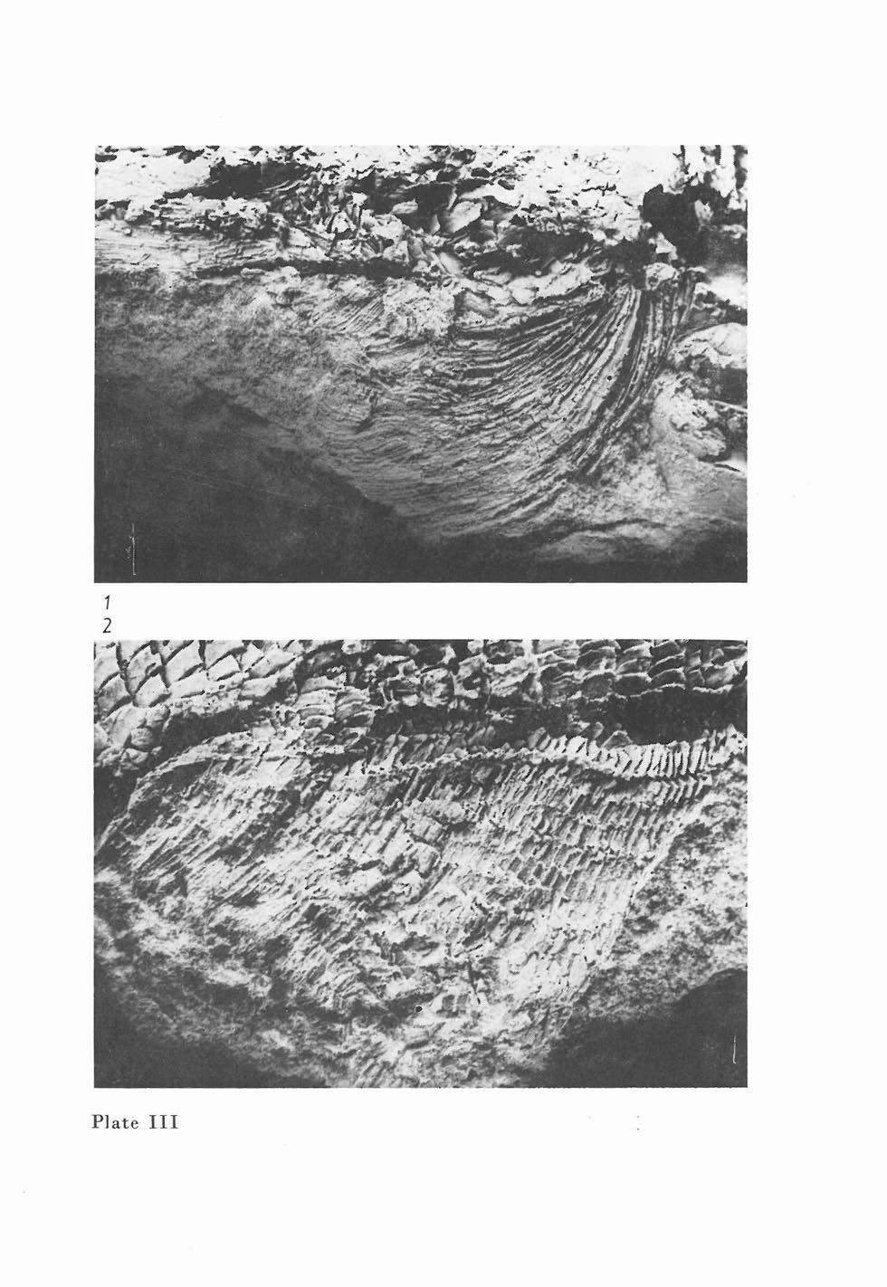
83 Pl ate
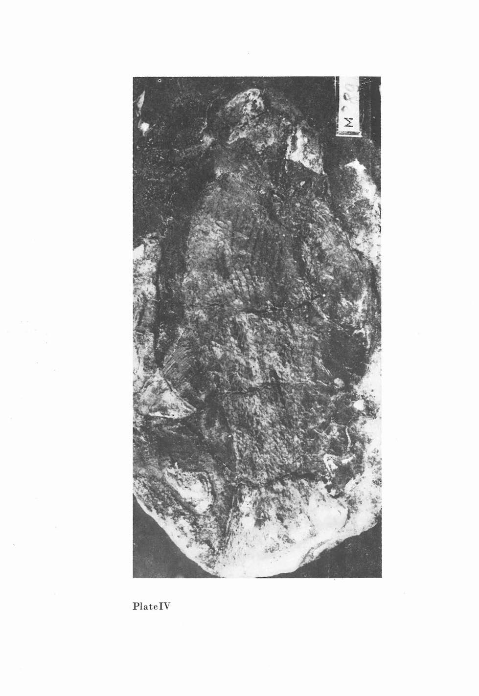
84 PlateV

85 1 2 Plate V
86 pq r fr no P la te V
87 Plate V
88 1 2 Plate V
89 2 Plate X

90 sop po - ptf - sci - - mx ---'-- sci fr Plate X
91 2 3 Plate X
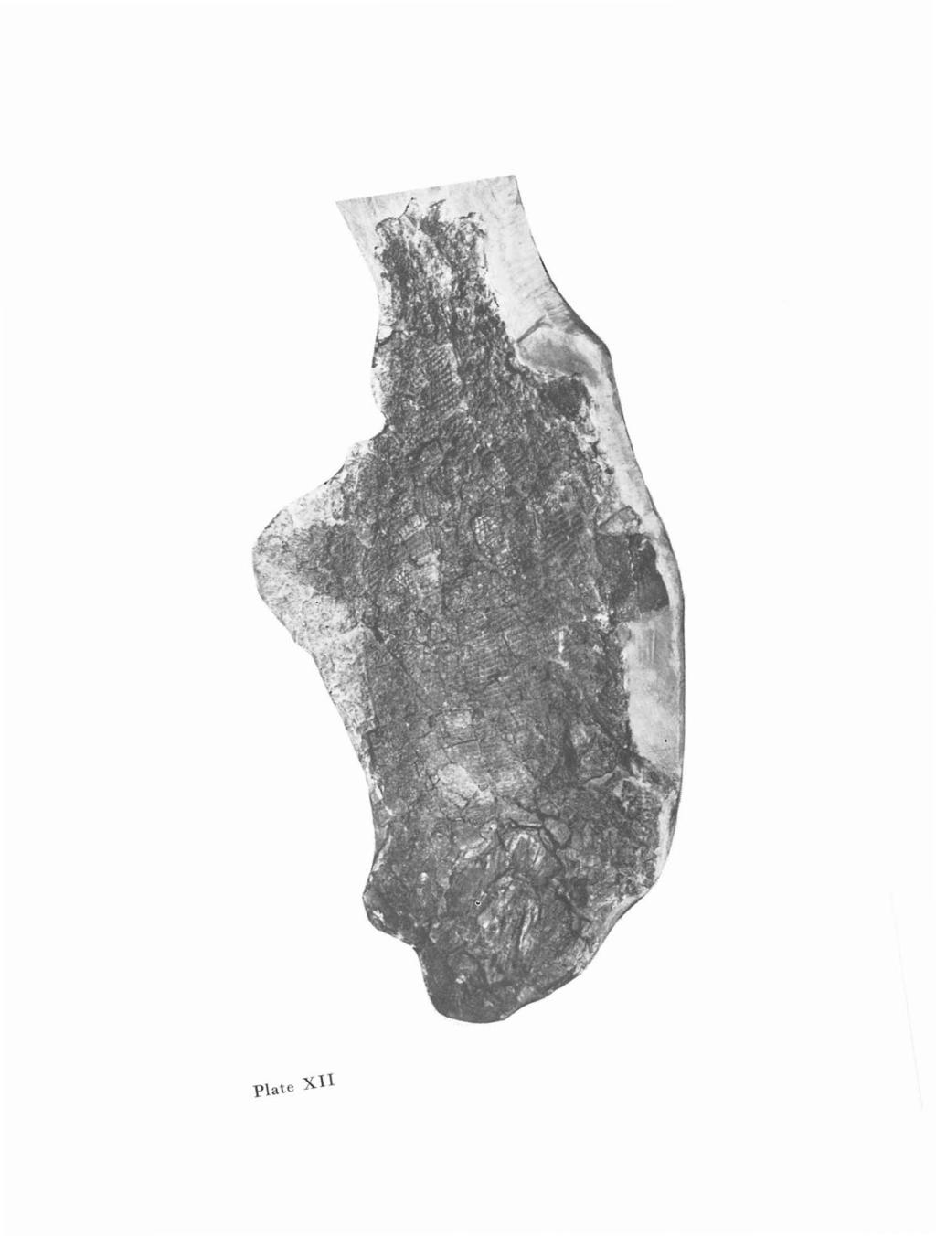
92 -Plate -X

93 .., , n a Plate X
94 1 3 2 Plate XV
95 1 2 Plate XV

96 ,( r dpt - - soc po po.,.,...~...jl'ii'jll p.t ext.t Plate XV

97 - - pop! ~~.f op m x mg ~ s - - ms Mr=" Plate XV

98 1 ju psph mx 2 1 r Plate XV
99 2 Plate XX
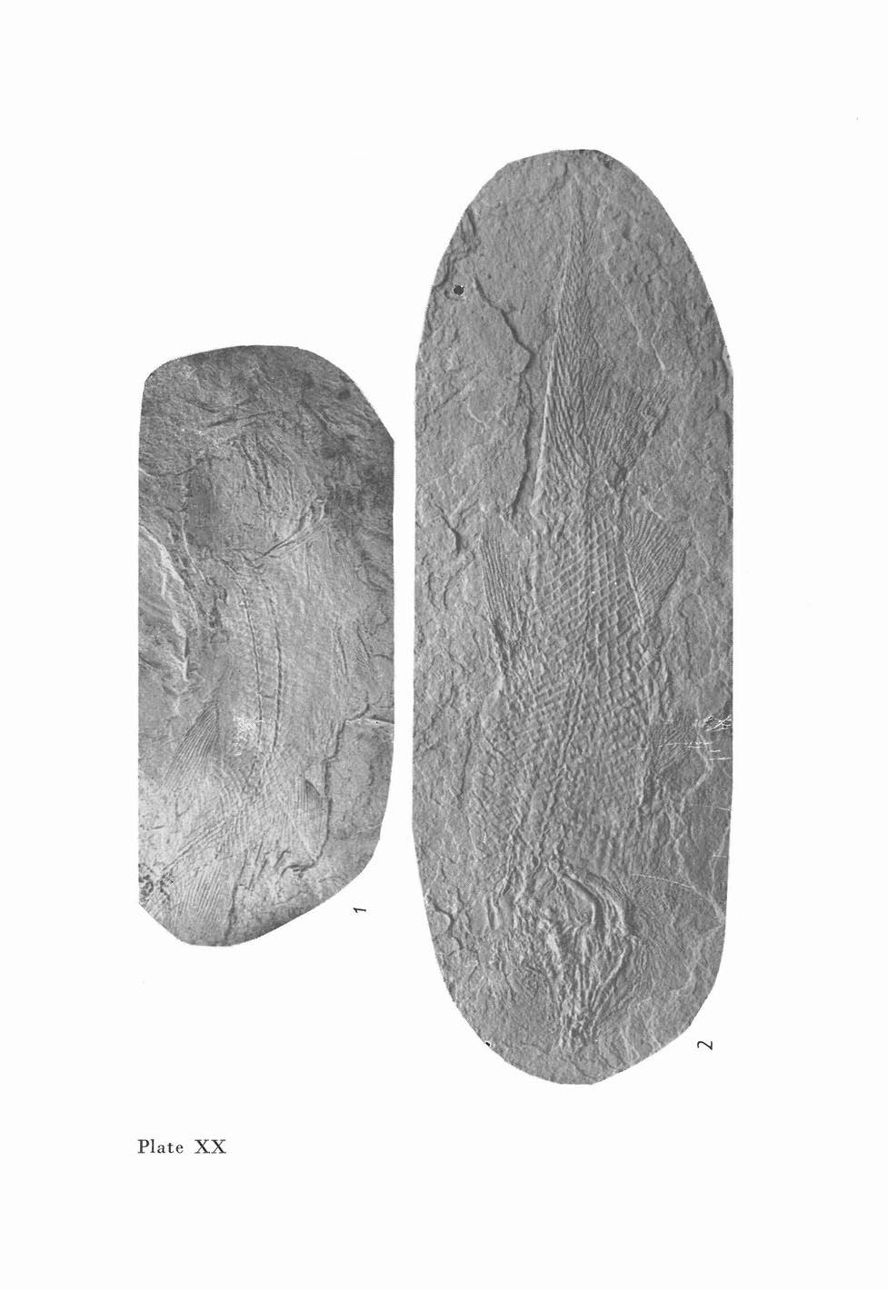
100 Plate XX
101 Plate XX
102 Plate XX 2 3
103 sci -- pop _ ~-..~1u. ~~:Hl'!"' ~1l'4~ mx sop -- r. - P late XX

104 2 Plate XXV
Jurassic Fish and their Stratigraphic significance from the Hengyang Region of Lingling Co., Hunan Province
 Jurassic Fish and their Stratigraphic significance from the Hengyang Region of Lingling Co., Hunan Province Nianzhong Wang Institute of Vertebrate Paleontology and Paleoanthropology, Academia Sinica Vertebrata
-8- spinous. nape caudal fin. body depth. pectoral fin. anus. total length Fig. 4
 click for previous page -8-1.3 Illustrated Glossary of Technical Terms and Measurements External Morphology and Measurements spinous dorsal fin soft nape caudal fin interorbital body depth snout lateral
2. SYSTEMATIC CATALOGUE
 click for previous page 15 2. SYSTEMATIC CATALOGUE 2.1 General Aids to Identification 2.1.1 Diagnostic Features of the Family Caesionidae Oblong to fusiform, moderately compressed, medium-sized to small
LIBRARY. Class\ V"^ A *Ii:T_
 LIBRARY Class\ V"^ A *Ii:T_ ^ Publications OP FIELD MUSEUM OF NATURAL HISTORY ZOOLOGICAL SERIES Volume X Chicago, U. S. A. 1909-1923 7/,3 ^Issued September 18, 19 12. 69 NEW SPECIES OF FISHES FROM
A new aeduellid actinopterygian from the Lower Permian of the Krkono e Piedmont Basin (Bohemian Massif) and its relationship to other Aeduellidae
 and its relationship to other Aeduellidae") A new aeduellid actinopterygian, Neslovicella elongata sp. nov., is described from the Lower Permian of the Krkonoše Piedmont Basin (Bohemian Massif). The new taxon is based upon a collection of specimens
A new aeduellid actinopterygian, Neslovicella elongata sp. nov., is described from the Lower Permian of the Krkonoše Piedmont Basin (Bohemian Massif). The new taxon is based upon a collection of specimens
* A New Species of Cichlid Fish From Lake Malawi. Pseudotropheus tursiops, \(I75 Tropical Fish Hobbyist a'l (3) : 8 L-? 0. ,$ IOU.
 : 8 L-? 0. ,$ IOU.") ,$ IOU. \(I75 Tropical Fish Hobbyist a'l (3) : 8 L-? 0. * 2.37 Pseudotropheus tursiops, A New Species of Cichlid Fish From Lake Malawi by Warren E. Burgess and Dr. Herbert R. Axelrod Among the cichlid
,$ IOU. \(I75 Tropical Fish Hobbyist a'l (3) : 8 L-? 0. * 2.37 Pseudotropheus tursiops, A New Species of Cichlid Fish From Lake Malawi by Warren E. Burgess and Dr. Herbert R. Axelrod Among the cichlid
NEW DATA ON THE OSTEOLOGY OF THE ACTINOPTERYGIAN FISH AMBLYPTERUS AND THE RELATIONSHIP BETWEEN AMBLYPTERUS AND PARAMBLYPTERUS
 SBORNÍK NÁRODNÍHO MUZEA V PRAZE ACTA MUSEI NATIONALIS PRAGAE Řada B Přírodní vědy sv. 69 2013 čís. 3 4 s. 183 194 Series B Historia Naturalis vol. 69 2013 no. 3 4 pp. 183 194 NEW DATA ON THE OSTEOLOGY
- 7 - DESCRIPTION OF SPECIES
 I - 7 - DESCRIPTION OF SPECIES./' Anguilla bicolor McClelland ' Level-finned eel (Figs.i & 2) Length of head 6-8 times in length of body; Diameter of eye 8-10 times, Inter-orbital length 2-2.5 times, Gape
Natural History of Vertebrates Characters Used in Fish Identification (modified )
") Natural History of Vertebrates Characters Used in Fish Identification 1-9-03 (modified 20050118) This lab is designed to familiarize the student with characters used in the identification of fishes. Only
Natural History of Vertebrates Characters Used in Fish Identification 1-9-03 (modified 20050118) This lab is designed to familiarize the student with characters used in the identification of fishes. Only
Landmarking protocol
 Landmarking protocol Jonathan Chang Introduction You will be marking key points on images of fish, which will help determine the shape of different fishes and how that affects their performance in the
FAO SPECIES IDENTIFICATION SHEETS MUGILOIDIDAE. (Parapercidae of some authors) Sandsmelts, sandperches, grubfishes
 Sandsmelts, sandperches, grubfishes") click for previous page MUGILO 1983 FAO SPECIES IDENTIFICATION SHEETS FISHING AREA 51 (W. Indian Ocean) MUGILOIDIDAE (Parapercidae of some authors) Sandsmelts, sandperches, grubfishes Body elongate, subcylindrical,
click for previous page MUGILO 1983 FAO SPECIES IDENTIFICATION SHEETS FISHING AREA 51 (W. Indian Ocean) MUGILOIDIDAE (Parapercidae of some authors) Sandsmelts, sandperches, grubfishes Body elongate, subcylindrical,
click for previous page D E
 click for previous page D E DREP FAO SPECIES IDENTIFICATION SHEETS 1974 FISHING AREAS 57, 71 (E Ind. Ocean) (W Cent. Pacific) DREPANIDAE Sicklefishes (placed by some authors, together with the Platacidae,
Field Identification of Tunas from Indian Waters
 3 Field from Indian Waters Subal Kumar Roul and Retheesh T. B. Pelagic Fisheries Division The Family Scombridae is one of the largest and most economically important fish family which comprises of most
Larvae of two Indo-West Pacific anthiine fishes, Giganthias immaculatus and Serranocirrhitus latus (Perciformes: Serranidae)
") Larvae of two Indo-West Pacific anthiine fishes, Giganthias immaculatus and Serranocirrhitus latus (Perciformes: Serranidae) Yoshinobu Konishi, Chiyuki Sassa and Makoto Okamoto Seikai National Fisheries
Larvae of two Indo-West Pacific anthiine fishes, Giganthias immaculatus and Serranocirrhitus latus (Perciformes: Serranidae) Yoshinobu Konishi, Chiyuki Sassa and Makoto Okamoto Seikai National Fisheries
FAO SPECIES IDENTIFICATION SHEETS CONGIOPODIDAE* Horsefishes
 click for previous page CONGIO 1983 FAO SPECIES IDENTIFICATION SHEETS FISHING AREA 51 (W. Indian Ocean) CONGIOPODIDAE* Horsefishes Bottom fishes, with large heads and strongly compressed bodies. Snout
FAO SPECIES IDENTIFICATION SHEETS KUHLIIDAE * Flagtails, daras
 click for previous page KUH 1983 FAO SPECIES IDENTIFICATION SHEETS FISHING AREA 51 (W. Indian Ocean) KUHLIIDAE * Flagtails, daras Body oblong, compressed. Maxilla mostly exposed, without supramaxilla;
Materials: Field notebook and pencil INTRODUCTION:
 Field Methods of Fish Biology 2014 Exercise 1: Basic Anatomy and Finding and Measuring Characters *Labs modified from Caillet et al. 1986 and Eric Schultz s Biology of Fishes lab Materials: Field notebook
TWO NEW SPECIES OF ARGULUS MULLER (CRUSTACEA: BRANCHIURA) FROM RIVER CAUVERY WITH A KEY TO INDIAN SPECIES
 FROM RIVER CAUVERY WITH A KEY TO INDIAN SPECIES") TWO NEW SPECIES OF ARGULUS MULLER (CRUSTACEA: BRANCHIURA) FROM RIVER CAUVERY WITH A KEY TO INDIAN SPECIES M. M. THOMAS AND M. DEVARAJ Central Marine Fisheries Research Institute Regional Centre, Mandapam
TWO NEW SPECIES OF ARGULUS MULLER (CRUSTACEA: BRANCHIURA) FROM RIVER CAUVERY WITH A KEY TO INDIAN SPECIES M. M. THOMAS AND M. DEVARAJ Central Marine Fisheries Research Institute Regional Centre, Mandapam
FAO SPECIES IDENTIFICATION SHEETS ACROPOMATIDAE. (= "Percichthyidae") Glow-bellies and splitfins
 Glow-bellies and splitfins") click for previous page ACRO 1983 FAO SPECIES IDENTIFICATION SHEETS FISHING AREA 51 (W. Indian Ocean) ACROPOMATIDAE (= "Percichthyidae") Glow-bellies and splitfins Body oblong, more or less compressed.
click for previous page ACRO 1983 FAO SPECIES IDENTIFICATION SHEETS FISHING AREA 51 (W. Indian Ocean) ACROPOMATIDAE (= "Percichthyidae") Glow-bellies and splitfins Body oblong, more or less compressed.
Slide 1. Slide 1. Next. 5:30:08 AM
 Slide 1 Slide 1 http://www3.utep.edu/leb/mosquito/larvslide1.htm10/27/2004 5:30:08 AM Slide 1 Slide 2 Recognition that the specimens are mosquito larvae is a prerequisite to identification of the genera.
Microbrotula randalli Cohen and Wourms, Samoa and Vanuatu at 30 to 38 m near reef-sand interface. Rare.
 click for previous page Ophidiiform Fishes of the World 107 Diagnosis and description: Body completely covered with small imbricate scales; head partly naked; snout depressed; eyes small, more than 6 times
FAO SPECIES IDENTIFICATION SHEETS SYNODONTIDAE. Lizardfishes
 click for previous page SYNOD 1474 FAO SPECIES IDENTIFICATION SHEETS FISHING AREAS 57,71 (E Ind. Ocean) (W Cent. Pacific) SYNODONTIDAE Lizardfishes Body elongate, usually cylindrical and with adipose fin.
NOVITATES PUBLISHED BY THE AMERICAN MUSEUM OF NATURAL HISTORY CITY OF NEW YORK APRIL 27, 1954 NUMBER 1655
 AtMERIICAN MUSEUM NOVITATES PUBLISHED BY THE AMERICAN MUSEUM OF NATURAL HISTORY CITY OF NEW YORK APRIL 27, 1954 NUMBER 1655 Review of the Deep-Sea Fishes of the Genus Asquamiceps Zugmayer, With Descriptions
oxfitates AMiiiui?can JMllselIm Threadfin from New Guinea BY J. T. NICHOLS A New Blenny from Bali and a New
 AMiiiui?can JMllselIm oxfitates PUBLISHED BY THE AMERICAN MUSEUM OF NATURAL HISTORY CENTRAL PARK WEST AT 79TH STREET, NEW YORK 24, N.Y. NUMBER i68o JUNE 30, 1954 A New Blenny from Bali and a New Threadfin
soft dorsal-fin and caudal-fin margin pale small embedded scales on maxilla
 click for previous page 82 FAO Species Catalogue Vol. 16 13a. Juveniles brown, with 7 dark brown bars on body, the last covering most of caudal peduncle, its upper half black; distance between fourth and
OCCASIONAL PAPERS OF THE MUSEUM OF ZOOLOGY UNIVERSITY OF MICHIGAN
 OCCASIONAL PAPERS OF THE MUSEUM OF ZOOLOGY UNIVERSITY OF MICHIGAN NOTES ON THE GOBIOID FISHES OF CALIFORNIA, WITH DESCRIPTIONS OF TWO NEW GENERA These notes result from our collecting along the Californian
TWO NEW SPECIES OF COPEPODS JAPANESE FISHES. Author(s) Yamaguti, Satyu; Yamasu, Terufumi.
 Yamaguti, Satyu; Yamasu, Terufumi.") Title TWO NEW SPECIES OF COPEPODS JAPANESE FISHES PARASIT Author(s) Yamaguti, Satyu; Yamasu, Terufumi Citation PUBLICATIONS OF THE SETO MARINE BIO LABORATORY (1960), 8(1): 137-140 Issue Date 1960-05-30
Title TWO NEW SPECIES OF COPEPODS JAPANESE FISHES PARASIT Author(s) Yamaguti, Satyu; Yamasu, Terufumi Citation PUBLICATIONS OF THE SETO MARINE BIO LABORATORY (1960), 8(1): 137-140 Issue Date 1960-05-30
Richard LUND 18 Hillside road, Mount Holly, New Jersey (USA)
") Three new palaeoniscoid fishes from the Bear Gulch Limestone (Serpukhovian, Mississippian) of Montana (USA) and the relationships of lower actinopterygians Kathryn E. MICKLE University of Kansas, Department
Three new palaeoniscoid fishes from the Bear Gulch Limestone (Serpukhovian, Mississippian) of Montana (USA) and the relationships of lower actinopterygians Kathryn E. MICKLE University of Kansas, Department
30 a. Allothunnus fallai Fig b.
 click for previous page - 18-30 a. Jaw teeth tiny, 40 to 55 on each side of upper and lower jaws; gillrakers fine and numerous, total of 70 to 80 on first arch; body elongate; distance from snout to second
FAO SPECIES IDENTIFICATION SHEETS FISTULARIIDAE. Cornetfishes, flutemouths
 click for previous page FIST 1982 FAO SPECIES IDENTIFICATION SHEETS FISHING AREA 51 (W. Indian Ocean) FISTULARIIDAE Cornetfishes, flutemouths Body elongate and depressed. Mouth small, at end of a long
BONY FISHES TECHNICAL TERMS
 previous page 1 TECHNICAL TERMS Principal Measurements Used (shortest distance between the points marked) preorbital length head length eye standard length postorbital length 1st dorsal fin base total
Class Myxini Order Myxiniformes Family Myxinidae (hagfishes)
") Class Myxini Order Myxiniformes Family Myxinidae (hagfishes) Lacks jaws Mouth not disk-like barbels present Unpaired fins as continuous fin-fold Branchial skeleton not well developed Eyes degenerate 70-200
Class Myxini Order Myxiniformes Family Myxinidae (hagfishes) Lacks jaws Mouth not disk-like barbels present Unpaired fins as continuous fin-fold Branchial skeleton not well developed Eyes degenerate 70-200
THEJ FIRST ZOEA OF PORCELLANA. By W K. BROOKS and E. B. WILSON. With Plate* VI and VII.
 7 "83?* {Jn-fJatoj 11 "T THEJ FIRST ZOEA OF PORCELLANA. By W K BROOKS and E. B. WILSON. With Plate* VI and VII. 7z O THE FIRST ZOEA OF PORCELLANA. By W K. BROOKS and E. B. WILSON. With Plates VI and VII.
FAO SPECIES IDENTIFICATION SHEETS CAESIONIDAE. Fusiliers
 click for previous page CAES FAO SPECIES IDENTIFICATION SHEETS FISHING AREA 51 (W. Indian Ocean) CAESIONIDAE Fusiliers Lutjanoid fishes, moderately deep-bodied to slender and fusiform, laterally compressed.
THREE NEANURID COLLEMBOLA FROM THE MARQUESAS ISLANDS
 Pacific Insects 9 (2) : 265-270 20 June 1967 THREE NEANURID COLLEMBOLA FROM THE MARQUESAS ISLANDS By Riozo Yosii YOSHIDA COLLEGE, KYOTO UNIVERSITY, JAPAN Abstract: Three neanurid species described by Carpenter
Chlorurus japanensis (Bloch, 1789) (Plate VIII, 57 and 58)
 (Plate VIII, 57 and 58)") click for previous page Perciformes: Labroidei: Scaridae 3477 Chlorurus japanensis (Bloch, 1789) (Plate VIII, 57 and 58) En - Palecheek parrotfish; Sp - Loro rostro pálido. Maximum standard length about
click for previous page Perciformes: Labroidei: Scaridae 3477 Chlorurus japanensis (Bloch, 1789) (Plate VIII, 57 and 58) En - Palecheek parrotfish; Sp - Loro rostro pálido. Maximum standard length about
34. Records of the Zoological Surt'ey 01 India
 34. Records of the Zoological Surt'ey 01 India study, the occurrence and density of different groups seems to be influenced by the temperature. The temperature variance causes presence of less population,
Remarks: Allen & Swainston (1988) have erroneously recorded this species from northwestern Australia.
 have erroneously recorded this species from northwestern Australia.") click for previous page 71 Literature: Masuda et al. (1975, 1984); Kyushin et al. (1977); Smith (1977); Randall et al. (1978, as P. townsendi); Rau & Rau (1980); Kyushin et al. (1982); Fischer & Bianchi
click for previous page 71 Literature: Masuda et al. (1975, 1984); Kyushin et al. (1977); Smith (1977); Randall et al. (1978, as P. townsendi); Rau & Rau (1980); Kyushin et al. (1982); Fischer & Bianchi
THE GENUS PHOTIS (CRUSTACEA: AMPHIPODA) FROM THE TEXAS COAST WITH THE DESCRIPTION OF A NEW SPECIES, PHOTIS MELANICUS
 FROM THE TEXAS COAST WITH THE DESCRIPTION OF A NEW SPECIES, PHOTIS MELANICUS") THE GENUS PHOTIS (CRUSTACEA: AMPHIPODA) FROM THE TEXAS COAST WITH THE DESCRIPTION OF A NEW SPECIES, PHOTIS MELANICUS L. D. McKinney Texas A&M University at Galveston, Moody College of Marine Technology,
THE GENUS PHOTIS (CRUSTACEA: AMPHIPODA) FROM THE TEXAS COAST WITH THE DESCRIPTION OF A NEW SPECIES, PHOTIS MELANICUS L. D. McKinney Texas A&M University at Galveston, Moody College of Marine Technology,
A NEW SPECIES OF ASELLOTE MARINE ISOPOD, MUNNA (UROMUNNA) HAYESI (CRUSTACEA: ISOPODA) FROM TEXAS
 HAYESI (CRUSTACEA: ISOPODA) FROM TEXAS") A NEW SPECIES OF ASELLOTE MARINE ISOPOD, MUNNA (UROMUNNA) HAYESI (CRUSTACEA: ISOPODA) FROM TEXAS Philip B. Robertson Biology Department, Lamar University, Beaumont, Texas 77710 A new species of asellote
A NEW SPECIES OF ASELLOTE MARINE ISOPOD, MUNNA (UROMUNNA) HAYESI (CRUSTACEA: ISOPODA) FROM TEXAS Philip B. Robertson Biology Department, Lamar University, Beaumont, Texas 77710 A new species of asellote
64 FAO Species Catalogue Vol. 18
 click for previous page 64 FAO Species Catalogue Vol. 18 Epetriodus Cohen and Nielsen, 1978 Type species: Epetriodus freddyi Cohen and Nielsen, 1978 by original designation. Fig. 61 Epetriodus freddyi
Sphagemacrurus Fowler, MACROUR Spha. S. decimalis Fig Genus with Reference : Sphagemacrurus Collett, 1896, by original designation).
.") click for previous page 287 Sphagemacrurus Fowler, 1925 MACROUR Spha Genus with Reference : Sphagemacrurus Collett, 1896, by original designation). Fowler, 1925, Am.Mus. Novit. 162:3 (type species Macrurus
click for previous page 287 Sphagemacrurus Fowler, 1925 MACROUR Spha Genus with Reference : Sphagemacrurus Collett, 1896, by original designation). Fowler, 1925, Am.Mus. Novit. 162:3 (type species Macrurus
GROUP II : 2a. Dorsal fin high, spinous second ray about as long as, or longer than, head length
 click for previous page 123 GROUP II : 1a. A prominent blackish blotch, sometimes ocellated, above and behind pectoral fin base; anus slightly removed from anal fin; underside of snout ; anterolateral
Freshwater actinopterygians of the Los Rastros Formation (Triassic), Bermejo Basin, Argentina
, Bermejo Basin, Argentina") Fossil Record 9(2) (2006), 238 258 / DOI 10.1002/mmng.200600011 Freshwater actinopterygians of the Los Rastros Formation (Triassic), Bermejo Basin, Argentina Adriana López-Arbarello*, 1, Raymond Rogers
Fossil Record 9(2) (2006), 238 258 / DOI 10.1002/mmng.200600011 Freshwater actinopterygians of the Los Rastros Formation (Triassic), Bermejo Basin, Argentina Adriana López-Arbarello*, 1, Raymond Rogers
FAO SPECIES IDENTIFICATION SHEETS ALBULIDAE. Bonefishes
 click for previous page ALBU 1983 FAO SPECIES IDENTIFICATION SHEETS FISHING AREA 51 (W. Indian Ocean) ALBULIDAE Bonefishes Elongate, fusiform fishes possessing a distinctive conical snout projecting beyond
Elasmobranches Found in the Bay of Bengal from Pelagic Longline and Drift Gill Net Fishing
 Elasmobranches Found in the Bay of Bengal from Pelagic Longline and Drift Gill Net Fishing Tassapon Krajangdara 1, Reangchai Sujittosakul 2 and Md. Jalilur Rahman 3 1 Andaman Sea Fisheries Research and
Description of the Immature Stages of Galindomyia leei Stone and Barreto, 1969l. Abdiel J. Adames* and Pedro Galindo*
 132 Description of the Immature Stages of Galindomyia leei Stone and Barreto, 1969l Abdiel J. Adames* and Pedro Galindo* The genus and species Galindomyia Zeei, a member of the tribe Culicini, was described
Key to the Species of Anamixis
 Key to the Species of Anamixis (Except A. stebbingi*) 1. Article 2 of gnathopod 2 with 3 or more long setae on anterior margin (1)...2 1. Article 2 of gnathopod 2 lacking long setae on anterior margin...3
PROCEEDINGS i j OF THE BIOLOGICAL SOCIETY OF WASHINGTON
 e3 if Vol. 86, No. 27, pp. 333-338 28 September 1973 PROCEEDINGS i j OF THE ' BIOLOGICAL SOCIETY OF WASHINGTON A NEW SPECIES OF SESARMA, S. (HOLOMETOPUS) RUBINOFFORUM, FROM THE PACIFIC COAST OF PANAMA
FAO SPECIES IDENTIFICATION SHEETS. FAMILY: LETHRINIDAE FISHING AREA 51 (W. Indian Ocean) Lethrinus conchyliatus (Smith, 1959)
 Lethrinus conchyliatus (Smith, 1959)") click for previous page LETH Leth 9 1983 FAO SPECIES IDENTIFICATION SHEETS FAMILY: LETHRINIDAE FISHING AREA 51 (W. Indian Ocean) Lethrinus conchyliatus (Smith, 1959) OTHER SCIENTIFIC NAMES STILE IN USE:
click for previous page LETH Leth 9 1983 FAO SPECIES IDENTIFICATION SHEETS FAMILY: LETHRINIDAE FISHING AREA 51 (W. Indian Ocean) Lethrinus conchyliatus (Smith, 1959) OTHER SCIENTIFIC NAMES STILE IN USE:
Coryphaenoides longifilis (Günther, 1877) Fig MACROUR Cory 9
 Fig MACROUR Cory 9") click for previous page 211 First dorsal fin with 2 spines and 9 to 11 rays; teeth along leading edge of spinous first dorsal ray slender, sharp, and prominent; pectoral fin rays i18 to i21; pelvic fin
click for previous page 211 First dorsal fin with 2 spines and 9 to 11 rays; teeth along leading edge of spinous first dorsal ray slender, sharp, and prominent; pectoral fin rays i18 to i21; pelvic fin
Tips for Identifying Common Fish Species in the Bush River
 Tips for Identifying Common Fish Species in the Bush River Juvenile Largemouth bass Spot Threadfin shad Juvenile White perch The Herrings (Family Clupeidae) Alewife American shad Blueback herring Atlantic
DESCRIPTION OF A BATHYPELAGIC FISH, LESTIDIUM BLANCI SP. NOV. (FAMILY PARALEPIDIDAE) FROM THE ARABIAN SEA*
 FROM THE ARABIAN SEA*") J.: mar. biol. Ass. India, 10, 12 (1 & 2): 146-150 DESCRIPTION OF A BATHYPELAGIC FISH, LESTIDIUM BLANCI SP. NOV. (FAMILY PARALEPIDIDAE) FROM THE ARABIAN SEA* K. N. RASACHANDRA KARTHA Central Marine Fisheries
J.: mar. biol. Ass. India, 10, 12 (1 & 2): 146-150 DESCRIPTION OF A BATHYPELAGIC FISH, LESTIDIUM BLANCI SP. NOV. (FAMILY PARALEPIDIDAE) FROM THE ARABIAN SEA* K. N. RASACHANDRA KARTHA Central Marine Fisheries
FAO SPECIES IDENTIFICATION SHEETS. FAMILY: SIGANIDAE FISHING AREA 51 (W. Indian Ocean) Siganis rivulatus Forsskål, 1775
 Siganis rivulatus Forsskål, 1775") click for previous page SIGAN Sigan 1 1983 FAO SPECIES IDENTIFICATION SHEETS FAMILY: SIGANIDAE FISHING AREA 51 (W. Indian Ocean) Siganis rivulatus Forsskål, 1775 OTHER SCIENTIFIC NAMES STILL IN USE : Teuthis
click for previous page SIGAN Sigan 1 1983 FAO SPECIES IDENTIFICATION SHEETS FAMILY: SIGANIDAE FISHING AREA 51 (W. Indian Ocean) Siganis rivulatus Forsskål, 1775 OTHER SCIENTIFIC NAMES STILL IN USE : Teuthis
-G\Su---c9 ~ ~ THE KANSA$ $CHOOL NATURALIST THE CARP A MANUAL STRESSING OBSERVATION. Kansas State Teachers College Emporia, Kansas. No.3. Vo l.
 THE CARP A MANUAL STRESSNG OBSERVATON -G\Su---c9 ~ ~ THE KANSA$ $CHOOL NATURALST Kansas State Teachers College Emporia, Kansas Vo l. 22 No.3 February 1976 The Ka nsa s School N aturalist Published by Emporia
SYNISOMA WETZERAE, A NEW SPECIES AND THE FIRST RECORD OF SYNISOMA FROM THE NEW WORLD (CRUSTACEA: ISOPODA: VALVIFERA: IDOTEIDAE) Bernard Ormsby
 Bernard Ormsby") . ^ - $. ^ tf\ &r*>fy ft( 18 December 1991 PROC. BIOL. SOC. WASH. 104(4), 1991, pp. 758-763 SYNISOMA WETZERAE, A NEW SPECIES AND THE FIRST RECORD OF SYNISOMA FROM THE NEW WORLD (CRUSTACEA: ISOPODA: VALVIFERA:
. ^ - $. ^ tf\ &r*>fy ft( 18 December 1991 PROC. BIOL. SOC. WASH. 104(4), 1991, pp. 758-763 SYNISOMA WETZERAE, A NEW SPECIES AND THE FIRST RECORD OF SYNISOMA FROM THE NEW WORLD (CRUSTACEA: ISOPODA: VALVIFERA:
Fish. Water Dwelling Animals
 Fish Water Dwelling Animals Class Agnatha (Jawless fish) They are believed to be the most primitive and oldest vertebrates. Lamprey and hagfish are the only 2 living members of this class and are placed
click for previous page 245 General Remarks STOMATOPODS by M. Tavares, Universidade Santa Úrsula, Brazil
 click for previous page 245 General Remarks STOMATOPODS by M. Tavares, Universidade Santa Úrsula, Brazil 246 Stomatopods TECHNICAL TERMS AND MEASUREMENTS antennule eye carapace thoracic stomites 5 6 7
Goldfish Varieties Ranchu By Peter J. Ponzio
 Goldfish Varieties Ranchu By Peter J. Ponzio In the original article for this series, we defined a number of characteristics common to all goldfish, and introduced the concept of goldfish varieties, or
Haemulon chrysargyreum Günther, 1859
 click for previous page 1538 Bony Fishes Haemulon chrysargyreum Günther, 1859 Frequent synonyms / misidentifications: Brachygenys chrysargyreus (Günther, 1859 ) / None. FAO names: En - Smallmouth grunt;
Handbook for the identification of yellowfin and bigeye tunas in fresh, but less than ideal condition
 0 WCPFC SC1 FT IP 1 1st Meeting of the Scientific Committee of the Western and Central Pacific Fisheries Commission WCPFC SC1 Noumea, New Caledonia 8 19 August 2005 Handbook for the identification of yellowfin
Etmopterus decacuspidatus Chan, 1966 SQUAL Etmo 8
 click for previous page - 75 - Etmopterus decacuspidatus Chan, 1966 SQUAL Etmo 8 Etmopterus decacuspidatus Chan, 1966, J.Zool., Proc.Zool.Soc.London, 148:220, fig. 1, P1. 1a. Holotype: British Museum (Natural
Dogfish Shark Dissection Introduction 1. What are two reasons why spiny dogfish are used for study in laboratories?
 Dogfish Shark Dissection Introduction 1. What are two reasons why spiny dogfish are used for study in laboratories? 2. Someone who studies fish is called an. 3. Sharks and fish belong to the Phylum a.
Apogon abrogramma Fraser and Lachner, 1985
 Apogon abrogramma Fraser and Lachner, 1985 English Name: Faintstripe cardinalfish Family: APOGONIDAE Local Name: Ehrongu boadhi Order: Perciformes Size: Common to 7cm; max. 10cm Specimen: MRS/0203/88 Distinctive
Lubbockichthys myersi, a new species of dottyback fish from Guam (Pseudochromidae: Pseudoplesiopinae)
") Zootaxa : 43 48 (2006) www.mapress.com/zootaxa/ Copyright 2006 Magnolia Press ISSN 1175-5326 (print edition) ISSN 1175-5334 (online edition) Lubbockichthys myersi, a new species of dottyback fish from
Zootaxa : 43 48 (2006) www.mapress.com/zootaxa/ Copyright 2006 Magnolia Press ISSN 1175-5326 (print edition) ISSN 1175-5334 (online edition) Lubbockichthys myersi, a new species of dottyback fish from
Thresher Shark Alopias vulpinus
 Thresher Shark Alopias vulpinus 30 Upper tooth Lower tooth Diagnosis The only Alaska species of shark with a distinctly elongate upper caudal-fin lobe. Dentition Teeth small, somewhat triangular, without
SOME ADDITIONAL NOTES ON EXTERNAL FEATURES AND ON THE JAW MUSCLES OF ORTHRAGORISCUS MOLA (L.)
") SOME ADDITIONAL NOTES ON EXTERNAL FEATURES AND ON THE JAW MUSCLES OF ORTHRAGORISCUS MOLA (L.) by J. M. VAN ROON In 1939 J. M. van Roon and J. J. ter Pelkwijk published some data on two specimens of Orthragoriscus
SOME ADDITIONAL NOTES ON EXTERNAL FEATURES AND ON THE JAW MUSCLES OF ORTHRAGORISCUS MOLA (L.) by J. M. VAN ROON In 1939 J. M. van Roon and J. J. ter Pelkwijk published some data on two specimens of Orthragoriscus
FAO SPECIES IDENTIFICATION SHEETS PSETTODIDAE. Spiny turbots
 click for previous page PSET FISHING AREA 51 PSETTODIDAE Spiny turbots A single species in the area; see species sheet for: Psettodes erumei (Schneider, 1801) PSET Pset 1 Prepared by J. Nielsen, Zoologisk
FAO Names : En - Longsnout dogfish; Fr - Squale-savate à long nez; Sp - Tollo trompalarga.
 click for previous page - 69 - FAO Names : En - Longsnout dogfish; Fr - Squale-savate à long nez; Sp - Tollo trompalarga. Field Marks : Extremely long snout, no anal fin, grooved dorsal fin spines, compressed
2a. Uropodal peduncle with asymmetric terminal projection 3 b. Uropodal peduncle terminally truncate... Leptognathia sp. B
 Leptognathia Species la. Uropodla uniramous 2 b. Uropodla blramous 5 2a. Uropodal peduncle with asymmetric terminal projection 3 b. Uropodal peduncle terminally truncate... Leptognathia sp. B 3a. Uropodal
Seale 154 TYSON R. ROBERTS AND JUNYA JUMNONGTH AI Km. Figure 1.
 154 TYSON R. ROBERTS AND JUNYA JUMNONGTH A 0 100 Km. Seale Figure 1. M iocene intermontane lacustrine basins of Thailand. The Phetchabun intermontane basin is number 1 L. Fossil fish also have been found
Myripristis adusta Bleeker, 1853
 Myripristis adusta Bleeker, 1853 English Name: Shadowfin soldierfish Family: HOLOCENTRIDAE Local Name: Hiyani dhanbodu Order: Beryciformes Size: Common to 25 cm; max. 32 cm Specimen: MRS/0322/88 Distinctive
The Formation and Fate of the Operculum and Gill-chambers in the tadpole of Rana temporaria.
 The Formation and Fate of the Operculum and Gill-chambers in the tadpole of Rana temporaria. By Gwendolen T. Brock, M.Sc, D.PM1. (Oxon.). With 16 Text-figures. THIS work has been undertaken with the object
PHYLLOSOMAS OF SCYLLARID LOBSTERS FROM THE ARABIAN SEA* Central Marine Fisheries Research Station, Mandapam Camp
 /. Mar. biol. Ass. India, 1960, 2 (2): 241-249 PHYLLOSOMAS OF SCYLLARID LOBSTERS FROM THE ARABIAN SEA* By R. RAGHU PRASAD AND P. R. S. TAMPI Central Marine Fisheries Research Station, Mandapam Camp INTRODUCTION
Pterygotrigla macrorhynchus Kamohara, 1936
 click for previous page 2376 Bony Fishes Pterygotrigla macrorhynchus Kamohara, 1936 Frequent synonyms / misidentifications: Parapterygotyrigla macrorhynchus (Kamohara, 1936); Dixiphistes macrorhynchus
NATIONWIDE: GOLDFISH STANDARDS
 The NATIONWIDE: GOLDFISH STANDARDS of the United Kingdom Nationwide Goldfish Standards of the United Kingdom Revised & Printed January 16 Nationwide Goldfish Societies UK Comprising: Association of Midland
Using a Dichotomous Classification Key to Identify Common Freshwater Fish of New York State
 D3()O!PPPPPPPPPPPPPPPPPPPPPPPPPPPPPPPPPP!Q3*)!&'(N7)*)LO!PPPPPPPPPPPPPPPPPPPPPPPPPPPPP! &73,,O!PPPPPPPPPPPP!-34!#.06*),O!PPPPPPPPPPPPPPP!C)3B?)$O!PPPPPPPPPPPPPPPPPPPPPPPPPPPPPPP! Using a Dichotomous Classification
Internal Anatomy of Fish
 Internal Anatomy of Fish The Systems of a Fish Skeletal System Muscular System Respiratory System Digestive System Circulatory System Nervous System Reproductive System Special Organs Skeletal System
122 FAO Species Catalogue Vol. 18
 click for previous page 122 FAO Species Catalogue Vol. 18 Diagnosis and description: Skin over head and fin bases thick, less so over body, body covered with tiny scales arranged in regular rows; small
Revision 7/2/02 MEASURING WRAVMA DRAWING DESIGNS WITH SCORING TEMPLATES
 Revision 7/2/02 MEASURING WRAVMA DRAWING DESIGNS WITH SCORING TEMPLATES -these suggested criteria supplement the criteria in the WRAVMA manual *Note item designs depicted here are representational and
STUDIES IN ANIMAL LOCOMOTION
 39 1 STUDIES IN ANIMAL LOCOMOTION III. THE PROPULSIVE MECHANISM OF THE WHITING (GADUS MERLANGUS) BY J. GRAY. (From the Laboratory of Experimental Zoology, Cambridge.) {Received ^th February, 1933.) (With
FAO SPECIES IDENTIFICATION SHEETS NOMEIDAE. Man-of-war fishes, also driftfishes
 click for previous page NOM 1983 FAO SPECIES IDENTIFICATION SHEETS FISHING AREA 51 (W. Indian Ocean) NOMEIDAE Man-of-war fishes, also driftfishes Slender to deep, laterally compressed fishes (in Psenes
The Cephenniini of China. I. Neseuthia SCOTT of Fujian Province (Coleoptera: Scydmaenidae)
") Genus Vol. 16(2): 171-175 Wroc³aw, 30 VI 2005 The Cephenniini of China. I. Neseuthia SCOTT of Fujian Province (Coleoptera: Scydmaenidae) PAWE JA OSZYÑSKI Os. Wichrowe Wzgórze 22/13, 61-678 Poznañ, Poland,
Genus Vol. 16(2): 171-175 Wroc³aw, 30 VI 2005 The Cephenniini of China. I. Neseuthia SCOTT of Fujian Province (Coleoptera: Scydmaenidae) PAWE JA OSZYÑSKI Os. Wichrowe Wzgórze 22/13, 61-678 Poznañ, Poland,
FAO SPECIES IDENTIFICATION SHEETS NEMIPTERIDAE. (including Scolopsidae of authors) Threadfin breams, monocle breams and dwarf monocle breams
 Threadfin breams, monocle breams and dwarf monocle breams") click for previous page NEMIP 1983 FAO SPECIES IDENTIFICATION SHEETS FISHING AREA 51 (W. Indian Ocean) NEMIPTERIDAE (including Scolopsidae of authors) Threadfin breams, monocle breams and dwarf monocle
click for previous page NEMIP 1983 FAO SPECIES IDENTIFICATION SHEETS FISHING AREA 51 (W. Indian Ocean) NEMIPTERIDAE (including Scolopsidae of authors) Threadfin breams, monocle breams and dwarf monocle
SKRIFTER OM SVALBARD OG ISHAVET CTENASPIS
 DET KONGELIGE DEPARTEMENT FOR HANDEL, SJ0FART, INDUSTRI, HANDVERK OG FISKERI NORGES SVALBARD- OG ISHAVS-UNDERS0KELSER LEDER: ADOLF HOEL SKRIFTER OM SVALBARD OG ISHAVET Nr. 33 johan KIlER CTENASPIS A NEW
A New Species of Caenis Stephens (Ephemeroptera: Caenidae) from Taiwan
 from Taiwan") PRIVATE LIBRARY OF WILLIAM L. PETERS q:i~~fi. 16: 55-59( 1996) Chinese J. Entomol. 16: 55-59(1996) A New Species of Caenis Stephens (Ephemeroptera: Caenidae) from Taiwan Shih-Chang Kang* and Chung-Tu Yang
PRIVATE LIBRARY OF WILLIAM L. PETERS q:i~~fi. 16: 55-59( 1996) Chinese J. Entomol. 16: 55-59(1996) A New Species of Caenis Stephens (Ephemeroptera: Caenidae) from Taiwan Shih-Chang Kang* and Chung-Tu Yang
Perciformes: Stromateoidei: Nomeidae 3771 NOMEIDAE. Driftfishes (cigarfishes)
") click for previous page Perciformes: Stromateoidei: Nomeidae 3771 NOMEIDAE Driftfishes (cigarfishes) by P.R. Last Diagnostic characters: Small to medium-sized (to 1 m) fishes with a compressed, slender
click for previous page Perciformes: Stromateoidei: Nomeidae 3771 NOMEIDAE Driftfishes (cigarfishes) by P.R. Last Diagnostic characters: Small to medium-sized (to 1 m) fishes with a compressed, slender
YALE PEABODY MUSEUM OF NATURAL HISTORY. Number 71 November 14, 1962 New Haven, Conn.
 YALE PEABODY MUSEUM OF NATURAL HISTORY Number 71 November 14, 1962 New Haven, Conn. SCOPELOGADUS (?) CAPISTRANENSIS, A NEW FOSSIL MELAMPHAID (PISCES: TELEOSTEI) FROM CAPISTRANO BEACH, CALIFORNIA ALFRED
Oreoglanis infulatus, a new species of glyptosternine catfish (Siluriformes: Sisoridae) from central Vietnam
 from central Vietnam") Journal of Fish Biology (2001) 59, 1164 1169 doi:10.1006/jfbi.2001.1730, available online at http://www.idealibrary.com on Oreoglanis infulatus, a new species of glyptosternine catfish (Siluriformes: Sisoridae)
Journal of Fish Biology (2001) 59, 1164 1169 doi:10.1006/jfbi.2001.1730, available online at http://www.idealibrary.com on Oreoglanis infulatus, a new species of glyptosternine catfish (Siluriformes: Sisoridae)
NATIONAL BIORESOURCE DEVELOPMENT BOARD Dept. of Biotechnology Government of India, New Delhi. General Category: Vertebrata (Zooplankton) Fish larvae
 Fish larvae") NATIONAL BIORESOURCE DEVELOPMENT BOARD Dept. of Biotechnology Government of India, New Delhi For office use: MARINE BIORESOURCES FORMS DATA ENTRY: Form- 1(general) Fauna: Flora Microorganisms General Category:
NATIONAL BIORESOURCE DEVELOPMENT BOARD Dept. of Biotechnology Government of India, New Delhi For office use: MARINE BIORESOURCES FORMS DATA ENTRY: Form- 1(general) Fauna: Flora Microorganisms General Category:
Colour: no distinctive markings. swimbladder ventral view
 click for previous page SCIAEN Penn 1 1974 FAO SPECIES IDENTIFICATION SHEETS FAMILY: SCIAENIDAE FISHING AREAS 57,71 (E Ind. Ocean) (W Cent. Pacific) Pennahia argentata (Houttuyn, 1782) SYNONYMS STILL IN
C. Lukhaup & R. Pekny
 Cherax (Cherax) holthuisi, a new species of crayfish (Crustacea: Decapoda: Parastacidae) from the centre of the Vogelkop Peninsula in Irian Jaya (West New Guinea), Indonesia C. Lukhaup & R. Pekny Lukhaup,
Pleuronectiformes: Pleuronectidae 3863 PLEURONECTIDAE. Righteye flounders
 click for previous page Pleuronectiformes: Pleuronectidae 3863 PLEURONECTIDAE Righteye flounders by D.A. Hensley Diagnostic characters: Body oval-shaped or elongate, strongly compressed (size to about
FEDERATION CYNOLOGIQUE INTERNATIONALE (AISBL) SECRETARIAT GENERAL: 13, Place Albert 1 er B 6530 Thuin (Belgique) FCI-Standard N 296 /
 SECRETARIAT GENERAL: 13, Place Albert 1 er B 6530 Thuin (Belgique) FCI-Standard N 296 /") FEDERATION CYNOLOGIQUE INTERNATIONALE (AISBL) SECRETARIAT GENERAL: 13, Place Albert 1 er B 6530 Thuin (Belgique) FCI-Standard N 296 / 28.10.2009 / GB COLLIE ( SMOOTH) 2 ORIGIN : Great Britain. DATE OF
FEDERATION CYNOLOGIQUE INTERNATIONALE (AISBL) SECRETARIAT GENERAL: 13, Place Albert 1 er B 6530 Thuin (Belgique) FCI-Standard N 296 / 28.10.2009 / GB COLLIE ( SMOOTH) 2 ORIGIN : Great Britain. DATE OF
Contribution to the knowledge of the fossil mammals from the ancient Tertiary terrains of Paraná. F. Ameghino * Tapiroidea
 Contribution to the knowledge of the fossil mammals from the ancient Tertiary terrains of Paraná F. Ameghino * Tapiroidea General characteristics: Upper molars with 2 transverse crests on the crown, each
Neatness 0 1 Accuracy Completeness Lab Class Procedure Total Lab Score
 New Paltz High School Name:... Due Date:... Science Department Lab Title: Identifying NY s Gamefish Lab #... Lab Partners: Your Lab Score will be based on the following: Neatness: All labs must be well-written
Longnose Skate Raja rhina
 Longnose Skate Raja rhina 42 dorsal ventral egg case Nuchal Orbital Dorsal diagram showing thorns on disc. Diagnosis The only Alaska species of Raja with an elongate snout and deeply notched pelvic fins.
A New Cottid Species, Artediellus neyelovi, from the Southeastern Coast of the Oshima Peninsula, Hokkaido, Japan
 Japan. J. Ichthyol. 41 (3): 275-280, 1 9 94 41 (3): 275-280, 1 9 94 A New Cottid Species, Artediellus neyelovi, from the Southeastern Coast of the Oshima Peninsula, Hokkaido, Japan Fumihito Muto, Mamoru
A new killifish of the genus Melanorivulus from the upper Paraná river basin, Brazil (Cyprinodontiformes: Rivulidae)
") Senckenberg Gesellschaft für Naturforschung, 2013. 63 (3): 277 281 20.12.2013 A new killifish of the genus Melanorivulus from the upper Paraná river basin, Brazil (Cyprinodontiformes: Rivulidae) Wilson
Senckenberg Gesellschaft für Naturforschung, 2013. 63 (3): 277 281 20.12.2013 A new killifish of the genus Melanorivulus from the upper Paraná river basin, Brazil (Cyprinodontiformes: Rivulidae) Wilson
Chapter 3: General Characteristics
 Chapter 3: General Characteristics Chapter 3 of the Bettas4all Standard describes the general characteristics that all show betta should possess regardless of their fin and/or color variety. General remark: