Vol. 3, No. 3, pp
|
|
|
- Cory Gerald Clarke
- 5 years ago
- Views:
Transcription

1 University of Nebraska - Lincoln DigitalCommons@University of Nebraska - Lincoln Journal of Parasitology Archives Parasitology, Harold W. Manter Laboratory of Vol. 3, No. 3, pp Journal of Parasitology Follow this and additional works at: Part of the Parasitology Commons Parasitology, Journal of, "Vol. 3, No. 3, pp " (1917). Journal of Parasitology Archives This Article is brought to you for free and open access by the Parasitology, Harold W. Manter Laboratory of at DigitalCommons@University of Nebraska - Lincoln. It has been accepted for inclusion in Journal of Parasitology Archives by an authorized administrator of DigitalCommons@University of Nebraska - Lincoln.
2 The Journal of Parasitology Volume 3 MARCH, 1917 Number 3 ON THE SPOROZOON PARASITES OF THE FISHES OF WOODS HOLE AND VICINITY I. FURTHER OBSERVATIONS ON MYXOBOLUS MUSCULI FROM FUNDULUS C. W. HAHN My knowledge of several points relating to the life history, structure, and habits of M. musculi as described in a previous paper was incomplete. More recent studies have supplied interesting additions to and confirmation of previous observations. The new matter relates to the method of infection, the pathological effects, the mode of attack. the distribution of the disease within the species, and certain obscure stages of the life cycle. DISTRIBUTION OF THE PARASITE IN NATURE Hitherto my observations have been made upon fish that had been captive for one or more days. Since a very large proportion of them were found to be infected with both bacteria and Myxosporidia, there seemed to be good grounds for expecting fish at large to be infected in rather large numbers. But this does not seem to be the case. When Fundulus are carefully examined immediately after reaching the laboratory, the number of fish having lesions of any kind are surprisingly few. In one catch of one thousand fish, only eleven had pathological abnormalities. One of these eleven fish was infected with Myxobolus iimuscili. From another catch of one hundred and seventy-five fish, Myxosporidia were found only in three fish which had lesions in the integument, there being no other fish having injuries. In a third catch o'f sixty-five fish the integument had typical lesions containing the parasites in but two cases. All of these counts were made when the water was at a temperature lower than the maximum in the vicinity of Woods Hole. The earliest count made was in July of a remarkably cold season. The proportion having Myxobolus was 4.4 per cent, while those taken in the latter part of the month of August had as low as 0.1 per cent infected. The temperature is no doubt an important factor. The water of the indoor aquaria is warmer than that of the ponds and bays where
3 NOTES ON THE CERCARIAE OF lthe BITTER ROOT VALLEY, MONTANA * ERNEST CARROLL FAUST Before the work of Cort (1915) only isolated accounts of North American Cercariae had been published. Most of these lacked the detail necessary to distinguish species of the same groups which bear a superficial resemblance to one another. The general idea which Charles Sedgwick Minot voiced is just as true for larval trematodes as for any other larvae, when he said "It is not true that embryos are alike; on the contrary, they show class, ordinal, and generic differences from one another." This has been the case with the trematode forms that have come under the observation and consideration of the writer. Some details of difference are visible from a study of the living animals. Other points of differentiation require a specific, altho not unusual technic. During a two-years residence at Missoula, Montana, the writer became acquainted with the fauna of the Bitter Root Valley. One of the striking features of its fauna is the small number of species, altho the number of individuals of each species is large. In contrast with this fact is the large number of parasites found in the aquatic fauna of the region. Thru the courtesy of Mr. Bryant Walker of Detroit, Michigan, who has identified shells from some fifteen collections, the writer has ascertained that the gasteropod fauna of the vicinity consists of three species, Lymnnaea proximca Lea, Physa gyrina Say, and Planorbis trivolis Say. Thirteen species of cercariae have been secured from these snails, species embracing three groups of Digenea. In addition, a fourteenth species, a Diplostornulumn, has been found in the squawfish, Ptychocheilus oregonensis Richardson. Of these fourteen, two are Monostomata, two are Holostomata, and the remaining ten belong to the Distomata. The writer wishes to take this opportunity to thank Professor Henry B. Ward for many valuable suggestions, and to express his gratitude to Mr. Norbert Sager for faithful collection of material during the summer and fall of Collections were made along the Bitter Root River for a stretch of some fifty miles from Hamilton to the confluence of the Bitter Root and Missoula Rivers. One collection came from Rattlesnake Creek, Missoula. From this latter collection, made in November, 1916, were * Contributions from the Zoological Laboratory of the University of Illinois under the Direction of Henry B. Ward, No. 80.
4 106 THE JOURNAL OF PARASITOLOGY secured one species common to several localities along the Bitter Root stream, and one new species not found in the Bitter Root Valley. All of the species examined have proved to be new. Preliminary studies have been made on living specimens to note the general behavior of the organism and to work out the details of the excretory system. Other organs and tissues have come out more advantageously from preserved and stained mounts. Extremely satisfactory results have been obtained from material fixed in Gilson's fluid and stained with Delafield's and Ehrlich's acid hematoxylin. MONOSTOMATA Three monostome cercariae previously described for North America are: Cercaria hyalocauda Haldeman 1842; C. (Glenocercaria) lucania (Leidy) 1877; and C. urbanensis Cort Two new species are contributed from the Bitter Root Valley, Cercaria pellucida and C. konadensis. CERCARIA PELLUCIDA nov. spec. [Figure 1] A light infection of Cercaria pellucida was found in the liver tissue of Physa gyrina Say, collected near Fort Missoula. A much heavier infection of the same species was secured from Lymnaea proxima Lea at Corvallis, Montana. The cercariae have an average length of 0.4 to 0.7 mm., and a width of 0.18 to 0.2 mm. The tail measures 0.5 mm. in length by 0.07 mm. at the base. The anterior end of the body tends to be bluntly fusiform, while the posterior part is elongate ovoid, with two symmetrically placed projections where the pair of locomotor pockets extend posteriad and ventrad. The pigmentation is prominent, centering around the two paired, lateral eye-spots, and a third which is single and median. The pigmentation proceeds posteriad along six lines, two lateral, two dorsal and two ventral. A single sucker at the anterior extremity is aided in locomotion by the paired lateral pockets, situated at the posterior extremity of the body somewhat lateral to the junction of trunk and tail. The animal has a typical "measuring worm" movement, produced by the action of the oral sucker and the locomotor pockets, accompanied by alternate contraction of longitudinal and transverse muscles. The "turbine movement" of the tail also pushes the animal forward thru the medium. The body of the cercaria is smooth, covered with a heavy integument, and the inside of the pharynx has a spinose lining. The cercaria is produced in a redia with a tough integument and well developed musculature. The redia measures 2.2 by 0.5 mm., and has a long single gut-pouch 2.0 by 0.3 mm. The gut is filled with reddish orange pigment caused by the digestive action of the organ on
5 FAUST-CERCARIAE OF BITTER ROOT VALLEY 107 the liver tissue of the host. The pharynx is strongly muscular and possesses a four-lobed evertible prepharynx with a spinose covering. The rhythmic driving of this organ against any object with which it comes in contact constitutes the most characteristic movement of the redia. The germinal epithelium is situated in the posterior part of the redia. The cercaria usually remains in the redia until it is mature and ready to seek a new host. A slight pressure on the redia causes it to burst at the anterior end, and the rent allows the mature cercariae to escape. They do not remain long in the surrounding medium (water or liver tissue) before encystment. This process is extremely rapid. A mucoid is first poured out around the wriggling worm and within this a granular area which acts as a liquid cushion for the cercaria. During the process of encystment the cercaria has coiled upon itself so that the resulting cyst is spherical. Encystment is so rapid that the tail is not dropped until the completion of the cyst. Cercaria pellucida is characterized superficially by no special feature that would differentiate it from any of the larger species of monostome cercariae with three pigment eye-spots. Internal characters, however, readily distinguish this form from previously described species. The locomotor pockets are smooth internally, differentiating the species from C. imbricata Looss (Looss, 1896) and C. ephemera Nitzsch (Ssinitzin, 1905). The tail has no central gland cells, so that the common median excretory tube is surrounded by the ordinary parenchyma cells. The excretory system of the trunk consists of a complete circuit with the anterior extremity just posterior to the median pigment eye, and the posterior limit of the system in the bladder. When contracted, the bladder is superficially triangular with the excretory pore caudad. The system is filled with many large granules of a high refractive index. They are probably of a derived protein nature and disappear on application of strong acids and alkalies. However, after treatment with a non-acid fixing agent (mercuric chlorid), they are acid-resistant. The eye-spots consist of a terminal ganglion cell for each spot, surrounded by a pigment cup. The eyes open dorso-laterad. They have a direct connection with the brain. Most specific of all the systems are the genital organs of Cercaria pellucida. The germaria (ovary and two testes) are situated in the posterior reaches of the trunk just anterior to the excretory vesicle. The ovary is median; the minute testes are lateral to the ovary. No Laurer's canal has been found. The vitellaria consist of five pairs of glands within the confines of the excretory circuit and three pairs situated more laterad. They cover a considerable area of the ventral surface. The glands are filled with fine granules closely associated.
6 108 THE JOURNAL OF PARASITOLOGY The ducts from the glands traverse the region on each side intermediate between the inner and outer series, and meet one another in the region of the ovary, emptying thru a common duct into the ootype. From this region there proceeds forward along a median line the long slender uterus, which enlarges just behind the median eye to form the vagina. To the left of the uterus is the common vas deferens which has resulted from the junction of the vasa efferentia just anteriad to the ovary. It runs parallel to the uterus, and at its anterior end expands into a cirrus pouch. The body is crowded with cystogenous gland cells filled with rhabditiform granules. Crowded in between these cells are the parenchyma cells and connective tissue. The cystogenous cells with their contents give to the worm a milky translucent appearance. The digestive ceca are given off from the esophagus just behind the plane of the paired eye-spots and extend to the posterior extremity of the body. Their lumina are filled with a jelly in which are granular inclusions. CERCARIA KONADENSIS nov. spec. [Figure 2] From the same individuals of Lymnaea proxima Lea at Corvallis, Montana, from which Cercaria pellucida was obtained, there were found in lesser numbers specimens of another new species of monostome cercariae for which I propose the name Cercaria konadensis, the specific designation of which is derived from the Indian term for Bitter Root. This species is very unlike Cercaria pellucida. It is considerably smaller, having a length of 0.4 to 0.46, and a width of 0.1 to 0.16 mm. The tail is equally long and has a transverse diameter of 0.03 to 0.04 mm. at the base. The anterior end is lanceolate, as is also the extremity of the tail. Superficially, the most striking feature of this cercaria is the lack of the median pigment eye, accompanied by a lesser amount of pigmentation in the posterior half of the body. This stands in contrast with the trioculate monostome cercariae where the pigmentation is more extensive. In this respect Cercaria konadensis bears similarity to C. urbanesis Cort (Cort, 1915), which has only two true eyes and a median condensation of pigment, and contrasts with C. ephemera Nitzsch (Ssinitzin, 1905; Lebour, 1905), and C. imbricata Looss (Looss, 1896). The germinal epithelium of the redia in which the cercaria develops is modified into a central rachis which is proliferated from the posterior part of the body. This redia is much smaller than that of the trioculate cercariae, with a length measurement of 1.7 mm., and a diameter in cross section of 0.35 mm. The pharynx is comparably
7 FAUST-CERCARIAE OF BITTER ROOT VALLEY 109 smaller and the gut extends only about three-fifths the way to the posterior extremity. It is about 1.0 mm. long and about 0.1 mm. in diameter. There is no oral armature. The walls of the redia are muscular and fairly well covered with integument. Superficially, Cercaria konadensis cannot be distinguished from C. urbanensis Cort, altho the writer believes it is on the average more attenuate than that species. On the other hand, there are internal characters that readily allow a differentiation. The posterior locomotor pockets are lined on their inner faces with a group of ten to twelve gland cells which probably pour out a secretion for attachment of these organs to the surface of the contact objects. There exist six paired groups of glands in the tail, lying just laterad to the caudal excretory tube. Each group constitutes a rachis, with the broadened end of mature cells directed toward the trunk, and the acute end of proliferating cells directed distad. This paired series of twelve groups of glands in the tail constitutes the readiest mark of distinction for the species. It separates it from C. pellucida, on the one hand, and on the other from C. urbanensis Cort, in which the writer has found six pairs of glands in the tail, each gland composed of a single polygonal cell. The exact number of these cells is not stated by Cort (1915), altho he mentions their presence. The excretory system is not dissimilar on the whole to that of other monostome species. The bladder is small, 14 to 151A thru the longitudinal axis of the larva and 16 to 17,u in breadth. The excretory tubes empty into the bladder from the extreme antero-lateral angles. The general aspect of the vesicle is a strongly compressed spheroid. The excretory pore is dorsal. The genital fundaments are hardly as clearly outlined in Cercaria konadensis as they are in C. pellucida. The ovary is just anterior to the excretory bladder, is pyriform in shape and lies dorsad to the ootype. It has a distinct Laurer's canal. The testes are slightly postero-lateral to the ovary. The cells of these glands are poorly defined, altho the efferent duct of each is indicated by a double row of cells. These ducts lead into a common efferent duct anterior to the ovary, and this runs forward to the right of the uterus, ending in a bulbous cirrus pouch a slight distance behind the vagina. As is usual for monostome cercariae, the yolk glands consist of a paired inner series of five glands and a paired outer series of three glands. They are very diffuse, dendritic, and are readily traced to the common lateral ducts which proceed posteriad, and, finally, turning mesad in the plane of the ovary, empty thru a common vitelline duct into the ootype. The uterus reaches from the region of the ootype to the plane of the anterior vitelline glands, where it enlarges into the vagina. This loca-
8 110 THE JOURNAL OF PARASITOLOGY tion of the genital atrium is considerably behind the two eye-spots, so that this feature distinguishes it from that of C. pellucida. The eye-spots are two in number situated superficially to the right and left of the roots of the posterior dorsal nerve trunks from which they receive innervation. Pigmentation is centered around the brain and its immediate nerve trunks. Cystogenous glands fill the greater portion of the connective tissue complex. Encystment is rapid. This species is precocious in that it encysts frequently within the tissues of the snail. HOLOSTOMATA Few Holostomes, either in larval or adult condition, have been described for North America. Diplostomum cuticuila, D. grande, D. volvens, and Tetracotyle typica, all Old World forms, have been reported for North America, and Stafford (1904) has described a new species, Diplostomum parvulumn. The descriptions of these forms are not detailed, and it is doubtful if they are sufficient for exact determination of the species. An undescribed hemistome larva occurring in the vicinity of Urbana, Illinois, has features common to all of these, and especially characteristic for Diplostomum cuticula; yet it is undoubtedly a new species, as determined by internal structure. An isolated case of a holostome larva yet unnamed has been recorded by Rettger (1897). The writer has found two holostomate larvae in the Bitter Root fauna, one a representative of the Hemistomes, and one a representative of the Holostomes. CERCARIA PTYCHOCHEILUS nov. spec. [Figure 3] This species was found in very large numbers (several thousand) in the agamic stage in the mesentery of Ptychocheilus oregonensis Richardson, caught at Stevensville and Carlton, Montana, in April, As a hemistome larva encysted in a vertebrate, it is technically a Diplostomulum. The stage in the mollusc has not been secured. The larvae were included in large vesicular cysts of a translucent consistency. The cyst was attached to the mesentery by a disc. Upon transfer to normal saline or Ringer's solution the end of the cyst was split and the larva emerged. Cercaria ptychocheilus is conspicuous because of its abbreviated posterior portion and its elongate patelliform anterior region. The body averages from 0.48 to 0.63 mm. in length by 0.17 to 0.37 mm. in width. There is a medium-sized pharynx. The ceca extend to the
9 FAUST-CERCARIAE OF BITTER ROOT VALLEY 111 acetabular region. The oral sucker is small and the acetabulum somewhat larger. The excretory system consists of a bellows-shaped bladder into which leads a single median excretory trunk. In the mid-acetabular region it divides into three trunks, one branch proceeding forward and one each directed laterad. The lateral trunks form anastomoses both anteriad and posteriad. The median trunk continues its course unbranched until it approaches the region of the forking of the digestive tract, where it branches. The branches bend laterad right and left, and join the anterior anastomoses of the lateral trunks. Thus the system is bisymmetrical and constitutes a double circuit for the conduction of the excretory products. All of the tubules are. filled with granules. The genital system is typically holostomate, with the genital pore posterior. A muscular organ anterior to the acetabulum represents the original genital pore, which has lost its connection with the genitalia. The ovary is a club-shaped organ lying transversely posterior to the acetabulum and continuous mesad with the oviduct on the left side. The vitellaria are diffuse, ventro-lateral. No uterus is present in the larva. Two testes are situated on the right side, ventral and posterior to the ovary. No vasa efferentia or vas deferens is present. The genital pouch lies ventral to the excretory bladder. It is muscular and has paired groups of glands emptying into it. CERCARIA FLABELLIFORMIS I0OV. spcc. [Figure 4] This holostome larva possesses a right and left sucking disc in addition to the oral and ventral suckers; in consequence it belongs to the group designated as tetracotyle larvae. The species was found in the livers of a large percentage of Physa gyrina Say from three collections in the vicinity of Corvallis, Montana, in October, Some of the parasites were encysted, others were free in the liver tissue, and still others were in the redia. No mature cercariae were found in the rediae. The animal is broadly spatulate from ventral aspect. The length of the mature cercaria is 0.48 to 0.56 mm., and the width, 0.44 mm. The redia measures about 0.5 mm. in length and 0.16 mm. in width. The rhabdocoel gut is short. A pharynx is present. The birth-pore is situated on the ventral side, slightly lateral. Within the redia the germ balls are developed from the germinal epithelium localized at the posterior end. These balls develop into other rediae or tetracotyle larvae, both within the same redia. Characteristic of the younger Cercaria flabelliformis is the tetracotyle suctorial apparatus, consisting of the oral and ventral suckers
10 112 THE JOURNAL OF PARASITOLOGY and two lateral sucking disks. Behind the acetabulum are two transversely plicated lappets. The lateral sucking disks are modified as the larva matures so that they become lappets and come to lie within a cupshaped hollow. Even at an early stage the larva is encysted. The excretory system consists of a very truncate common vesicle and two long vesicular tubes. In the region of the transverse lappets these trunks give off a transverse tube which joins the two lateral systems. On its anterior side are given off tubules in fan-shaped arrangement. Lateral to the transverse trunk, and extending posteriad, are numerous anastomoses. The genital cell-masses bespeak a typical holostome system. The yolk glands consist of paired tubular chords extending from the forking of the gut to the testes. They have large vesicular cells. Thick ducts lead into the ootype which is ventral to the ovary. The uterus leads obliquely from the right side of the ovary posteriad into the genital pouch. The testes are large pyriform glands, lying at the sides of the genital pouch. They open into the cone near the genital pore. DISTOMATA Distome cercariae may be grouped according to certain larval characters, which, altho not holding over to the adult trematode, are coexistent with other characters that are more deep seated. The Bitter Root species of the distome larvae consist of six xiphidiocercariae, two echinostome cercariae, and two furcocercariae. Among the xiphidiocercariae were found six species. CERCARIA CRENATA nov. spec. [Figures 5 and 10] A heavy infection of this species was found in 13.6 per cent. of Lymnnaea proxima Lea collected at the springs at Fort Missoula, Montana, in October, It is a minute larva, oblong-ovate in contour. The length of the trunk is 0.25 mm. and the width 0.13 mm. A weak tail, 0.15 to 0.16 mm. in length by 0.02 to 0.03 mm. in cross section at the base, is inserted into an aspinose caudal pocket, just posterior to the excretory vesicle. The larva possesses a very acute stylet fastened into the dorsal roof of the oral sucker, about 30/t long and 5,u broad at the base (Fig. 10). The cercaria develops from the germ balls proliferated from the localized germinal epithelium within the very simple oval sporocyst. The mature sporocyst measures about 0.5 by 0.25 mm. It is nonmuscular and has no organs of attachment. It depends for movement on the movement of the cercariae developing within it. The prominent muscular parts of the larva.are the large oral sucker, 60,u in diameter, the small acetabulum, 30t min diameter, the small but
11 FAUST-CERCARIAE OF BITTER ROOT VALLEY 113 powerful pharynx with a median transverse constriction, and the crenate muscular excretory bladder. Above the vesicle the excretory trunks diverge as a U from a single stem, each arm giving off a posterior and two anterior tubules. The digestive system consists of a filiform esophagus and two ceca in the form of a typical furculum. The oral pocket anterior to the esophagus is large and deep. The pharynx sphincter surrounds the posterior half of the esophagus. When at rest the ceca end at the posterior margin of the acetabulum. Salivary glands consist of two series, an outer group of eight small cells and an inner group of five large cells. These groups empty into the oral cavity thru separate ducts. The genitalia are represented by cell masses in the acetabular and postacetabular regions of the body. Antero-sinistral is Laurer's canal and proceeding forward is the coiled uterus-vagina fundament, ending in the genital pore mesad and just anteriad to the acetabulum. The testes are elongate pyriform bodies, extending postero-laterad at a 40? angle. The vitellaria are poorly developed, altho a few follicles and three main ducts are visible as they proceed mesad toward the ootype. The vitellaria are probably limited in the adult to the third quarter of the body. CERCARIA GLANDULOSA lnov. spec. [Figures 11 and 16] This species was obtained from a heavy infection of liver tissue of Physa gyrina Say from the vicinity of Hamilton, Montana, in October, The cercaria is moderately small, with a length of 0.45 mm. and a width of 0.2 mm. The tail has a length of 0.35 mm. and is 0.05 to 0.06 mm. in trans-section at the base. It is set into a caudal pocket, with locomotor spines in the lateral pockets. The stylet is placed in the roof of the oral sucker. It has a blunt point and measures 39,/ in length by about 5ju in width (Fig. 11). The sporocyst is extraordinarily simple in structure with a delicate epidermal wall. It is obovoid and measures 0.34 mm. in long diameter by 0.17 mm. in short diameter. The cercaria is proliferated from a localized germinal epithelium. The cercaria is characterized by an unusual supply of glands. Cystogenous glands fairly crowd the other body structures. A paired series of nine glands of salivary nature empties into the oral cavity. In addition, the entire digestive tract is covered with gland cells, especially in the region of the muscular pharynx, so that the alimentary tract simulates superficially a cluster of grapes. The esophagus and the crura are short, just clasping the anterior margin of the acetabulum.
12 114 THE JOURNAL OF PARASITOLOGY The oral sucker is somewhat larger than the acetabulum; the former measures 86,/ in diameter and the latter 66/,. The excretory system consists of a compressed vesicle and two cornua, each of which receives a single posterior tube and a single anterior tube. The anterior tube has three tributaries in the region of the acetabulum. Posterior to this region it receives several transverse tributaries (Fig. 16). The genitalia are typically Plagiorchid. The ovary is situated dorsal to the acetabulum and merges into a large uterus-vagina fundament. Laurer's canal is prominent, arising from the vicinity of the ovary and turning dorso-sinistrad. The testes are not distinguishable at this time. The vitelline follicles extend from the extreme oral region to the extreme posterior region. Vitelline ducts run mesad toward the region of the ovary. CERCARIA DIAPHANA n1ov. spec. [Figures 12 and 17] The species Cercaria diaphana is a delicate larva of such a beautiful gray as to remind one of a mere shadow. It is extremely transparent. It occurred as a heavy infection in the liver tissues of Lymnaea proxinia Lea obtained from the Bitter Root River, Corvallis, Montana, in October, When contracted the larva is compressed ovoid, and measures 0.2 to 0.26 mm. in length and 0.1 to 0.12 mm. in width, but it is capable of extraordinary expansion. The tail is lanceolate, 0.15 mm. in length by 0.04 mm. in trans-section at the base, where it is included within the spinose caudal pocket. The sporocyst in which the cercaria develops, is oblong, measuring 0.35 by 0.15 mm. One end may be drawn out as a sort of club-shaped process. An extremely simple germinal epithelium produces the cercariae. It is non-localized and lines the whole body cavity. No external organs of attachment or movement are present. The oral sucker of Cercaria diaphana measures 441x in cross section, and the acetabulum only 32,u. The tail is deeply sunken at the base into the posterior caudal pocket. There are a few (eight to ten) long spines at the dorsal edges of the pocket. A unique stylet is located in the dorsal roof of the oral cavity. It measures 39tj in length by 5t in breadth at the base. Its anterior reinforcement is confined to two dorso-lateral plates at the anterior end. Between these lies a minute spine 51/ in length by 0.5,t in diameter (Fig. 12). The excretory system consists of a highly muscular, compressed vesicle, from which there extends anteriad a long median protuberance. This trunk forks to form two trunks slightly posteriad to the acetabulum. Just postacetabular each trunk becomes constricted and connects
13 FAUST-CERCARIAE OF BITTER ROOT VALLEY 115 with a common lateral tubule. The tubule receives three main branches, two from the cephalic region and one from the caudal portion (Fig. 17). The digestive system consists of a long slender esophagus and crura of equal length. The latter are broadly furculate. A small muscular pharynx is provided with., an immense mass of gland cells. The pharynx itself measures about 15/,u in cross section, while the gland complex includes a sphere of 65,/ diameter. In addition, there are the paired salivary glands, eight in each paired group, small and poorly developed, emptying into the oral pocket. The genital cell masses are typically Plagiorchid. Vagina, Laurer's canal and ovary are situated dorsad to the acetabulum. Testes are not yet visible. Vitelline follicles extend from the posterior margin of the oral hood to the base of the caudal pocket. Ducts arise from the ovary posterior and lateral, and are directed antero-mesad. CERCARIA DENDRITICA niov. spcc. [Figures 13 and 18] The species Cercaria den dritica was obtained from the liver tissues of highly infected Lymnnaea proximta Lea, collected from the sloughs of the Bitter Root River at Fort Missoula, Montana, in October, The larva is an extremely muscular individual, altho the tail is weak and of questionable value in movement. The cercaria performs a characteristic "measuring worm" movement as it travels forward. It is about 0.38 mm. long by 0.15 mm. wide, and has a tail 0.16 by 0.04 mm. at the base, inserted into a spinose caudal pocket. The oral and ventral suckers are large and well developed. They measure 62~x and 60/1, respectively, in diameter. The tail is included at its base within a caudal pocket provided with stout spines thruout the entire lining. The stylet in the roof of the oral cavity measures 44y/ in length by 14/, in breadth thru its basal knob. The quill is triangular, scutate, and is joined to the shaft by a median and a pair of lateral reinforcements (Fig. 13). The sporocyst is well developed. It consists of an elongate ovoid body provided with an oral sucker 80,/ in diameter and is well supplied with muscular elements. The sporocyst itself measures 0.38 by 0.11 mm. The germ cells are situated at the posterior end. The cercaria is obovate, possibly due in part to the extreme muscular development of the oral sucker. The excretory system deserves special emphasis. The sub-spherical crenate vesicle is remarkably muscular and the two cornua which are anterior are equally muscular. At the extreme anterior reaches of each cornu three tubules flow into it, two from the anterior portion and one from the posterior extremity. The tubules are dendritic (Fig. 18).
14 116 THE JOURNAL OF PARASITOLOGY The digestive tract consists of a large pharynx 30,t in transection and 36,u long, a short esophagus of about two-thirds the length of the pharynx, and extremely rudimentary crura, hardly as long as the nonmuscular portion of the esophagus. Salivary glands, eight in number on each side, arise from the region just anterior to the oral cavity. The genital organs are well-defined. Ovary and uterus lie on the right side over the acetabulum. On the left side is the definitely outlined Laurer's canal, and just caudad to the acetabulum are the testes. Yolk glands consist of a pair of rather slender racemes arranged in zigzag fashion all along the lateral reaches of the cercaria, from the extreme ends of the trunk. The vitelline ducts lead into the ootype from a posterior angle. Cystogenous cells fill all of the mesenchyme spaces of the body. They are large, white, oval bodies. All of the cercariae reach maturity almost synchronously. They are mature when they break thru the wall of the sporocyst and swim out into the surrounding medium. The tail is soon cast off. In fact, the animal travels much more rapidly without the tail than with it, for it can then use the spines of the caudal pocket. Encystment is slow; the cyst is a thin oval membrane within which the larva is coiled. CERCARIA MICROPHARYNX t0ov. spec. [Figures 14 and 19] This species was secured from the liver tissues of Lymnaea proxima Lea obtained from the Rattlesnake Creek, Missoula, Montana, in November, The cercaria is oval, minute, measuring 0.18 mm. in length by 0.09 mm. in width. The tail is 0.14 mm. long by 0.03 mm. in width at the base. It is fairly active. Anteriad is the stylet organ, superficially set in the oral roof, so that its leverage is poor. The organ is rounded at the point, and reinforced all around the margin. Across the top is a thin translucent mucoid velum. The stylet is 34/z long and 5tz in breadth along the shaft (Fig. 14). The tail is inserted proximally into the caudal introvert provided with spinose projections. The entire body is covered with minute spines arranged in diamond pattern and decreasing in size from the anterior to the posterior margin. The excretory system is entirely non-muscular. The vesicle is subspheroid and laterally compressed, and the two cornua which arise antero-laterad are likewise sub-spherical. Each receives three tubules, a small posterior, a large outer, and a small inner anterior tubule (Fig. 19). The digestive tract is diminutive. It consists of a minute pharynx around the middle portion of the esophagus, and small vesicular crura.
15 FAUST-CERCARIAE OF BITTER ROOT VALLEY 117 Paired groups of salivary glands, each with eight cells in the group, are found in the acetabular region. The pre-pharynx is provided with a large spheroidal group of small gland cells. The genital cell masses consist of a non-differentiated band of tissue just dorsal and posterior to the acetabulum, in the neighborhood of the future ootype, a uterus-vagina cell mass running cephalad over the acetabulum, and in addition broad bands of yolk follicles extending along the margins from the pharynx region to the caudal pocket. The beginning of the tests are not yet distinguishable. Laurer's canal is definitely set off to the right of the uterus. The sporocyst is ovoid, measuring 0.24 by 0.18 mm. It is remarkably simple, with a single layer of epidermal cells constituting the body wall. The germinal epithelium is non-localized. There is an intercellular complex of excretory channels in which are found many excretory calculi. When the germinal epithelium has been exhausted, the cercariae maturing last drop off their tails and encyst within the sporocyst. The cercaria is provided with many minute subspherical cystogenous cells thruout the parenchyma. CERCARIA RACEMOSA tiov spec. [Figures 15 and 20] This ornate cercaria was found in the liver tissues of Lyninaea proxima Lea obtained from the sloughs at Fort Missoula, Montana, in October, It is oblong-spatulate, with a delicate quill stylet and a fluted tail. The body measures 0.29 mm. in length by 0.11 mm. in width, while the tail is 0.22 mm. in length by 0.04 mm. in width at the base. Cercaria racemosa is found developing in rhomboidal sporocysts about 0.93 mm. long and 0.56 mm. in trans-section, with a poorly defined attachment pocket at one end. At the antipodal end is the localized germinal epithelium from which the cercariae develop. The cercariae grow to maturity within the sporocyst. The body of the cercaria is aspinose. The slender stylet measures 12/4 in length by 24 in width at the base (Fig. 15). This is reinforced only at the pointed tip. It is advantageously set in the roof of the oral cavity so as to give a good leverage. A pair of non-pigmented eye-spots are present superficially in the region of the brain ganglia. The excretory organs consist of a truncate vesicle, a median tube anterior to the vesicle, and two fusiform cornua which receive racemose tubules at their anterior extremities. The vesicle contains two groups of three cells each, probably glandular, attached to the anterior margin of its inner wall (Fig. 20).
16 118 THE JOURNAL OF PARASITOLOGY The digestive tract consists of a small muscular pharynx, a long slender esophagus, and short crura clasping the anterior margin of the acetabulum. Paired salivary glands, eight in each group, are situated in the acetabular region. Their long ducts open into the oral cavity. The genital cell masses are restricted to the acetabular and postacetabular portion of the cercaria. A vagina and a Laurer's canal are discernible. Vitelline glands are confined to the region just posterolateral to the ootype. No testes can be made out. The tail is of considerable power in swimming and is not readily detached. No encystment occurs for some time after the cercaria is placed in a watch glass of normal saline solution. ECHINOSTOME CERCARIAE Echinostome cercariae possess a circum-oral collar with spines and usually contain three large flame cells in the anterior portion of the excretory system. The further criteria added by Cort (1915: 37). namely, an excretory system opening on each side of the anterior part of the tail, and "tail powerful, longer than body," may or may not be typical of individual species: they are not family characters. Of the two species of this family that have come under the writer's observation, only one has a tail longer than the trunk, while neither one has the excretory system opening on each side of the anterior part of the tail. The two species described by Cort (1915) as echinostome cercariae, C. trivolvis and C. rubra, with the probable echinostome larva, C. refiexae, constitute the only species of this group previously described from North America. Two new species are contributed from the Bitter Root collection. CERCARIA TRISOLENATA niov. spec. [Figure 6] This species is an echinostome larva of unusual features. It is considerably more slender than the usual species in this group. The tail is short and lanceolate. The acetabulum is studded with spines. The body is 0.45 mm. long and 0.1 mm. wide when the animal is at rest. The tail measures about 0.2 mm. in length and mm. in section at the base. The acetabulum is a third larger in diameter than the oral sucker which measures 30u. Around the dorsal margin of the collar and extending a short distance ventrad is a ring of spines, 36 in number, in a single, altho somewhat irregular line. These spines are aciculate, yet blunt at the base and at the extreme tip. The cercaria is developed in rediae found in the liver tissues of two snails, Physa gyrina Say and Planorbis trivolvis Say, collected along the entire course of the Bitter Root River. It is one of the two dis-
17 FAUST-CERCARIAE OF BITTER ROOT VALLEY 119 tinctly cosmopolitan species of the valley. While the infection of the Physa was heavy (22 to 100 per cent of all Physas examined) and the Planorbis infection was 50 per cent, the infection of the individual Planorbis was much heavier than that of the individual Physa. The redia when mature measures 1.0 mm. in length by 0.25 mm. in cross-section. It is provided with a small pharynx, 55/, in trans-section, and a large rhabdocoel gut extending the entire length of the body cavity. The locomotor "feet" occupy a position about one-third the body distance from the oral opening. Proliferation of germ balls occurs from the posterior end. The rhythmic movement of the redia is due to its own muscular action and that of the daughter cercariae. The excretory system of the cercaria consists of a small obtruncate bladder and the lateral canals which remain unbranched until they reach the cephalic region. Here each forms a single deltoid anastomosis and end in three flame cells. The tubules are filled with excretory granules. The caudal tube is single, median, and unbranched thruout the entire course. The digestive tract consists of a long esophagus with a small pharynx mid-way along its length, and a pair of long crura extending posteriad to the subterminal region. Soon after the crura arise from the esophagus they cross under the excretory trunks and run parallel to them externally all the way posteriad. The genitalia are not well developed in the larva. They consist of an ovarian mass some distance behind the acetabulum, a vagina to the right and just anterior to the acetabulum, and two testes, one behind the other in the posterior extremity of the trunk. Encystment starts with the rejection of the tail and later the slow formation of a semi-membranous cyst capsule from. the abundance of glandular material with which the cercaria is filled. The cyst is very transparent, but extremely resistant to mechanical and chemical disturbances. The trisolenate arrangement of the excretory tubules cephalad is clearly seen thru the cyst membrane. CERCARIA BIFLEXA nov. spec. [Figure 7] This form is broadly wedge-shaped at the cephalic margin and rounded posteriad, with long powerful tail, large groups of salivary glands and marked bodily activity; the cercaria is extraordinarily destructive to the host which harbors it. It was found in a small percentage of Physa gyrina Say from the vicinity of the Buckhouse Bridge near Fort Missoula in November, The collar spines are elongate-ovoid, 10 in length, 42 in number. The acetabulum is situated in the posterior third of the trunk. It is 60/, and the oral sucker 50tt in diameter. The body measures 0.45 to
18 120 THE JOURNAL OF PARASITOLOGY EXPLANATION OF PLATE Fig. 1.-Dorsal view of Cercaria pellucida; specimen partially contracted, showing eye-spots, excretory and genital systems. X 80. Fig. 2.-Dorsal view of Cercaria konadensis; specimen relaxed, showing eyespots and anterior pigmentation, excretory and genital systems, and gland cells of tail. X 105. Fig. 3.-Ventral view of Cercaria ptychocheilus; specimen freed from cyst, showing digestive, excretory and genital systems. X 80. Fig. 4.-Ventral view of Cercaria flabelliformis; young specimen within cyst, showing digestive ceca, excretory system and lateral suctorial cups. X 50. Fig. 5.-Dorsal view of Cercaria crenata; digestive, excretory and genital systems shown; salivary glands in two series, inner and outer, empty intc oral cavity thru long ducts; cystogenous cells not shown. X 170. Fig. 6.-Ventral view of Cercaria trisolenata; digestive and excretory systems shown. X 150. Fig. 7.-Ventral view of Cercaria biflexa; excretory and genital systems shown. X 105. Fig. 8.-Posterior two-thirds of Cercaria gracillima; specimen shows genital cell masses; testicular follicles proliferated from the posterior end. X 270. Fig. 9.-Dorsal view of Cercaria tuberistoma; excretory system and salivary glands shown. X 170. Fig. 10.-Stylet organ of C. crenata. X 540. Fig. 11.-Stylet organ of C. glandulosa. X 370. Fig. 12.-Stylet organ of C. diaphana. X 540. Fig. 13.-Stylet organ of C. dendritica. X 250. Fig. 14.-Stylet organ of C. micropharynx. X 540. Fig. 15.-Stylet organ of C. racemosa. X 333. Fig. 16.-Excretory vesicle of C. glandulosa. X 75. Fig. 17.-Excretory vesicle of C. diaphana. X 170. Fig. 18.-Excretory vesicle of C. dendritica. X 113. Fig. 19.-Excretory vesicle of C. micropharynx. X 270. Fig. 20.-Excretory vesicle of C. racemosa. X 150. Reference line in Figs. 1-9 and 16-20, 50/t long; in Figs , 10A/ long. Lines in Fig. 6 indicate important regions not discussed in this paper.

19 - / PLATE 93F 16 y- I i\t Il '^
20 FAUST-CERCARIAE OF BITTER ROOT VALLEY mm. in length by 0.13 to 0.15 mm. in width. The tail is of equal length to the body and 0.06 mm. in cross-section at the base. The redia of Cercaria biflexa measures 0.4 mm. in length and 0.09 mm. in cross section. The locomotor "feet" are found in the posterior third of the body. The pharynx is moderately large, 40Mu in cross section, and well developed. On the other hand, the rhabdocoel gut is short, extending only thru the cephalic fourth of the body. The posterior margin is characterized by a number of small integumentary spines. Cercariae are produced from a localized germinal epithelium in the posterior part of the body. The excretory system of the cercaria consists of an elongate vesicle and a U-shaped trunk system leading into it anteriorly. Lateral tributaries are received by these two branches thruout the body tissues. At the anterior end cephalad to the collar prominence, the main tube on each side becomes attenuated, loops back on itself as far as the collar region, then turns again anteriad and ends in three flame cells. The excretory tube in the tail is single, two-fifths of the distance distad. There it forks, altho the bifurcations never open laterad. The digestive tract consists of a very long esophagus, extending to the acetabulum, and a pair of ceca arising just preacetabulad and extending nearly to the posterior margin of the body. An inner and an outer series of salivary glands, fifty to sixty in each series, occupies the larger part of the body ventral to the excretory trunks. They empty thru united lateral ducts into the oral cavity. The genital cell masses are fairly well developed in the cercaria. An ovary posterior to the acetabulum, vitelline ducts and a uterine duct have a common center at the ootype. The uterus proceeds anterodextrad around the acetabulum and ends in a large muscular vagina anterior to the acetabulum. Two testes are observable posteriad to the vitelline ducts, one almost on top of the other. The animal encysts readily. While no encystment was noticed within the redia, it may take place as soon as the cercaria escapes from the mother. Most of the specimens were found encysted in the tissue of the host. THE FURCOCERCARIAE This group of larval trematodes is characterized by a forked tail and, as far as the writer knows, the absence of a true pharynx. However, glands in the pharyngeal region may lead one to consider the mass a pharynx, which is evidently the error Looss (1896) has made in his study of Cercaria zviax Sons. The apharyngeal furcocercariae are undoubtedly larval Schistosomidae, as demonstrated by the experimental work of Leiper (1916) and by a close comparative study which the writer has made on larvae and adults. Two new furocercous larvae
21 122 THE JOURNAL OF PARASITOLOGY have been obtained from the Bitter Root Valley. These, in addition to Cercaria douthitti (Cort, 1915), constitute the only described forms of North American Schistosome larvae. CERCARIA GRACILLIMA nov. spec. [Figure 8] This is an extremely slender tho wiry individual. It has a body length of 0.13 to 0.16 mm. and a width of 0.02 to 0.03 mm. The tail is approximately twice as long as the body and is equally divided between the simple and bifurcate portions. This cercaria is of common occurrence in the Bitter Root Valley, altho it is most abundant in the lower part of the valley. It was found abundantly in liver tissues of Physa gyrina Say, and in Lymnaea proxima Lea, along with a large infection of Cercaria micropharynx. The body is provided with an oral sucker covered with spines; the ventral sucker measures about 12/x. The oral sucker can be drawn into the esophagus. Vestiges of non-pigment eye-spots are found dorsally in close proximity to the brain. The cercariae develop in sporocysts from a localized germinal epithelium. The proximal end is provided with an attachment disk. The sporocyst is about 0.5 mm. long at maturity and mm. wide. It has no musculature and depends on the cercariae within for its motility. The cercariae escape thru a rent in the wall of the sporocyst. The excretory system includes a common non-muscular vesicle at the posterior margin of the trunk, and two lateral canals which anastomose frequently and characteristically in the anterior two-thirds of the body. Flame cilia are present in a restricted region of the main tubes in the posterior third of the body. The junction of body and tail is accompanied by an "eyelet anastomosis," commonly found in furcocercous larvae. The common tube of the anterior unbranched region of the tail, branches into the rami of the tail. The digestive system consists of a long esophagus which branches to form the ceca just anteriad to the acetabulum. The ceca end at the posterior margin of the acetabulum. Paired salivary glands, four to each series, lying in the posterior third of the body, open by long ducts into the oral cavity. Anterior to the acetabulum are the ovary-uterus cell mass on the right and that of the cirrus on the left. Posterior is the male germinal epithelium from which is proliferated a large number of testicular follicles. Ventro-lateral are lines of vitelline glands which empty their products thru ducts into the ootype anterior to the acetabulum. Encystment has not been noted in the species.
![FAUST-CERCARIAE OF BITTER ROOT VALLEY 123 CERCARIA TUBERISTOMA nov. spec. [Figure 9] Two prominent tubercles are present at the anterior end of the spineless body of this species.](/docs-images/91/105142767/images/22-0.jpg "The chamber for the oral sucker occupies the core of the anterior third of the worm. The body is about 0.2 mm. long by 0.05 to 0.06 mm. wide. The tail is about 0.32 mm.")
22 FAUST-CERCARIAE OF BITTER ROOT VALLEY 123 CERCARIA TUBERISTOMA nov. spec. [Figure 9] Two prominent tubercles are present at the anterior end of the spineless body of this species. The chamber for the oral sucker occupies the core of the anterior third of the worm. The body is about 0.2 mm. long by 0.05 to 0.06 mm. wide. The tail is about 0.32 mm. long, of which the unbranched portion constitutes approximately onehalf. It measures 35,u at the base. The ventral sucker measures 301u. The larva was found in Physa gyrina Say at Corvallis, Montana, in October, The infection was light. The cercaria develops in sporocysts, which are about 0.5 mm. long and 0.05 mm. in trans-section. At one end is a sucking disk, and at the other end is the broad attachment organ. The germinal epithelium is localized at this latter end. The excretory system consists of a small muscular maliform bladder situated posteriad, and slender lateral trunks which receive occasional branches more anteriad. No flame cell areas have been made out. The "eyelet anastomosis" at the junction of the body and tail is muscular. A slender median caudal canal divaricates just anterior to the bifurcation of the tail. At the proximal end of the tail are given off a pair of lateral tubules which are recoiled on themselves. The digestive system is of the usual type for the furcocercariae. The genital anlagen have not been worked. Encystment has not been observed in the species. REFERENCES CITED Cort, W. W Some North American Larval Trematodes. Ill. Biol. Monogr., 1: ; 8 pis. Lebour, Marie V Larval Trematodes of the Northumberland Coast. Trans. Nat. Hist. Soc. Northumberland, n. s., 1:437-54; 5 pls. Leiper, R. T On the Relation Between Terminal-Spined and Lateral- Spined Eggs of Bilharzia. Brit. Med. Jour., 1:411. Rettger, L. J Some Additions to Our Knowledge of the Anatomy and Embryology of the Holostomidae. Proc. Ind. Acad. Sci. for 1896: Ssinitzin, D. Th Distomes des poissons et des grenouilles des environs de Varsovie. Materiaux pour l'histoire naturelle des Trematodes. Mem. Soc. Nat. Varsovie, sect. biol., 15; 210 pp.; 6 pls. Stafford, J Trematodes from Canadian Fishes. Zool. Anz., 27:
23 92 THE JOURNAL OF PARASITOLOGY the fish are commonly caught. This, in part, accounts for the larger percentage of infection in fish that have been confined a day or two. But there are two other factors. It has already been demonstrated (Hahn, 1913: 193) that injuries to the integument encourage the entrance of the Myxobolus. An examination of the gills of a number of Fundulus has recently revealed the fact that M. musculi is far more common on the gills of fish that are apparently healthy than it is in the integument and muscle. Fish having injuries and confined in aquaria are therefore exposed to infection from the gills of a comparatively large number of previously infected fish. These facts explain the discrepancy in the distribution of the parasites as found in captive fish and in free fish. EXPERIMENTAL TRANSMISSION OF THE M. MUSCULI AND THE CONDITIONS FAVORABLE FOR RECOVERY In order to confirm the results of previous experiments along this line, two experiments were undertaken. Twelve Fundulus were placed in one aquarium jar having a capacity of at least 5 gallons and supplied with running water. Six fish were put into a second jar for the purpose of a control. The six controls had incisions cut in the integument in exactly the same manner as the fish which were inoculated, but a sterile scalpel was used. Bits of tissue known to contain the myxospores of M. musculi were inserted into pockets made with a clean scalpel under the scales of the opercle and head of six of the twelve fish above mentioned. Similar bits of tissue were inserted into incisions made in the integument of the remaining six fish so as to be in contact with the body muscle. By the second day after the operation, all of the eighteen fish were still active. The wounds of all had developed into open infected sores, due, no doubt, to the bacteria which enter from the water. But there was far greater activity in the wounds of the twelve fish which had received infected tissue. The adjacent integument was rough, swollen and the scales were loosened. In some the flesh was exposed for a distance around the incision and a thick layer of white flaky flesh was about ready to fall out of the wound. This condition is unmistakably due to the destructive work of the Myxosporidia. Those fish which had received infection in the head region had more or less inflammation in the vicinity of the lesion and in some cases it had spread under the jaw and to the opposite side of the head. In one case the roof and floor of the mouth were found later to be highly infected with Myxobolus. This fish and one of the controls died on the second day of the experiment. The latter had a bad wound which proved to have numerous myxospores. They probably entered the wound from the
24 HAHN-SPOROZOON PARASITES 93 water or found their way in some way from the gills of one of the controls. As stated before, recent observations have shown that M. musculi is rather common in the gills of fish that show no signs of disease. The same conditions apply to a second control which died on the third day. The other four controls recovered and lived throughout the period of observation. By the sixth day five of the inoculated fish died from the effects of the Myxobolus. The parasite was found in the infected tissues in each case. Altogether, eight of these fish died, three escaped, and after twenty-three days the remaining fish had apparently recovered. The three that escaped were seriously afflicted when last seen. This experiment was repeated with some slight modifications for the purpose of gaining more light upon the natural immunity of the host. Infected material was introduced under the integument of four Fundulus as follows: (1) Fragments of tissue containing myxospores were placed under the integument of the operculum; (2) the same material was introduced under the integument of another fish on the dorsal side just between the eyes; (3) infected material was pushed into slits cut into the integument around the mouth; (4) the infected tissue was introduced into the flesh on the left side of the body. These four fish were given plenty of food and fresh water. They had been confined for thirteen days so that it was safe to assume that there were no well developed infections at the beginning of the experiment. No controls were kept. The locus of the infections all developed into conspicuous lesions. The fourth fish developed a large open sore, three-fourths of an inch in diameter, with white opaque flesh. It died on the sixth day. The muscle around the area over which the integument remained unbroken was rich in the trophic stages of the Myxobolus, including some propagative stages. In the tissue used to infect this fish there were few, if any trophoblasts of either propagative or multiplicative stages. Myxospores were very abundant and other propagative stages were probably present. It seems likely that the new host was infected by the latter. The rapid hypertrophy of the tissues is characteristic of the disease and tends to show that the fish has little or no defence when muscle tissue is attacked. In Fish No. 1 the muscle of the back and sides was involved by some means, probably by the spread of the disease to the dorsal side of the operculum. Here again a typical lesion was developed and resulted fatally. The fate of the other two fish was very different. After twenty-six days both were alive and their wounds were healing rapidly. At first. both these fishes appeared to have wounds sufficiently serious to cause their death. But the thin subdermal connective tissue over the skull
25 94 THE JOURNAL OF PARASITOLOGY either does not conduct the parasites beyond the reach of immunizing agents as in the case of the body muscle, or saprophytic bacteria and their toxins have not the favorable conditions to poison the host that are provided when the infection occurs in body muscle. Inasmuch as there is ample evidence that M. musculi does attack epidermis and connective tissue, one must conclude that in this case either the defense of the fish was sufficient to destroy the parasite before it spread to the body muscle or that the parasite passed through its trophic stages and had become non-virulent. In the fish which received infection through the muscles of the lower jaw, there was nothing to limit the spread of the virulent stages into muscles where it would be fatal, such as the eye muscles. One is therefore inclined to the view that the parasites pass into a comparatively inactive condition. This would require a very simple explanation, namely, that the trophic stages develop simultaneously into sporogenic stages. Such was doubtless the case with most of the parasites in the primary host. In the latter the disease never at any time assumed very injurious conditions. Yet I have observed cases of infection in the head region which resulted fatally. This particular fish lived for over a month after the disease was first observed on the middle of the opercle. It did not spread beyond the border of the opercle, and when last observed at the end of the season the wasted tissues were rapidly regenerating. At the start, myxospore and sporoblast stages alone were encountered in large numbers. All of the parasites seem to have developed into sporoblasts and eventually myxospores so that the host was safe for the season unless the spores germinated again. In the two fish mentioned above, the transfer of the myxospores to another host apparently supplied the necessary stimulus, or there were still a number of trophoblasts of the propagative cycle. The conditions of the recovery in these three cases were chiefly the location of the primary infection. Had the fish not been well fed, they would doubtless have died, as have many others having infected jaws, eyes, opercles, etc. But food alone will not explain their recovery, because I had here two and have had at other times many other fish with infections in the body muscle which nearly always kill the fish. Recovery in the barbel when afflicted with abscesses caused by M. pfeifferi, is possible when there is no external lesion or when no vital organ is involved. Usually these are the conditions when the body muscle alone is infected. According to de Drouin de Bouville (1908), phagocytosis then prevents the fatal accumulation of atrophied tissue. As has been already observed, the conditions are just the contrary in the Fundulus. When M. musculi invades the body muscle it is rarely checked and when the attack is superficial as in the head region, the chances of recovery are good. In this conclusion I have assumed that
26 HAHNT-SPOROZOON PARASITES 95 the myxospores of M. musculi are not capable of germinating in the tissues where they have matured. Mercier (1906) has established this as a frequent method of multiplication in M. pfeifferi of the barbel. Altogether, the evidence that the myxospores of M. musculi may germinate in the original host is negative. The fact that numerous myxospores were observed unaccompanied by other stages for such a long period in the case above mentioned is, in itself, a sufficient proof that, in this case at least, the necessary stimulus for germination of the myxospore was lacking. In regard to the propagation of M. musculi from fish to fish, it may eventually prove that the myxospores may enter the tissues both through lesions as is indicated by the above experiments, and through the gills and through the digestive tube. Since it has been shown that M. pfeifferi is taken into the barbel with its food, the latter mode of infection for M. musculi seems the more probable, especially when it is recalled that the relations of both parasites to their host are so very similar. The attack upon the muscle fibers is almost identical in the two species. The myxospores of M. inequalis which causes the disease known as carp pox, are also transmitted to new hosts by means of the food (Wierzejsky, 1898). Contrary to my expectation, there is absolutely no evidence that Fundulus ever suffers from an internal infection by M. musculi, unless it be about the mouth and gill region. In the summer of 1915 I again inoculated fish with Myxosporidia. In these experiments the ultimate object was to discover if the species of Myxobolus hitherto commonly encountered in Fundulus heteroclitus would grow and produce the same typical pathological conditions in F. majalis and F. diaphanus, and to see if the parasite could be recovered in the same host in one of its characteristic stages. A Fundulus heroclitus which proved by examination of stained tissues to have typical large schizonts in considerable numbers was first secured. From two typical Myxobolus lesions in the lateral region of the body, bits of flesh about 1 by 3 mm. in size were removed by means of sharp sterilized forceps. The subjects were confined in clean aquaria, with running sea water for F. majalis and F. heteroclitus, and fresh water for F. diaphanus. They were fed regularly each day. Inasmuch as it has been shown that lesions free from Myxosporidia in fish which are well cared for rapidly recover, no controls were provided. This was partly due to the fact that one cannot be sure that the water is free from Myxosporidia, since the gills of many Fundulus may be infected and presumably disseminate the germ. The results of operations upon thirteen fish are summarized in the following table:
27 TABLE Species NCatalogue Length in Time of Time of Period of Dead or Killed Condition of SpecCes Number inches Inoculation Examination Growth o ile (gross exam Futndulus ma /24/15 8/26/15 19 hrs. Died; water Large sore on sit jalis 12 a. m. 7 a. m. supply failed sion Fundulus ma /24/15 8/26/15 19 hrs. Died; water Large lesion, adv jalis 12 a. m. 7 a. m. supply failed Fundulus ma /24/15 8/26/15 19 hrs. Died; water Moderate sized l jalis 12 a. m. 7 a. m. supply failed Funduluhs ma /24/15 8/26/15 19 hrs. Died; water Large lesion, adva jalis 12 a. m. 7 a. nm. supply failed Fundulus ma /24/15 9/ 6/15 13 days Killed... Lesions /4"xl/s", jalis 12 a. m. shallow, white All except one produced serious lesions. Fundulus di /24/15 8/26/15 19 hrs. Died... Moderate lesion... aphanus 12 a. m. 7 a. m. Fundulus di /24/15 8/26/15 19 hrs. Died... Large lesion; ope aphanus 12 a. m. 7 a. m. purulent flesh Fundulus di /24/15 8/26/15 19 hrs. Died from the Large, with in aphanus 12 a. m. 7 a. m. infection extending to pe Fundulus di /24/15 8/26/15 19 hrs. Died from the Moderately develo aphanus 12 a. m. 7 a. m. infection Fundulus di /24/15 8/27/15 3 days Died from the Large area with aphanus 12 a. m. infection tion extending o * Schizonts sporulating. Extensive lesions developed in all. Schizonts present in all but rather rare.
28 TABLE Kind of Fish Catalogue Length in Date of Date of Period of Dead or Killed Condition of Number Inches Innoculation Examination Growth Fundulus eroclitus Fundulus eroclitus het- het- Fundulus heteroclitus Fundulus majalis Fundulus majalis Cyprinodon variegatus /31/15 12 a. m. 8/31/15 12 a. m. 8/31/15 12 a. m. 8/31/15 12 a. m. 8/31/15 12 a. m. 8/31/15 12 a. m. 9/1/15 10 a. m. 9/1/15 3 p. m. 9/2/15 9 a. m. 9/6/15 9/6/15 9/1/15 10 a. m. 22 hrs. 27 hrs. 45 hrs. 7 days 7 days 22 hrs. Died, probably shock Died, probably from infection Died... Alive... Alive... Died... head Lesion almost unc Gills diseased; hea infected; lesion Badly infected ab eye and opercles ing; lesion not c Wound almost inv evidence of dise Wound still open dence of infectio Lesion not develo Cyprinodon vari egatus Cyprinodon variegatus /31/15 12 a. m. 8/31/15 12 a. m. 9/1/15 3 p. m. 9/2/15 27 hrs. 45 hrs. Died... Died... Slight infection ab lesion but little d Inflammation on ous; swollen eye cle; incision not much
29 98 THE JOURNAL OF PARASITOLOGY The right-hand column of the above table indicates the kind and number of Myxobolus in the hypertrophied tissues, especially muscle, of the operated fish. In twelve out of thirteen fish the Myxobolus was recovered after being introduced. In all cases it had multiplied and was growing in a perfectly normal way. There is no evidence that the change of host has modified the usual course of the life cycle. Considering the last two columns together, one may conclude that the parasite encountered a favorable medium for growth in all three of the species concerned. In F. diaphanus there is a marked 'difference in the abundance of the Myxobolus as compared with either F. majalis or F. heteroclitus. In two cases of F. majalis, one is justified in assuming that there were large numbers of parasites, though they were not actually seen, because in one case the fish died of the disease and the slide preparation of its tissues was in some way lost; in the other case the extremely degenerate condition of the tissue justifies one in the expectation that no parasites will be found. Had the slide included muscle near the edge of the lesion, it is certain, on the basis of previous observations, that a large number of parasites would have been found. One may conclude so far as this experiment goes that F. diaphanus is less favorable to the growth and multiplication of the Myxobolus. By reference to Columns 6 and 7, it is clear that, notwithstanding the smaller number of parasites, the disease is equally if not more destructive, having produced extensive necrotic sores and killed all specimens of F. diaphanus in three days. The unfortunate failure of the sea water at the end of nineteen hours prevented an interesting comparison of the endurance of the three species with reference to this parasite. These observations prove beyond doubt that there is a succession of multiplicative cycles, and that large trophoblasts do not pass directly into the propagative condition. The propagative stages are distinctive and easily recognized both by their habit and staining qualities. It is now certain that some considerable multiplication in the multiplicative individuals involving several cycles must intervene before the propagative trophoblasts are produced. The objection may be made that the culture utilized in the abovementioned experiments was not pure, since one fish known as proved to be afflicted with both Chloromyxum funduli and M. musculi. It is necessary to admit that one could not with precision distinguish the trophoblasts of the Chloromyxum from those of the Myxobolus unless conditions happened to be very favorable. This is not the case, however, if either of these parasites are in the propagative cycle. In this case all the stages are distinctive for the two genera. There are besides this two very good reasons for believing that the fish from which these primary cultures were taken did not harbor Chloromyxum to the exclusion of Myxobolus: (1) The Fish is the second case


30 HAHN--SPOROZOON PARASITES 99 of Chloromyxum funduli which I have observed in the tissues of many hundred infected Fundulus; (2) no recognizable stages of C. funduli could be found in the material available in any of the other twelve fish mentioned above. One would hardly expect this particular combination of circumstances which would provide only one example of a parasite in the propagative cycle when they usually advance simultaneously from stage to stage, and at the same time that the initial infection be of rare occurrence, one which is encountered about one time in two hundred. The inoculation experiments which follow are of a similar character to the above, and give support to and throw additional light upon some of the conclusions mentioned above. The purpose, however, was to aid in solving two questions which arise from the following circumstances. I have observed slight differences in the size of the myxospores from the gill and from the flesh of the Fundulus. In the gill I have encountered a range of variability in length from 13.4 to 12u, Figure 1 Figure 2 Figure 3 Fig. 1.-Cyst from gill filaments of Fundulus containing four myxospores of M. musculi. The cytoplasm around the myxospores is unstained. In this particular gill there were a number of these cysts. Fig. 2.-Cyst from gill filaments of Fundulus containing a small number of myxospores of M. musculi. A conspicuous granular cyst plasm with definite outer wall characterizes this common type of encystment in the gill. Fig. 3.-Cyst from filaments of Fundulus containing a large number of myxospores of M. musculi which have been assembled without any evidence of surrounding cyst plasm. There is, however, a definite limiting membrane. 68 by 67As. and in width from 10.4 to 6,/. For those seen in the flesh we have recorded elsewhere an average length for apparently mature myxospores of 14.3,/ and an average thickness of 6.7/x. For obviously immature myxospores the dimensions average about 12 by 7.5,u. The size difference is therefore rendered invalid as an evidence of difference by the element of age. Another possible specific difference is suggested by the occurrence of myxospores both singly and in sporocysts of different sizes (Figs. 1, 2, and 3) in the gills, whereas in the flesh they
31 100 THE JOURNAL OF PARASITOLOGY are usually isolated in our smear preparations. This difference can scarcely be due to the process of making smear preparations, because one should at least find the myxospores clustered if not occasionally in pseudocysts. It is very probable in view of what follows that the myxospores are either mechanically aggregated in the gills or if normally so related, they are mechanically distributed by the action of muscular contraction. In order to finally settle this question of identity it was planned to introduce some of the myxospores of the gill, and if it so happened, some of their related trophic stages, into the body muscle. If the species were not identical, one would expect a marked difference in the pathological conditions and general habit of the parasite, if indeed it would grow at all. Some entire gill filaments of F. heteroclitus, 1098, which contained the myxospores of a Myxobolus in large clusters. singly and in sporocytes having four myxospores in each (Fig. 1), and large multiplicative or possibly propagative trophoblasts, were introduced under the integument oi a F. heteroclitus 6.5 inches long. In four days the infected fish was dying. The mouth was gaping and there was an acute inflammation around the mouth and head. A large lesion had developed around the incision and the adjacent flesh under the unbroken swollen integument was a purulent mass. It was a typical myxosporidian wound. The muscle fibers of the fish were abundantly infected with numerous small multiplicative trophoblasts, many large trophoblasts and also large masses of multinucleated sporoblasts. Unfortunately the water with which these fish were supplied was exposed to contamination by other infected fish. The head infection was doubtless due to direct contamination by handling or to the infected water. But I believe the flesh to have received its deep-seated and profound infection from the fragment of gill which was introduced. The contaminated water made it necessary to repeat the experiment. As a number of Cyprimodon 'ariegatus were available it was planned to test the possibility that M. lintoni and M. musculi are one and the same species (Hahn, 1913: 206). The gill filaments containing one or more large pseudocysts composed of apparently mature myxospores of the genus Myxobolus were removed from F. majalis. After carefully isolating a single filament it was introduced under the integument overlying the body muscle. The details of the experiment with summary of the observations will be found in Table 2. It should first be noted that Fish died in less than a day, and thereupon in Column 8 the visible injury is found to be slight. The same condition prevails in , but in , , , and no reason can be given for the non--development of a typical lesion.
32 HAHN-SPOROZOON PARASITES 101 If one considers Column 8 it is impossible to deny that in some cases, at least, typical lesions do develop; but the evidence is not conclusive. The regular occurrence of one or more stages of the parasite in the flesh as indicated by Column 9 certainly forbids the conclusion that the myxobolus of the gill will not grow in the flesh. Allowing for the fact that one does not always happen to include in a smear prepartion Myxosporidia when present, it may be assumed that all the tissues reported in Column 9 contain multiplicative trophoblasts. No propagative stages were encountered. In those fish that lived fortyfive hours and seven days were found large trophoblasts and stages which I have considered practically mature, i. e., schizonts. This fact harmonizes with the assumption that the transplanted myxospores have given rise to the new infection. When compared with Columns 8 and 9 of Table 1, Columns 8 and 9 of Table 2 are not strikingly different, especially if one takes into consideration the period of development (twenty-two to forty-five hours), and a possibly longer time required for a myxospore to germinate. One must also consider the relative numbers of individual parasites represented in a bit of flesh containing hundreds of individuals and a bit of gill filament with only one or two pseudocysts like that in Figure 3. Obviously far more significance must be attributed to the presence of parasites at all, as indicated in Column 9, than is at first apparent. Considering the fragile nature and the relative size of myxospores which vary at different stages of development, and the difference in the nature of pseudocysts which may be either mechanical or due to too limited observations, I feel justified in taking the view that there is but one species of Myxobolus in Fundulus, and that it can be transplanted both by myxospores and trophic stages. The case of the identity of M. musculi and M. lintoni is more perplexing. Since M. musculi grew readily (see Table 2) in F. diaphanus from fresh water, it might be supposed that it would grow more or less in the flesh of C. variegatus. If, on the other hand, the growth in C. variegatus had produced a typical tumor and the large type of myxospore had been recovered (Hahn, 1913: 206) we might find in the above observations evidence of the identity of the two species. It should be recalled that the M. lintoni, described by Linton (1891) and Hahn (1913), produced in all cases a very characteristic dermal tumor, which caused the death of the fish, according to Hahn, in a period of from two to three days. Such tumors are never encountered in the Fundulus, and nothing suggesting them was produced in the Cyprinodon of this experiment. On the other hand, M. musculi produces a typical ulcer in every way comparable to that in Fundulus. It is worthy of note that, though the number of cases is small, there was an apparent difference between the number of parasites found in
33 102 THE JOURNAL OF PARASITOLOGY F. heteroclitus and in either F. majalis or C. variegatus. This, together with the slight difference in the degree of development of the lesion, indicates that the parasite grows more readily in the muscle of F. majalis and C. variegatus than in F. heteroclitus. The number and maturity of the myxospores introduced must be taken into account. The conclusion is therefore not positive. The above experiment, of which Table 2 is a summary, furnishes a minor contribution to the life-history of M. musculi. It would appear that if a pure myxospore culture were used in the inoculations. and if after seven days large schizonts are found in the second host, that not more than seven days is required for the parasite to pass from sporoplasm to schizont. Reference to Columns 6 and 9 of the table shows that such was the case in Fishes and But Fish had many large schizonts which must have developed in a forty-five-hour period. At least one cycle may therefore be completed in forty-five hours, and probably less, since the number of individual parasites in the twenty-two-hour cultures was far greater (1103.2) than the number of myxospores introduced. This conclusion is not absolutely certain inasmuch as a gill filament containing a pseudocyst might also contain other stages unseen, but it is very improbable if the myxospores are ripe. If trophoblasts were present, they were not numerous and the time relations above recorded would then apply to the period of a cycle starting with a multiplicative spore rather than a myxospore. The ease with which one can introduce either multiplicative spores and trophoblasts or myxospores and probably propagative trophoblasts into the tissues of a healthy fish provides a very plausible explanation of the way by which fish whose integument has been broken may pick up the Myxobolus from the water. Thus, though commonly in the gill and hlead region where it is comparatively harmless, it comes to react on the body muscle where it is oftentimes fatal. Rough handling and close confinement in aquaria tend to provide the ideal conditions for the infection of the muscle. A final solution of a question which confronted the writer during the first stages of his investigations of M. musculi (Hahn 1913: 199). namely the possible causative relation of certain bacilli to the pathological changes in F. musculi, is found in the inoculation experiments. When one can produce.at will the typical condition by the use of Myxobolus myxospores but fails to get it by laceration, one may conclude that the bacteria are purely secondary. Moreover, numerous preparations show that the vanguard of the infection is always a tissue comparatively free from bacteria. I am not prepared to say that bacteria do not poison and kill the host as secondary agents. They are probably saprophytic and the primary Myxosporidian parasite prepares the way for them.
34 HAHN-SPOROZOON PARASITES 103 SUMMARY OF RESULTS OBTAINED IN INOCULATICN EXPERIMENTS 1. M. musculi is communicable in all stages of its life-history. 2. Many multiplicative cycles are repeated before M. izmusculi passes into its propagative cycle. 3. The Myxobolus which is very common in the gills, where it is seldom destructive, is identical with that which occurs in the flesh. 4. Infection of lesions in the integument takes place upon the entrance of any stage of M. musculi from contaminated water. The water is presumably contaminated from the gills. 5. Transplanted M. imusculi may continue for some time in the same cycle in the new host. Or they may pass into the next cycle soon after the transfer. 6. Myxospores germinate when transplanted to another fish and produce schizonts in considerable less than one day. 7. The multiplicative cycle requires less than one day and probably takes place many times in this period. 8. The propagative cycle may be reached in 48 hours. 9. Recovery from infection with M. musculi is possible if the body muscle is not involved and if the fish are fed and supplied with oxygen. Eye muscles and possibly other parts of the head are also vital. 10. Recovery is probably possible if the infection occurs when the parasite is in or near the end of the propagative cycle even when the body muscle is involved. 11. Progress of the disease is slow in the integument. 12. The parasite almost invariably migrates from hypertrophied tissues. 13. Passage from stage to stage is approximately simultaneous. 14. Fundulus majalis, heteroclitus, and diaphanus and Cyprinodon variegatus are culture media for M. musculi.. C. variegatus is a little less favorable for its growth but is perhaps less immune to the toxic products evolved in this particular kind of a lesion. 15. The Myxobolus from the gill of Fundulus is identical to that which is common in the flesh. 16. There is no valid reason as yet to consider M. musculi and M. lintoni of the Cyprinodon variegatus as one and the same species. On the other hand the bulk of the evidence favors the opposite view. 17. The associated bacteria are either purely saprophytic or secondary parasites which gain entrance from the water and find the natural resistance of the tissues lowered by the Myxobolus. The latter invade the normal tissue and leave the atrophied ti.sues to the bacteria. Observations upon the multiplicative stages of M. imusculi will follow in an early number of THE JOURNAL OF PARASITOLOGY.

35 104 THE JOURNAL OF PARASITOLOGY REFERENCES CITED de Bouville, R. de Drouin Maladie des abces du Barbeau, Myxoboliasis tumerosa. Bull. soc. sci. Nancy, (3) 9: ; 1 pl. Hahn, C. W Sporozoon Parasites of Certain Fishes in the Vicinity of Woods Hole, Massachusetts. Bull. Bur. Fish., 33: ; 2 pl. Linton, E Notice of the Occurrence of Protozoan Parasites (Psorosperms) on Cyprinoid Fishes in Ohio. Bull. U. S. Fish Corn., 9: ; 1 pl. Mercier, L Contribution a l'etude du developpement des spores chez Myxobolus pfeifferi. C. R. soc. biol., Paris, (12) 9: Wierzejsky, A Ueber Myxosporidien des Karpfens. Bull. internat. acad. sci. Cracovie, pp
36 THE DEVELOPMENT OF GREGARINES AND THEIR RELATION TO THE HOST TISSUES: (I) IN STENOPHORA LACTARIA WATSON * MINNIE WATSON KAMM The object of this paper is the depiction of the stages through which the sporozoite passes in the species Stenophora lactaria Watson in becoming a free adult sporont. Whether or not the trophozoite at any stage in its development possesses an epimerite and the effect of the parasite upon the cell parasitized will also be discussed, conclusions reached affecting only the particular species under consideration. This effect upon cells parasitized will be reported in several other genera before conclusions can be stated as to the general influence upon the host-cells. Stages in the growth of the parasite from the sporozoite to the sporont have been described by many writers. Leger and Duboscq have studied the development of Pyxinia (1902), Stylorhynchus (1904), and Pileocephalus (1909a), and to some extent of Stenophora (1904); Laveran and Mesnil of Gregarina (1900); and Siedlecki, Brasil, Caullery and Mesnil, and Hesse of parasites in the tunicates and annelids. In very few instances has a complete series of stages been shown. To the writer's knowledge, consecutive stages from the incipient penetration to the vacation of the cell by the parasite have not been depicted for the genus Stenophora. I am able to offer nine stages. somewhat arbitrarily chosen, in the development of a single species, the species chosen being Stenophora lactaria from the milliped Callipus lactarius (Say), described by the writer (1915: 29-30; 1916; 72-4). The intestines of several hosts were removed intact, fixed with corrosive-acetic, and sectioned. Sections were cut 4,t thick and stained with Ehrlich's hematoxylin. All the intestines proved heavily infected. The lumen reveals parasites in the proventriculus and in the large intestine, but intracellular stages are generally lacking except in the first-named portion.1 In several instances parasites were found boring through the walls into the coelom. Successive movements have been traced from the penetration of the muscular layer of the digestive tract * Contributions from the Zoological Laboratory of the University of Illinois under the direction of Henry B. Ward, No. 88. 'The digestive tract of the milliped (Fig. 12) is, according to Leidy (1853), divided into (a) and (b) six salivary glands, (c) esophagus, (d) proventriculus, (e) two bile ducts, (f) broad opaque cuticular curtain, (g) ventriculus, (h) large intestine, (i) rectum.
37 KAMM-DEVELOPMENT OF GREGARINES 125 through the coelomic epithelial layer until the parasites are free in the coelom. This phenomenon was reported by the writer in a former paper (1916:29). The gregarine possesses no boring apparatus; it has no chitinous style and it even lacks an epimerite; thus, mechanical apparatus being absent, the hypothesis is made that the means used is chemical and that the body of the parasite secretes a fluid destructive to the cell epithelium. The cells in the immediate vicinity of the tubular opening are crowded back and disorganized and their nuclear material scattered. Both the nuclear and cytoplasmic protoplasm stain less deeply than the normal. This fluid present is either a secretion of the parasite available for purposes of penetration or the normal excretion of the parasite which is toxic to the adjacent tissues. After the cyst has been discarded from a host along with its feces and has dehisced, the spores are liberated and accidental parasitism occurs, the released spores being eaten by a host of the same or a nearly related species,2 even by the original host. The spore, upon reaching the alimentary canal, loses its sporocyst by the action of the digestive juices, releasing, in all the Eugregarinae, eight falciform sporozoites. The stages in development from the incipient to the mature parasite which have been studied are as follows: The liberated sporozoite, which is slightly motile, although it possesses neither cilia nor flagella, reaches the epithelium of the proventriculus and penetrates between the terminal cilia into the free end of one of the absorptive cells (Fig. 1). The sporozoite can be readily detected, although less than 5, in length, because of its intense coloration, staining darker than the cytoplasm of the cells. A nucleus can be discerned as it is still darker. The sporozoite loses its falciform shape and rounds off at one end, penetrating the cell by the remaining pointed extremity. It journeys down the cell past the nucleus, the pointed end preceding (Fig. 2).3 The parasite is small enough to make the passage without injuring the cell other than by the temporary crowding of the nucleus. It comes to rest at the end of the cell, next the muscular layer (Figs. 3 and 4, a). The sporozoite now becomes trophic, viz., a trophozoite. With the beginning of growth and consequent excretion, the parasite effects a chemical change upon the parasitized cell. The cytoplasm becomes slightly vacuolated and stains a little less deeply than normal, but its nucleus is not yet visibly affected. In the next stage, the parasite has lost its characteristic sporozoite shape and has become a subspherical body of larger dimensions and 2Leger and Duboscq (1902) have shown that if the spore is eaten by any other animal, it will not dehisce but passes intact through the digestive tract. 3 Leger and Duboscq record that in Stenophora acuieata the sporozoite pushes the cell nucleus ahead of it toward the muscular layer gradually absorbing it.
38 126 THE JOURNAL OF PARASITOLOGY with a conspicuous nucleus containing one karyosome, which is characteristic of the adult nucleus (Fig. 4, b). The host cell has become still further vacuolated and stains still less deeply. The nucleus is now affected, for it, too, stains more faintly. The trophozoite now becomes differentiated into protomerite and deutomerite separated by a septum (Fig. 4, c). The nucleus and karyosome have grown more rapidly than the parasite and are proportionally much larger than before. It is already apparent that the protomerite stains more intensely than does the deutomerite. This continues into the mature sporont stage. The young trophozoite generally orients itself so as to lie "head" downward, i. e., with the protomerite next the muscular layer, but exceptions occur. In some instances also the parasite lies at right angles to its usual position or in the antero-posterior plane of the host. In a few instances out of hundreds, the protomerite was seen to be directed toward the lumen. Leger and Duboscq record this in Stenophora aculeata (1904), and Mercier in Cephaloidophora talitri (1912). The former authors name the two possibilities, viz., that the sporozoite penetrated the cell posterior end first or else turned after entrance. The cytoplasm of the cell is still further destroyed. It may be vacuolate with the nucleus intact and seemingly but little changed (Fig. 5) or vacuolate with the nucleus already destroyed, its remaining chromatin massed at the base (Fig. 4, c). The protoplasm of the two divisions of the trophozoite is becoming differentiated (Fig. 6). In the protomerite it stains deeply and consists of small homogeneous granules, while in the deutomerite it is more coarsely granular and less closely packed together. The nucleus of the parasite has now begun to assume the shape of that in the adult sporont; it has become ellipsoidal and is now smaller in proportion to the size of the gregarine than it has been before. The protomerite has acquired a papillate apex which is retained in the adult. This is the only structure in this species which may be compared to an epimerite. It is not a true epimerite, for it performs no function. It may possibly be a vestigeal remnant from a lower group of gregarines which possesses and uses a true epimerite, but the relationship of the families of gregarines has been little discussed and the higher or lower position of the Stenophoridae is not determined. Some members of the genus appear to possess epimerites, as S. nematoides Leger and Duboscq (1904) and S. dipolcorpa Watson (1916); one species retains a minute apical style (S. aculeata Leger and Duboscq (1904)); but most species have no trace of an epimerite at any stage of development. The parasite has by this time acquired something of the normal sporont shape (Fig. 7). It has grown so as to absorb several adjoining cells besides that first parasitized, nuclear and cytoplasmic vestiges
39 KAMM-DEVELOPMENT OF GREGARINES 127 remaining at the base at one side of the apex of the protomerite. By growth and expansion the animal has laterally compressed the cells which border it, leaving a small opening into the lumen of the alimentary tract. It is my opinion that a part of the parasite's nourishment is acquired by absorption direct from the intestine through this opening. The stage shown in Figure 8 is very similar to the last. The space leading to the lumen is larger and the contiguous cells have been forced farther apart with a consequent compression of many cells, their subspherical nuclei having become very long and slender. It is to be noted that the deutomerite is growing faster than the protomerite and is forcing itself down over the former at the septum, while the protomerite is flattened against the muscular layer. The trophozoite has become capable of living free in the intestinal lumen (Fig. 9). It no longer receives sufficient nourishment from the epithelium and through the small opening into the lumen, and is forced out henceforth to lead a free existence in the lumen. Just what forces it out of the epithelium, I am unable to say. One would not be inclined to assume in it the power of volition with the ability to leave the cell at the critical moment; but, on the other hand, one cannot assume that the cells force it out by swelling and expressing it, for up to now they have been passively forced apart by the growing parasite. By whatever means, the animal has left, after absorbing nourishment from many cells which are not entirely destroyed but only distorted with their nuclei shrivelled. These cells are probably able to revive themselves and acquire new vigor, unlike the first few cells, which were totally destroyed. The animal has not straightened itself out yet from the cramped position when embedded, and the deutomerite still overlaps the protomerite. The liberated trophozoite has become a free living sporont. The young sporont (Fig. 10) must now receive all its nourishment from liquids in the alimentary tract and not indirectly through the media of cells. The animal is rotund in appearance and sluggish; the epicyte is seen to be thicker at the septum than elsewhere; the papillated apex of the protomerite is apparent, and there is visible for the first time a minute indentation in this apex which is frequently reported for this genus. The fully developed sporont is much more graceful (Fig. 11). The deutomerite has grown more rapidly than the protomerite, leaving the latter a small conoidal segment while the latter has elongated and become slender and tapering. The nucleus acquired its permanent shape in an early stage, and now remains elongate-ellipsoidal with one large karyosome.
40 128 THE JOURNAL OF PARASITOLOGY It is seen that in the species considered there is no epimerite. Nourishment, then, takes place by absorption from surrounding cells directly through the epicyte of the parasite. An epimerite would, moreover, be superfluous to an animal which is intracellular in development, useful only in a species in which one end only of the parasite is embedded in an epithelial cell. The parasitized cell is apparently unaffected until the sporozoite has begun to grow. Then the cytoplasm becomes reticulate, vacuolate, and resistant to the staining fluid. It commences to shrivel in length and in width; atrophy has set in. This may be due to the toxic influence of the parasite, but it is undeniably due in part at least to the absorption of its vital fluids by the rapidly growing parasite. No hypertrophy is noted from the first. The nucleus is affected less readily than the cytoplasm, but soon shows a resistance to the stain. For a time the chromosome count is unaltered, although the size of the individual chromosomes is reduced. The growing animal utilizes the space left by the contracting cell and compresses adjoining cells in its proximity, while the cells become elongate and wider at the uncompressed ends. The posterior end of the gregarine soon projects into the lumen, for it forces the cells so far apart that they are no longer able to overlap the end of the parasite. The latter now undoubtedly receives some of its nourishment from the lumen and the drain on the cells themselves is decreased. This probably accounts for the fact that frequently not more than two or three cells are actually destroyed, the others being compressed only during the occupancy of the parasite. When the latter departs, the cells return tc something like their former shape, and the space through which the parasite left almost immediately closes over. The cells surrounding the parasite always are separated a trifle from it, leaving in many instances a thin clear area around it. I think this is due to the fact that in fixing the parasite shrinks slightly more than does the host tissue, rather than to the fact that the parasite affects the cells toxically and causes them to draw back. The cells of the proventriculus lie in lobes (Figs. 1, 4, 7, and 9), the deep-seated lobes being the ones generally parasitized. Only in unusual cases do the outlying ones harbor gregarines. The deeper seated cell forms a safe harbor for the young sporozoite where it is not in danger of being swept along in the lumen by the undigested food masses and by the animal's movements. These shorter cells also afford easy exit when the parasite is ready to leave the epithelium, being about as long as the mature trophozoite when it leaves. The trophozoite always lies next the basement muscular layer, but not actually in contact with it; it never remains half way up the cell. As aforesaid, it is usually placed "head downward."
41 KAMM-DEVELOPMENT OF GREGARINES 129 That the gregarine is intra-cellular rather than inter-cellular is seen from the early stages when the cell itself contains the parasite; with later stages alone this could not be determined. Several writers have noted that in the species which they studied distinct hypertrophy of the parasitized cell occurred. Laveran and Mesnil (1900) and Leger and Duboscq (1909a) have shown that the parasitized cell decreased in length, but at the same time increased abnormally in width due to the influence of toxic excretions of the parasite. The latter authors think the cell is not killed, but assumes its normal shape and function after being relieved of its burden. They state, however, that the influence of the parasite upon the host cells is very different in different hosts, with different parasites, and even in different parts of the same host. The present research has shown that there is no hypertrophy of the cell, but that the originally parasitized cell shrinks from the start without widening and is destroyed, and that the two adjoining cells are in many instances also destroyed. Other nearby cells are temporarily compressed and elongated, later to return to their normal functions; their staining reaction is unaffected. SUMMARY Consecutive stages in the growth of Stenophora lactaria Watson are depicted. This species does not possess an epimerite. Development is intracellular and the parasitized cell is destroyed. No hypertrophy is indicated at any stage of development. REFERENCES CITED Laveran, A., and Mesnil, F Sur quelques particularites de l'evolution d'une Gregarine et la reaction de la cellule-h6te. C. R. soc. biol., 52: 554-7; 1 fig. Leger, L., and Duboscq, Les Gregarines et 1'epithelium intestinal chez les tracheates. Arch. parasit., 6: ; 5 pl Nouvelles recherches sur les Gr6garines et 1'epithelium intestinal des tracheates. Arch. Protistenk., 4: ; 2 pl Etudes sur la sexualite chez les Gregarines. Arch. Protistenk., 17: ; 5 pl. 1909a. Protistes parasites de 1'intestin d'une larve de Ptychoptera et leur action sur l'h6te. Acad. roy Belg., 1909: ; 4 pl. Leidy, J A Flora and Fauna within Living Animals. Smithson. Contrib. Knowl., Wash., v. 5, Art. 2:1-67; 10 pls. Mercier, L Cephaloidophora talitri, n. sp., Gregarine parasite du Talitre. C. R. soc. biol., 72:38-9; 1 fig. Watson, M. E Some New Gregarine Parasites from Arthropods. Jour. Parasit., 2:27-36; 2 pl Studies on Gregarines. Ill. Biol. Monogr., v. 2, no. 3, 258 pp.; 15 pl.
42 130 THE JOURNAL OF PARASITOLOGY EXPLANATION OF PLATES 1 AND 2 Fig. 1.-Sporozoite (s) beginning to penetrate an epithelial cell of the intestine. Fig. 2.-Sporozoite (s) ascending the cell. Fig. 3.-Sporozoite (s) at rest at the base of the epithelial cell. The line at the right represents 50 A. Fig. 4.-Three stages in the growth of the parasite: (a) same as shown in Fig. 3; (b) trophozoite with enlarged nucleus and one karyosome; (c) larger trophozoite with formed septum, the cell nucleus destroyed and cytoplasm somewhat vacuolated. The line at the right represents 50 A. Fig. 5.-Still larger trophozoite with cell nucleus yet intact altho the cytoplasm is affected. The line represents 25 A. Figs. 6, 7, and 8.-Stages in the growth of the trophozoite, cell nuclei in each instance destroyed, vestiges remaining at the "head" of the parasite. The line represents 50 A. Fig. 9.-Trophozoite leaving the epithelium and migrating into the intestinal lumen. The line represents 50 A. Fig. 10.-Sporont soon after emerging and still rotund in appearance. Fig. 11.-Mature sporont from lumen showing proportional growth of protomerite and deutomerite. Fig. 12.-A copy of Leidy's figure (1853, Plate VII, Fig. 21) of the digestive tract of Julus marginatus.

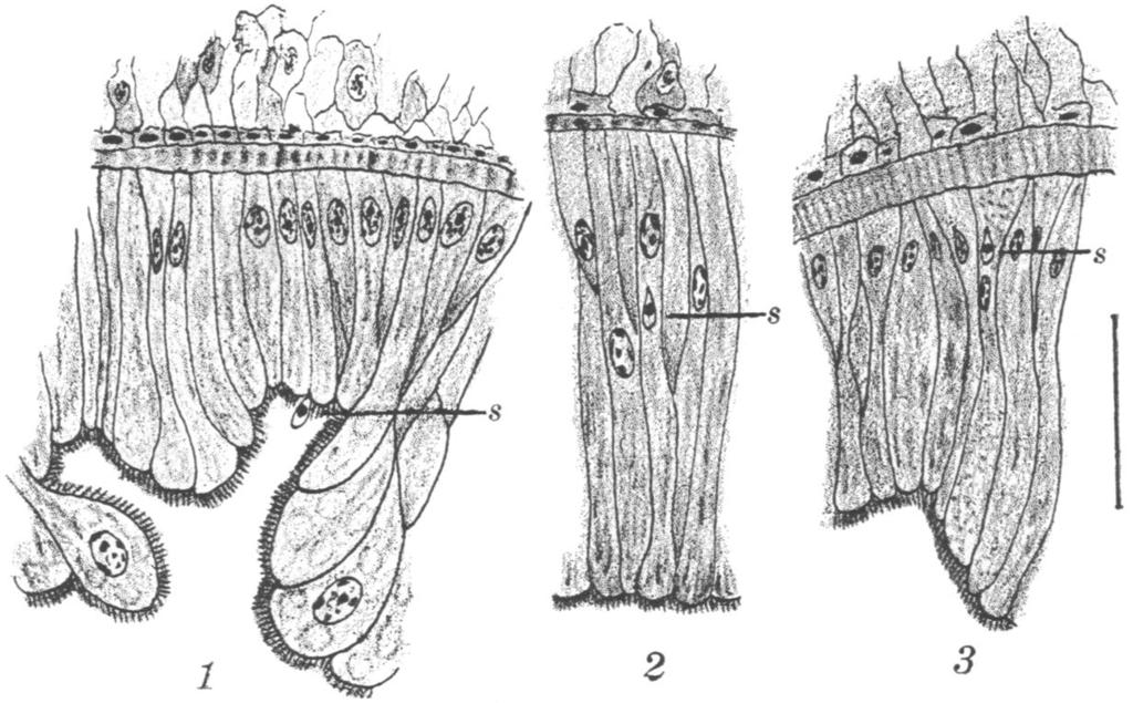
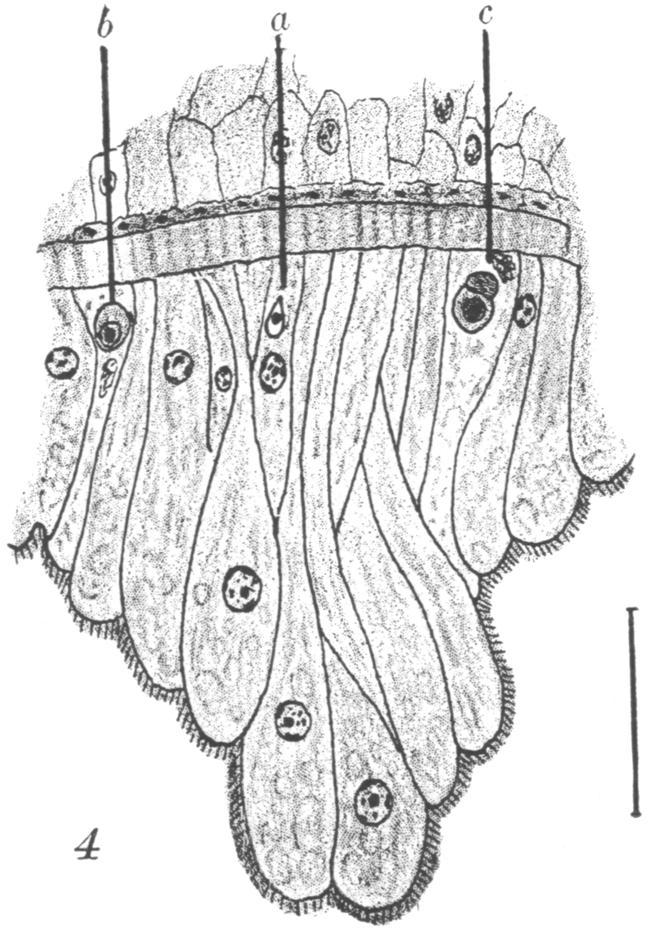
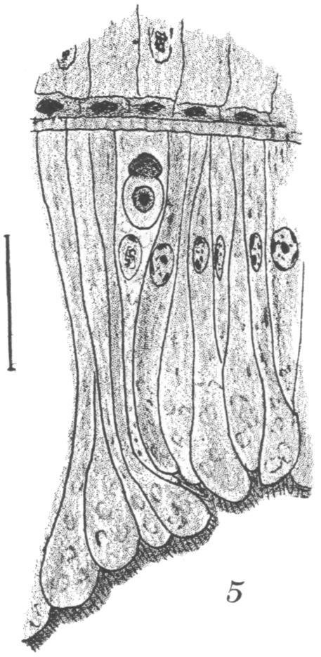
43 PLATE I

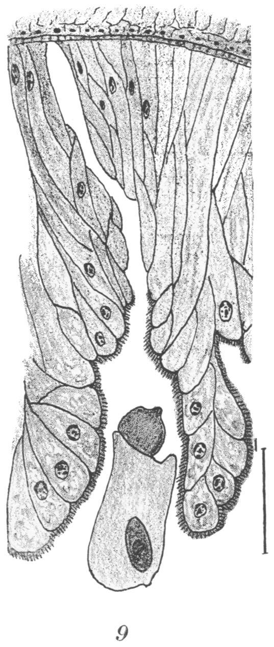
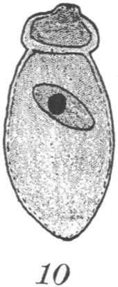
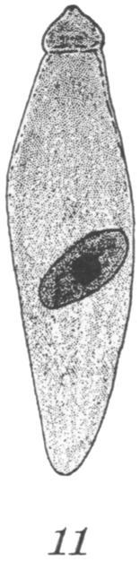
44 1 PLATE
45 THE CERCARIAE OF NATAL F. G. CAWSTON During the months of April, May, June, and July of 1916, I examined 1,500 molluscs from the rivers and fresh-water pools of Natal. They included several species. Limnaea natalensis is a common form with a dextral shell. Physopsis africana, a common mollusc amongst decomposing vegetation, has a blunt-pointed sinistral shell with a truncate columella. Planorbis pfeifferi is a common form with a round, flat shell. Planorbis leucocheilus is not unlike it, but is much smaller. Isidora tropica is fairly common; it has a blunt-pointed sinistral shell. Isidora forskali is rarer and has a conical shell. In one brickfield I found a large number of specimens of Ancylus (ferrissia) burnupi, which has a small oval shell. Two hundred eleven specimens harbored cercariae of various kinds. Infected specimens were most common in one brickfield at Durban and in a small pool along the course of the Umsindusi River at Pietermaritzburg, which had been formed as a result of an overflow of the river. Infected specimens were most frequently met with in May and June. All of the cercariae possessed a long, slender tail; those that were found in specimens of Physopsis africana more often than not possessed divided tails. The tail was easily detached from the body of a cercaria and continued to move for some time after becoming free. All of the cercariae were distomes; the oral sucker was terminal, and in a few specimens the posterior border of the sucker was incomplete; the acetabulum was situated slightly nearer the tail end of the body. None of the cercariae had spines or stylets, and there were no projections from the body or tail. A pharynx was noted in only one form, and eye-spots were present only in cercariae from one specimen. CERCARIA CATENATA A large cercaria, Cercaria catenata, from the Toll Gate brickfield at Durban, present in about 30 per cent of Planorbis pfeifferi and in fewer Limnaea natalensis and Physopsis africana developed in rediae. These rediae gave an orange color to the liver-substance of infected specimens. The rediae were very mobile and possessed two pairs of locomotor appendages. The posterior extremity was pointed. There were infoldings of outer cuticula at the extremities of the posterior extremity of the redia and of its appendages which were not unlike suckers. The rediae contained a somewhat distended intestine,
46 132 THE JOURNAL OF PARASITOLOGY a large amount of orange pigment, and several fully developed cercariae. On one occasion a cercaria was seen attempting to draw itself through the ruptured wall of the redia by means of its suckers. The head of the cercaria varied in appearance, but was often shaped like a leaf. It possessed a large oral sucker and a large acetabulum or ventral sucker; a chain of blackish granules, varying in number from about twenty-five to twenty-eight, lay on each side of the divided alimentary canal. The tail of the cercaria was as long or slightly longer than the body and tapered towards its extremity. HIypodermic injection of a large number of both rediae and cercariae into a guinea-pig threw no light whatever on the life history of this cercaria, which was the only form found to develop in rediae. "TADPOLE CERCARIAE Sixteen sporocysts, containing leptocercous distomes, or "tadpole" cercariae, were found in Physopsis africana from the Umsindusi. Similar sporocysts were present in two specimens of Limnaea natalensis from the same source. The sporocyst intersected almost the entire liver-substance of infected specimens, giving it a whitish appearance. The cercariae consisted of a body with two suckers and a tail which was about the same length as the body. There was a divided gut and an elementary bladder. Some forms presented a stumpy appearance, others were longer. Some of these cercariae found in Durban suggested the appearance of Schistosome cercariae in every respect except that their tail was not divided. No cercariae were found in specimens of Isidora (over fifty were examined from infected places), and furcocercous cercariae were found only in specimens of Physopsis africana. Some specimens of this latter mollusc harbored more than one form of cercaria. Occasionally one came across a specimen which harbored both the "tadpole" and furcocercous forms. Ninety-nine specimens of Physopsis africana harbored Bilharzia cercariae. These are characterized by the absence of a pharynx and by a divided tail. One specimen obtained from the Durban brickfields on May 9 harbored cercariae with long undivided tails, as well as a sporocyst containing an eye-spotted form of furcocercous cercaria. This is the only specimen of the kind I have seen, and, in consideration of its resemblance to the Egyptian form, I have suggested for it the name Cercaria oculata. The eye-spots had a crescentic appearance and were situated nearer the oral end of the body, on either side of the divided gut. They could be readily seen through the thin walls of the sporocyst in which the cercariae were well developed. No pharynx was discernible in the body of the cercariae. There was a long, slender tail which was divided into two short, fin-like prongs.

47 CAWSTON-CERCARIAE OF NATAL 133 CERCARIA SECOBIANA A common cercaria from the Umsindusi pool, for which I have suggested the name Cercaria secobiana, occurred in about seventy specimens of Physopsis africana. It was narrower and slightly smaller than the eye-spotted form. This distome had a long, slender tail which was divided into two prongs. The prongs were as long as the tail itself. When the tail moved, the prongs became bent to the form of a crescent, causing the cercaria to swim backwards- a form of locomotion which would seem to be common to furcocercous cercariae. The cercariae developed in a sporocyst which intersected the whole liversubstance of an infected specimen. They were found only in Physopsis africana from the Umsindusi River. At present, no light has been thrown upon the life-history of this cercaria, which has the appearance of an avian trematode. SCHISTOSOME CERCARIAE Cercariae which answered to the description of the Schistosome group were found in sporocysts from the liver-substance of twentythree specimens of Physopsis africana (15 per cent) from the brickfields at Durban. They were present in a lesser proportion of specimens of this same mollusc collected from the Umsindusi River. Bilharzia disease is common amongst bathers in both these places. Except for the absence of eye-spots, the cercariae were identical with the eyespotted form. The long, slender tail was divided into two short, finlike prongs. There was no pharynx to be seen. In the Medical Journal of South Africa for April, 1916, Dr. J. G. Becker reported that these distome cercariae occurred in Physopsis africana from a pool at Nijstroom in the Transvaal. I have seen the microscopic preparations he has made of them. He injected some hypodermically into a guineapig and, as I announced at a meeting of the Witwatersrand Branch of the British Medical Association two months later, succeeded in producing three adult male Bilharzia worms in the portal system. This confirmed the opinion that these cercariae, which have only been found in areas known to be infected with Bilharzia disease, are in reality the larval form of the Bilharzia parasite of man. On April 28, I added some water containing miracida obtained from the urine of a patient suffering from Bilharziasis to a vessel of water containing specimens of Physopsis africana from the Umsindusi River. At the end of a fortnight a small sporocyst containing undefined cercariae was seen throwing out branches throughout the liversubstance of one specimen, giving it a yellowish-white appearance. In another specimen a similar young sporocyst occurred; in this could be seen undeveloped cercariae with bifid tails. By the end of three weeks
48 134 THE JOURNAL OF PARASITOLOGY fourteen out of thirty-one specimens, or 45 per cent, harbored Bilharzia cercariae, while only 15 per cent of specimens obtained direct from the river were found to be infected at that period of the year. In another series of experiments, the addition of miracidia to the water in which specimens of Physopsis africana were kept, apparently increased the number of infected forms from 22 to 37 per cent, and from 23 to 27 per cent. Similar experiments with specimens of Planorbis pfeifferi and Limnaea ilatalensis proved entirely negative. SNAILS HARBORING "TADPOLE" CERCARIAE, 1916 Date Source Species No. Infected Percentage April... May... July... April... May... July... May... June... July... April... May... June... July... April... May... June... July... June... May... June... July... June... March... April... Umsindusi... (Pietermaritzburg).. (Pietermaritzburg).. (Pietermaritzburg).. (Pietermaritzburg).. (Pietermaritzburg).. Limnaea natalensis... Limnaea natalensis... Limnaea natalensis... Physopsis africana... Physopsis africana... Physopsis africana... Toll Gate...Limnaea natalensis... (Durban)... Limnaea natalensis... (Durban)... Limnaea natalensis... (Durban)... (Durban)... (Durban)... (Durban)... (Durban)... (Durban)... (Durban)... (Durban)... (Durban)... Umgeni (Durban)... Boshoff St... (Pietermaritzburg).. (Pietermaritzburg).. (Pietermaritzburg).. (Pietermaritzburg).. Physopsis africana:... Physopsis africana... Physopsis africana... Physopsis africana... Planorbis pfeifferi... Planorbis pfeifferi... Planorbis pfelfferi... Planorbis pfeifferl... Isadora tropica... Planorbis pfeifferl... Physopsis africana... Physopsis africana... Ancylus... Isidora forskali... Isidora forskali... 2 out of 88 0 out of 30 0 out of 6 12 out of out of out of 6 7 out of 47 1 out of 12 3 out of 21 I out of 7 7 out of 85 0 out of 13 4 out of out of out of out of 20 a out of 13 0 out of 56 5 out of 15 0 out of 20 0 out of 6 0 out of 20 0 out of 2 0 out of 1 I I ] I PHYSOPSIS HARBORING BILHARZIA OERCARIAE, 1916 Month Source Number Percentage April... Toll Gate... 1 out of 7 May... (Durban) out of June... (Durban)... 2 out of 13 July... (Durban)... 8 out of 131 April... Umsindusi out of 197 May... (Pietermaritzburg) out of June... (Pietermaritzburg)... 7 out of 30 July... (Pietermaritzburg)... 0 out of 6 June... Boshoff St. Pool... 0 out of 20 0 July... (Pietermaritzburg)... 0 out of 6
49 CAWSTON-CERCARIAE OF NATAL 135 With the exception of the Schistosome cercariae, we are at present entirely ignorant of the life-history of the various South African cercariae. Some of the "tadpole" forms may give rise to the flukes which occur in sheep and cattle in certain parts of the Transvaal, Natal, and Griqualand East. Others may produce the flukes which I am told are common in the lungs of frogs from some of the pools and rivers of Natal; but, as stated in a letter from Sir Arnold Theiler of the Agricultural Department, "Nobody has yet undertaken to work out the life-history of these flukes in South Africa, and the only reference is that given by Doctor Gilchrist in his book on South African Zoology." The importance of this study is emphasized by our need of a drug to destroy the adult forms of cercariae in the human host. Perhaps a drug which would destroy the liver-fluke in sheep would be equally efficacious in destroying the Schistosome parasite of man.
50 NOTE ON A SPECIES OF NOSEMA INFECTING ATTACUS CYNTHIA DRURY SHIGETANE ISHIWATA The Imperial Sericultural Experiment Station, Tokyo, Japan While working on dead larvae of Attacus cynthia Drury I met with. many individuals which were infected by a species of Nosema with spores characterized by shape and refraction of light sufficiently different from those of the silk-worm parasite, Nosema bombycis Nageli, to distinguish it from that species, although more closely allied to it than to any other species of the genus. The following note is meant to give a short description of the structure of the spores thus far observed, the details of the same as well as the life-history of the parasite being reserved for further investigations. The spores taken directly from the body of the worm were fixed in sublimate-alcohol and stained with Giemsa's solution. Heidenhain's and Delafield's hematoxylin were also used. For the protrusion of the polar filament I employed the method which Kudo (1913) tried with success for the spores of Nosema bombycis Nageli. The spores (Figs. 1 and 2) are ovoid, tapering toward each end. The refraction of light under the microscope is not so sharp as those of other species. On account of the taper they look long and narrow, like the spores of the species infecting Anthaerae pernyi Guer. and A. yamamai Guer., but measurements show that this is not the case. the length and the breadth of the spores of the present species being 3 to 3.5,/ and 2,u, respectively, like those of Nosema bombycis, the measurements of which given by Kudo (1916), were 3 to 4,u in length and 1.5 to 2,u in breadth, by Stempell (1909) 4y/ in length and 21A in breadth, and by Omori (1912) 2 to 4xu in length and 1 to 24u in breadth. The spore is covered by a thick membrane of a transparent and homogeneous substance like that of N. bombycis, but as mentioned above, the refraction of light is not so sharp as in that species. The inner membrane, observed for the first time by Kudo (1916), also exists in the present species and can be pressed out easily. The outer membrane, however, appears to be rather brittle, as it is liable to be crushed into two or more pieces during the process of pressing. With an India ink preparation (Burri's method) the protoplasm stains slightly black, as does that of the silk- worm Nosema (Figs. 1 and 2). The polar filament can be easily extruded from the end of the spore by the method used by Kudo (1913) for Nosema bombycis. It is somewhat shorter and thicker than that of the latter, being about 30 to 35,/ in length. The filament ends always in a round knob of special
51 ISHIWATA-NOTE ON A SPECIES OF NOSEMA 137 form (Figs. 3, 4, 5 and 6), which is most probably of a sticky nature. In some specimens of N. bombycis, the polar filament has a round end, but not in all, and this again is not so well pronounced as in the present species. Moreover, the filament is not coiled transversely within the spore as in that of N. bombycis or of N. anomalum Monz., as is assumed to be the case by Stempell in these species. Careful observation of the spore under a high power shows a clear line running longitudinally within the body, which in some appears to be placed in parallels (Fig. 7). These lines undoubtedly represent the filament coiled up within the polar capsule; and when the filament begins to uncoil and a part of it protrudes from the body of the spore, concentric 1 2 $ 4 5 ) 8 (All the figures are drawn with the help of an Abbe-Zeiss camera.) Figs. 1 and 2.-Spores with protoplasm stained slightly black. India ink preparation. In Figure 1 narrow and Figure 2 broad. X Figs. 3 and 4.-Spores with extruded polar filaments. The end of the filament has a round point. X1545. (Figure 3 shows some crushed pieces of outer membrane which are deeply stained.) Figs. 5, 6, and 7.-Spores containing coiled filament. In Figures 5 and 6 filament partially extruded. X Fig. 8.-Polar filament extruded from side of spore. X lines can often be seen within the latter (Fig. 6). Sometimes the filament extruded from the side of the spore takes the form of a ring, which is most probably caused by the sticking of the round end of the spore at the base of the filament (Fig. 8). Finally, I wish to express my hearty thanks to Prof. Dr. C. Ishikawa for his kind advice in the preparation of this paper. PAPERS CITED Kudo, R Eine neue Methode die Sporen von Nosema bombycis Nageli mit ihren ausgeschnellten Polfaden dauerhaft zu praparieren und deren Lange genauer zu bestimmen. Zool. Anz., 41: Contribution to the Study of Parasitic Protozoa, I. On the Structure and Life-History of Nosema bombycis Nageli. Bull. Imp. Sericult. Exp. Sta., Japan, 1: Omori, J Zur Kenntnis des Pebrine-Erregers Nosema bombycis Nageli. Arb. Kais. Gesundh. Amt., 40: , 1 pl Nihon Sanbyo Ron. Stempell, W Ueber Nosema anomalum Monz. Arch. Protistenk., 4: 1-42, 3 pls Ueber Nosema bombycis Nageli. Arch. Protist., 16: , 7 pl.
52 NOTES ON POROCEPHALUS GLOBICEPHALUS THESLE T. JOB AND A. R. COOPER Mary L. Hett of the London Zoological Society described and named Porocephalus globicephalus from a single mature female specimen, procured from the lung of an American specimen of "moccasin," Tropidonotus fasciatus (Linn.). In the Proceedings of the Zoological Society of London, 1915, pages , she gives the following characters: length, 50 mm.; annulations, about 50; hooks, simple and sharply curved; mouth, pear-shaped with pointed anterior end; head, globular; well marked neck; anus transverse slit on terminal segment. The above description, which is necessarily meager, is the only reference the writers can find to this species. In view of this condition the following data are herewith reported: A large black snake, Basscanion constrictor (Linn.), was received at the State University of Iowa in the fall of 1916 from Garrison-on- Hudson, New York. When the specimen was killed five males and five females of P. globicephalus Hett were found in the respiratory tract. Three males and three females were taken from the lung and two males and two females from the dorsal body wall of the air sac. The females were found with the head only embedded in the lung tissue, or (those in the air sac) in the musculature of the body, where a copious hemorrhage had been formed. The rest of the body of the parasites was free from attachments, hanging limply in the lumen of the lung or air sac. The heads of the males were not embedded in the tissues of the host, but only superficially attached to the walls of the lung or air sac by the hooks. The females vary from 82 to 96 mm. in length, being somewhat larger than the specimen described by Hett, while the males were from 14 to 30 mm. long. The color of the female is lemon yellow, the body wall being transparent, thus permitting easy observation of the mass of embryos and the movement of the intestine within. The male is pale cream in color and the body wall is opaque. The head is globose dorsally; ventrally it is slightly concave with four sharply curved hooks at the anterior edge of the concavity, two on either side of the pear-shaped mouth. The neck is markedly constricted; the body subcylindrical, slightly tapering to the posterior end which is blunt; the digestive tract is seen from the dorsal side; laterally an opaque band runs the full length of the body (this becomes transparent in specimens preserved in alcohol, while the rest of the body becomes opaque). There are about 50 annulations, 48 to 52 having been counted. The digestive tract, which is gorged with blood, is readily seen in the living specimen, and may be traced in preserved ones.
53 BOOK REVIEWS THE ANIMAL PARASITES OF MAN. H. B. Fantham, J. W. W. Stephens, F. V. Theobald. New York: William Wood and Company, xxxii pages. 423 figures. $ As the title page indicates, the work is adapted from the fourth German edition of Braun's well known treatise. It appeared simultaneously in London and New York just after the publication of the fifth German edition of Braun which was reviewed in the JOURNAL for June, A comparison of the views expressed in this work with those in the fifth edition of Braun is especially interesting as it shows the conclusions reached entirely independently by two groups of writers in the same field. It is not surprising that each book treats with greater fulness the work done by investigators of the same nation as the authors, and passes over more briefly the results achieved by workers in foreign nations. The plan of the work is ideal: the section on Protozoa was entrusted to H. B. Fantham, that on worms to J. W. W. Stephens, and that on the Arthropoda to F. V. Theobald. At the present day the work accomplished in each of these fields is so great and the questions under discussion so involved that no one man can cover them all with equal proficiency. Under the plan adopted here each field is assigned to an investigator who is qualified to write with ultimate authority on the problems in that field, and it would have been difficult to select three men anywhere who would measure up to the ideal better than those chosen. On the other hand, the time was not particularly propitious for a great work. Other things are in the air that make insistent demands on the attention of all men. There is no leisure for reflection, and concentration on a scientific problem must be well nigh impossible for a man working anywhere in Europe. In truth the book itself shows some evidence of present conditions in the world. It contains a wealth of information on little known topics. It has been brought thoroly up-to-date, even to the extent of two appendices including important material of later date than the general text, and further in having very recent items incorporated on slips bound in between the finished pages at the last moment. This makes the work appear confused and even in the text there are places where the same impression is given the reader. It seems as if the authors had been working under pressure and the finish one expects in a masterful production had been marred. The volume of scientific material presented to the worker is large and in every way equal to one's expectations, but it is not equally well assimilated at all points. The practice of adding paragraphs here and there incorporates new material at the expense of fluency and the text is not always easily read and understood. The bibliography is very extensive, covering some pages in fine type, but it lacks all recent items, being in fact but a reprint of the lists in the 1908 edition of Braun. Some references to the literature of the recent items in the text are given in footnotes; but in too many places the new facts are recorded without exact credit, or sometimes without even the name of the author, and the student is left to htint for himself the precise source of the information. This is least noticeable in the section on Protozoa where footnote references are particularly abundant. It is curious to note that even with such a large space devoted to bibliography one cannot find references to important recent papers by Leiper and other English workers; the authors have treated everyone impartially as not only other references are wanting but even some important papers of Stephens himself are not listed. The illustrations are numerous and well distributed. They include fewer relics of the past than one usually finds in so comprehensive a work. Most
54 140 THE JOURNAIL OF PARASITOLOGY of the figures are well done and satisfactorily reproduced. One can not help feeling a little disappointed, however, that some have crept in which are new and yet unfortunately inferior. The diagrammatic representations of the Echinococcus cysts on pages 352 and 353 are not well drawn and their reproduction on so large a scale is still more open to criticism. A reduction to one-half or one-third the present size would have made their sketchiness less conspicuous without the loss of any important details. In the opinion of the reviewer English and American works are distinctly inferior to continental publications in the character of their illustrations, and the present volume is undoubtedly less deserving of criticism in this respect than most of our works. Despite its evident minor imperfections this treatise is a valuable and usable work. No one can question that the splendid volume is easily the largest and most complete work on the subject which has appeared in the English language. The work of the printer has been well done and deserves especial commendation. Both paper and type are such as contribute to ease of reading and one lays the volume aside with the conviction that its authors and publishers alike deserve the thanks of scientific workers for the results they have achieved. JAPANESE MEDICAL LITERATURE, a review of current periodicals the initial number of which was noted in the Journal (3: 42), has completed its first volume, July to December, The General Index is well prepared and will be a real convenience even though complicated by the fact that it is paged after the China Medical Journal from which the reviews are reprinted, and not according to the reprints themselves. References to animal parasites are numerous and important. The value of the Japanese literature and its great inaccessibility make such reviews of unusual service and American investigators are deeply indebted to Dr. Mills and his colleagues for their work. The first number of the second volume has also come to hand, and scientific workers gladly look forward to the continuance of this very valuable serial which is without any competition in this difficult and important field.
55 NOTES "ECHINORHYNCHUS MONILIFORMIS" IN NORTH AMERICA * Among the Acanthocephala which are exclusively parasitic and highly specialized for the parasitic habit, only three species have been reported for the human host and even these are rare or doubtful. Gigantorhynchus hirudinaceus (= G. gigas) is said to occur in man in southern Russia but the statement is unconfirmed. Lambl found in the intestine of a boy a single parasite to which he gave the name of Echinorhynchus hominis. The third species, originally named Echinorhynchus moniliformis, has commanded especial attention by virtue of its relation to man. Grassi and Calandruccio found it in Sicily in the small intestine of field mice, rats, and marmots. They detected the intermediate host in Blaps mucronata and in some cases found as many as 100 larvae encysted in a single cockroach. They fed such larvae to a white rat and Calandruccio swallowed some at the same time. These developed well in both hosts. The authors identified eggs apparently of the same species in the feces of a young peasant but were unable to carry out a cure and confirm the diagnosis. Through the courtesy of Mr. G. E. Clark some material has been placed in my hands which belongs to a larger species of closely related type. These worms were taken from a squirrel in Illinois. They furnish the first record of this type for the North American continent. As noted above the European species has been grown experimentally in the human host and this species is likely to show the same power if the mature larvae are introduced by any chance into the human intestine. After extended study it may be said that the two species are both sufficiently characteristic in their resemblances to each other and in their differences from other known forms to constitute a new genus to which the name of Hormorhynchus may be given. H. mon4liformis (Bremser 1819) is designated as type and attention is called to the fact that Liihe believes there are several species in Europe all included under the one name. The American species is designated as H. clarki. Specimens measure 100 to 130 mm. in length. The proboscis is very small, being only mm. long by 0.12 mm. broad. The first ring is 5 mm. from the anterior tip. The rings begin faintly but distinctly; they are about 1 mm. long and little wider than the body. They increase rapidly up to 2 mm. in length and at the point of greatest length the individual rings are so swollen as to become markedly wider than the body. From this region they taper out very gradually. The last 15 mm. of the body shows no trace of rings and for the same distance anterior to it the rings are very faint. A full description of the species will be published elsewhere. HENRY B. WARD DIPTERA IN THE HUMAN INTESTINE On Sept. 30, 1916, Dr. W. C. King of Helena, Ark., sent some intestinal parasitic worms which he reported as coming from an adult woman, to the State Hygienic Laboratory connected with the Medical Department of the University of Arkansas for identification. Dr. A. C. Shipp, in charge of the Hygienic Laboratory, consulted with me about these worms. They were plainly annular with about thirteen segments. The shape was very nearly pyramidal with some seven or more papillae at the blunt posterior end. * Contributions from thle Zoological Laboratory of the University of Illinois, No. 91.

56 142 THE JOURNAL OF PARASITOLOGY These worms looked so much like Dipterous larvae that I suggested that we place them on nutrient agar for a few days to see if they would pupate. Pupation took place in about three days and after about five days more there was hatched a medium-sized black fly. Later others were hatched. Some of these were sent to Prof. James S. Hine of the Entomology Department of Ohio State University, who identified them as Sarcophaga assidua Walker. Our attention has been called to two or three other cases of similar occurrence of what seemed to be Dipterous larvae in the stools of persons this past fall and summer. In one case the sputum gathered in a sterile bottle under a physician's guidance showed what seemed on examination to be young Dipterous larvae. Unfortunately these larvae were killed in the overheating of our incubator. How these larvae gain access to the intestine unless through ingestion of food in the stage of the freshly laid eggs on exposed cooked food or in uncooked food, is a question. Whether they passed the gastric digestion in the stage of egg or larva is problematical. CHARLES BROOKOVER Department of Anatomy, University of Arkansas. Dr. E. Gonzalez reported to the Congress of Medicine at Maracaibo the occurrence of Leishmania brasiliensis Vianna in a servant woman brought to the clinic at San Fernando de Apure. The preparations were examined by members of the Yellow Fever Commission of the Rockefeller Institute and the diagnosis confirmed. This is the first case of cutaneous Leishmaniasis from Venezuela. Parasitologists will be interested in the circular of the Merchants Association of New York on the Dangerous House Fly. It is important to urge on every community the necessity of early action this year to eradicate this dangerous agency in the transmission of disease. Incidentally also laboratory teachers may be glad to know that in the flies which appear early in the season a flagellate (Crithidia sp.?) occurs abundantly in the gut. Material can be secured readily from this host that is well adapted to class study or demonstration of this group.
General Characters of Trematodes
 Parasitology Department General Characters of Trematodes By Hala Elwakil, MD Intended Learning Outcomes By the end of this lecture, the student will be able to know: 1. General morphology of trematodes
Mehratreraa polynemusinis, n.sp.
 TREMATODES FROM INDIAN MARINE FISHES Part III. Description of a New Species of the Genus Mehratrr (Family Monodhelminthidae Dollfus, 1937) BY B. S. CHAUHAN (College of Science, Nagpur, India) Received
Trematode Parasites of the Opossum, Didelphis virginiana, from Florida
 Proc. Helminthol. Soc. Wash. 46(2), 1979, pp. 207-212 Trematode Parasites of the Opossum, Didelphis virginiana, from Florida G. PREMVATI AND THOMAS D. BAiR1 Department of Zoology, University of Lucknow,
THEJ FIRST ZOEA OF PORCELLANA. By W K. BROOKS and E. B. WILSON. With Plate* VI and VII.
 7 "83?* {Jn-fJatoj 11 "T THEJ FIRST ZOEA OF PORCELLANA. By W K BROOKS and E. B. WILSON. With Plate* VI and VII. 7z O THE FIRST ZOEA OF PORCELLANA. By W K. BROOKS and E. B. WILSON. With Plates VI and VII.
ON A NEW GENUS PARAMACROLECITHUS (TRI~MATODA: ALLOCREADIIDAE) FROM A CYPRINID FISH, RASBORA RASBORA (HAMILTON)
 FROM A CYPRINID FISH, RASBORA RASBORA (HAMILTON)") ON A NEW GENUS PARAMACROLECITHUS (TRI~MATODA: ALLOCREADIIDAE) FROM A CYPRINID FISH, RASBORA RASBORA (HAMILTON) BY C. B. SRIVASTAVA AND R. K. GrlOSH (Zoological Survey of India, Calcutta-12, India) Received
ON A NEW GENUS PARAMACROLECITHUS (TRI~MATODA: ALLOCREADIIDAE) FROM A CYPRINID FISH, RASBORA RASBORA (HAMILTON) BY C. B. SRIVASTAVA AND R. K. GrlOSH (Zoological Survey of India, Calcutta-12, India) Received
Larval Bucephalids (Trematoda: Digenea) Parasitizing Bivalve Molluscs in the Galveston Bay Area, Texas
 Parasitizing Bivalve Molluscs in the Galveston Bay Area, Texas") J. Helminthol. Soc. Wash. 57(1), 1990, pp. 5-11 Larval Bucephalids (Trematoda: Digenea) Parasitizing Bivalve Molluscs in the Galveston Bay Area, Texas WILLIAM J. WARDLE Department of Marine Biology, Texas
J. Helminthol. Soc. Wash. 57(1), 1990, pp. 5-11 Larval Bucephalids (Trematoda: Digenea) Parasitizing Bivalve Molluscs in the Galveston Bay Area, Texas WILLIAM J. WARDLE Department of Marine Biology, Texas
SHAHNAZ BANO. Chemotherapy Division, C.D.R.I., Lucknow, V.P. (With 2 text-figures) I-DESCRIPTION. Lutztrema singbi sp. n.
 I-DESCRIPTION. Lutztrema singbi sp. n.") STUDIES ON HELMINTH PARASITES OF CROWS. TWO NEW SPECIES OF THE GENUS LUTZTREMA TRAVASSOS FROM CORVUS SPLENDENS (VIEILLOT) AND A DISCUSSION ON THE GENUS LUTZTREMA By SHAHNAZ BANO Chemotherapy Division,
STUDIES ON HELMINTH PARASITES OF CROWS. TWO NEW SPECIES OF THE GENUS LUTZTREMA TRAVASSOS FROM CORVUS SPLENDENS (VIEILLOT) AND A DISCUSSION ON THE GENUS LUTZTREMA By SHAHNAZ BANO Chemotherapy Division,
BIO Parasitology Spring 2009
 BIO 475 - Parasitology Spring 2009 Stephen M. Shuster Northern Arizona University http://www4.nau.edu/isopod Lecture 13 Important Orders a. Echinostomatiformes b. Strigeiformes c. Opisthorchiformes d.
Studies on Echinostomatidae (Trematoda) in Malaysia. XVI. The Life History of Echinostoma ilocanum (Garrison, 1908)*
 in Malaysia. XVI. The Life History of Echinostoma ilocanum (Garrison, 1908)*") OF WASHINGTON, VOLUME 40, NUMBER 1, JANUARY 1973 59 Studies on Echinostomatidae (Trematoda) in Malaysia. XVI. The Life History of Echinostoma ilocanum (Garrison, 1908)* LIE KIAN JOE AND S. NASEMARY Institute
OF WASHINGTON, VOLUME 40, NUMBER 1, JANUARY 1973 59 Studies on Echinostomatidae (Trematoda) in Malaysia. XVI. The Life History of Echinostoma ilocanum (Garrison, 1908)* LIE KIAN JOE AND S. NASEMARY Institute
TWO NEW SPECIES OF COPEPODS JAPANESE FISHES. Author(s) Yamaguti, Satyu; Yamasu, Terufumi.
 Yamaguti, Satyu; Yamasu, Terufumi.") Title TWO NEW SPECIES OF COPEPODS JAPANESE FISHES PARASIT Author(s) Yamaguti, Satyu; Yamasu, Terufumi Citation PUBLICATIONS OF THE SETO MARINE BIO LABORATORY (1960), 8(1): 137-140 Issue Date 1960-05-30
Title TWO NEW SPECIES OF COPEPODS JAPANESE FISHES PARASIT Author(s) Yamaguti, Satyu; Yamasu, Terufumi Citation PUBLICATIONS OF THE SETO MARINE BIO LABORATORY (1960), 8(1): 137-140 Issue Date 1960-05-30
Description of the Immature Stages of Galindomyia leei Stone and Barreto, 1969l. Abdiel J. Adames* and Pedro Galindo*
 132 Description of the Immature Stages of Galindomyia leei Stone and Barreto, 1969l Abdiel J. Adames* and Pedro Galindo* The genus and species Galindomyia Zeei, a member of the tribe Culicini, was described
Shark Lab Key. dorsal surface. click on picture for ventral surface
 Shark Lab Key Study this basic information about the spiny dogfish shark. Print this Shark Lab Report Guide. Pre-Lab Research Study this website. It provides several useful videos of large shark dissections.
Dogfish Shark Dissection
 Dogfish Shark Dissection Name Date Period Fun Facts: Materials: The teeth of sharks are modified scales embedded in the skin of its mouth Sharks have pits on their face used to detect electric fields Sharks
Lumbricus terrestris - preserved specimens for dissection
 Lumbricus terrestris - preserved specimens for dissection External Anatomy: Prostomium (observe under dissecting microscope for external sensory organs), peristomium, clitellum, setae (dissecting microscope),
UROMASTIX DIGESTIVE SYSTEM
 UROMASTIX DIGESTIVE SYSTEM by Dr. Rashmi Tripathi Department of Zoology Brahmanand College, Kanpur DIGESTIVE SYSTEM : The digestive system consists of (A) Alimentary canal and (B) Associated digestive
A New Species of Lissorchis (Trematoda: Lissorchiidae) from Creek Chubsuckers (Erimyzon oblongus) in the Big Thicket National Preserve, Texas, U.S.A.
 from Creek Chubsuckers (Erimyzon oblongus) in the Big Thicket National Preserve, Texas, U.S.A.") Comp. Parasitol. 77(1), 2010, pp. 1 5 A New Species of Lissorchis (Trematoda: Lissorchiidae) from Creek Chubsuckers (Erimyzon oblongus) in the Big Thicket National Preserve, Texas, U.S.A. MICHAEL A. BARGER
Comp. Parasitol. 77(1), 2010, pp. 1 5 A New Species of Lissorchis (Trematoda: Lissorchiidae) from Creek Chubsuckers (Erimyzon oblongus) in the Big Thicket National Preserve, Texas, U.S.A. MICHAEL A. BARGER
Materials and Methods
 158 ~ 40 (1971) this purpose the life history in the early stage of the gill trematodes must be made clear, especially in the form and ecology of the miarcidium as well as of the egg. This paper has been
Slide 1. Slide 1. Next. 5:30:08 AM
 Slide 1 Slide 1 http://www3.utep.edu/leb/mosquito/larvslide1.htm10/27/2004 5:30:08 AM Slide 1 Slide 2 Recognition that the specimens are mosquito larvae is a prerequisite to identification of the genera.
Chapter 30 Nonvertebrate Chordates, Fishes, and Amphibians Name
 Chapter 30 Nonvertebrate Chordates, Fishes, and Amphibians Name Lab Dissecting a Perch Background Information Fish are the largest group of vertebrates found in fresh and salt water. In fact, over 25,000
from a marine ancestor. In fact, haploporids are now known to be more numerous and varied in freshwater than in the sea, so perhaps the reverse
 TWO NEW HAPLOPORIDAE (TREMATODA) OF FISHES Vernon E. THATCHER* ABSTRACT Two new Haploporidae (Trematoda) are described from fishes of the State of Rondônia, Brazil. Rondotrema microvitellarum gen. et sp.
Heterophyiasis Heterophyes heterophyes. Samar N. El-Beshbishi Prof. of Medical Parasitology Mansoura Faculty of Medicine
 Heterophyiasis Heterophyes heterophyes Samar N. El-Beshbishi Prof. of Medical Parasitology Mansoura Faculty of Medicine 2 Objectives 1. Overview on heterophyiasis. 2. Geographical distribution. 3. Morphology
Studies on the Monogenea of Plymouth. Gastroootyle traohuri v. Ben. and Hesse, 1863.
 [ 227 ] Studies on the Monogenea of Plymouth. Gastroootyle traohuri v. Ben. and Hesse, 1863. By By E. Idris Jones, B.Sc. (Lond.) With 4 Figures in the Text. IN August, 1932, six specimens of this species
CHAPTER 22. Echinoderms 22-1
 CHAPTER 22 Echinoderms 22-1 Phylum Echinodermata: Diversity and Characteristics Characteristics All members have a calcareous skeleton Spiny endoskeleton consists of plates Unique water-vascular system
Exercise 18B Class Chondrichthyes Cartilaginous Fishes
 AP Biology Chapter 24 Exercise #18: Chordates: Fish Cartilaginous Fishes Lab Guide Exercise 18B Class Chondrichthyes Cartilaginous Fishes This group contains about 970 species that are characterized by
Chapter 12 Part 2. The Worms Platyhelminthes, Nematoda & Annelida
 Chapter 12 Part 2 The Worms Platyhelminthes, Nematoda & Annelida Phylum: Platyhelminthes Examples: Flatworms, Planaria sp., tapeworms and blood flukes Acoelomate, Invertebrate, Simplest critter w/ bilateral
TWO NEW SPECIES OF ARGULUS MULLER (CRUSTACEA: BRANCHIURA) FROM RIVER CAUVERY WITH A KEY TO INDIAN SPECIES
 FROM RIVER CAUVERY WITH A KEY TO INDIAN SPECIES") TWO NEW SPECIES OF ARGULUS MULLER (CRUSTACEA: BRANCHIURA) FROM RIVER CAUVERY WITH A KEY TO INDIAN SPECIES M. M. THOMAS AND M. DEVARAJ Central Marine Fisheries Research Institute Regional Centre, Mandapam
TWO NEW SPECIES OF ARGULUS MULLER (CRUSTACEA: BRANCHIURA) FROM RIVER CAUVERY WITH A KEY TO INDIAN SPECIES M. M. THOMAS AND M. DEVARAJ Central Marine Fisheries Research Institute Regional Centre, Mandapam
-8- spinous. nape caudal fin. body depth. pectoral fin. anus. total length Fig. 4
 click for previous page -8-1.3 Illustrated Glossary of Technical Terms and Measurements External Morphology and Measurements spinous dorsal fin soft nape caudal fin interorbital body depth snout lateral
Ree. zool. Surv. India, 91 (3-4) : , 1992
 : , 1992") Ree. zool. Surv. India, 91 (3-4) : 319-323, 1992 RECORDS OF TWO CERCARIAE, HETEROPHYES SP. AND HAPLORCHIS SP. FROM THE SNAIL, MELANO/DES TUBERCULATA (MULLER) FROM TAMIL NADU. N. VEERAPPAN AND H. N. ACHUTHAN
Ree. zool. Surv. India, 91 (3-4) : 319-323, 1992 RECORDS OF TWO CERCARIAE, HETEROPHYES SP. AND HAPLORCHIS SP. FROM THE SNAIL, MELANO/DES TUBERCULATA (MULLER) FROM TAMIL NADU. N. VEERAPPAN AND H. N. ACHUTHAN
External Anatomy Dissection Guide
 External Anatomy Dissection Guide Dissection is the cutting of a dead animal or a plant into separate parts for the purpose of careful and detailed examination and study. The external anatomy is as important
LIBRARY. Class\ V"^ A *Ii:T_
 LIBRARY Class\ V"^ A *Ii:T_ ^ Publications OP FIELD MUSEUM OF NATURAL HISTORY ZOOLOGICAL SERIES Volume X Chicago, U. S. A. 1909-1923 7/,3 ^Issued September 18, 19 12. 69 NEW SPECIES OF FISHES FROM
Phylum Platyhelminthes. You will need: five colours of pencil crayon or pen (preferably blue, green, red, orange and purple)
") Phylum Platyhelminthes You will need: five colours of pencil crayon or pen (preferably blue, green, red, orange and purple) Phylum Platyhelminthes bilaterally symmetrical have all 3 embryonic germ layers:
Phylum Platyhelminthes You will need: five colours of pencil crayon or pen (preferably blue, green, red, orange and purple) Phylum Platyhelminthes bilaterally symmetrical have all 3 embryonic germ layers:
 Echinoderms Name Means: Echino- Spiny Derm- Skin About 7,000 species No Head! No Brain! No ventral, dorsal, posterior, or anterior sides! Do have oral (mouth) and aboral sides. Symmetry: Larvae are
Gen Bio 2 Lab #10: Chondrichthyes and Osteichthyes
 Name: Date Gen Bio 2 Lab #10: Chondrichthyes and Osteichthyes Pre-Lab Reading: pages 687-690 Pre-Lab Vocabulary: 1) Ampullae of Lorenzini 2) Claspers 3) Lateral line 4) Ovoviviparous 5) Squalene 6) Viviparous
V. Subclass Digenea (Chapters 15-18, BLY ) A. Background 1. Flukes 2. Digenea refers to having an alternation of hosts in the life cycle B.
 A. Background 1. Flukes 2. Digenea refers to having an alternation of hosts in the life cycle B.") V. Subclass Digenea (Chapters 15-18, BLY 459 2009) A. Background 1. Flukes 2. Digenea refers to having an alternation of hosts in the life cycle B. Characteristics 1. Cuplike suckers without hooks a..
V. Subclass Digenea (Chapters 15-18, BLY 459 2009) A. Background 1. Flukes 2. Digenea refers to having an alternation of hosts in the life cycle B. Characteristics 1. Cuplike suckers without hooks a..
Classification. Phylum Chordata
 AP Biology Chapter 23 Exercise #17: Chordates: Urochordata & Cephalochordata Lab Guide Chordates show remarkable diversity. Most are vertebrates. All animals that belong to this phylum MUST, at some point
NATIONAL BIORESOURCE DEVELOPMENT BOARD Dept. of Biotechnology Government of India, New Delhi
 NATIONAL BIORESOURCE DEVELOPMENT BOARD Dept. of Biotechnology Government of India, New Delhi MARINE BIORESOURCES FORMS DATA ENTRY: Form- 1(general ) (please answer only relevant fields;add additional fields
2. SYSTEMATIC CATALOGUE
 click for previous page 15 2. SYSTEMATIC CATALOGUE 2.1 General Aids to Identification 2.1.1 Diagnostic Features of the Family Caesionidae Oblong to fusiform, moderately compressed, medium-sized to small
* A New Species of Cichlid Fish From Lake Malawi. Pseudotropheus tursiops, \(I75 Tropical Fish Hobbyist a'l (3) : 8 L-? 0. ,$ IOU.
 : 8 L-? 0. ,$ IOU.") ,$ IOU. \(I75 Tropical Fish Hobbyist a'l (3) : 8 L-? 0. * 2.37 Pseudotropheus tursiops, A New Species of Cichlid Fish From Lake Malawi by Warren E. Burgess and Dr. Herbert R. Axelrod Among the cichlid
,$ IOU. \(I75 Tropical Fish Hobbyist a'l (3) : 8 L-? 0. * 2.37 Pseudotropheus tursiops, A New Species of Cichlid Fish From Lake Malawi by Warren E. Burgess and Dr. Herbert R. Axelrod Among the cichlid
Natural History of Vertebrates Characters Used in Fish Identification (modified )
") Natural History of Vertebrates Characters Used in Fish Identification 1-9-03 (modified 20050118) This lab is designed to familiarize the student with characters used in the identification of fishes. Only
Natural History of Vertebrates Characters Used in Fish Identification 1-9-03 (modified 20050118) This lab is designed to familiarize the student with characters used in the identification of fishes. Only
TWO DIGENETIC TREMATODES (FAMILY HEMIURIDAE) OF MARINE. FISHES OF INDIA M. HAFEEZULLAH* Zoological Survey of India, Calcutta
 OF MARINE. FISHES OF INDIA M. HAFEEZULLAH* Zoological Survey of India, Calcutta") ljiill~ %0.0/. Surv. India, "( 1) 111-11S, 1981 TWO DIGENETIC TREMATODES (FAMILY HEMIURIDAE) OF MARINE FISHES OF INDIA M. HAFEEZULLAH* Zoological Survey of India, Calcutta ABSTRACT The paper deals with
ljiill~ %0.0/. Surv. India, "( 1) 111-11S, 1981 TWO DIGENETIC TREMATODES (FAMILY HEMIURIDAE) OF MARINE FISHES OF INDIA M. HAFEEZULLAH* Zoological Survey of India, Calcutta ABSTRACT The paper deals with
Cylicocyclus spp. Walls of the Buccal Capsule:
 Figure 8a Cylicocyclus ashworthi Figure 9a Cylicocyclus auriculatus Figure 0a Cylicocyclus brevicapsulatus Figure a Cylicocyclus elongatus Figure 3a Cylicocyclus insigne Figure 4a Cylicocyclus radiatus
Lesson 28. Function - Respiratory Pumps in Air Breathers Buccal Force Pump Aspiration Pump - Patterns of Gas Transfer in Chordates
 Lesson 28 Lesson Outline: Evolution of Respiratory Mechanisms - Air Breathers Form - Accessory Air Breathing Organs Facultative vs Obligate - Lungs Function - Respiratory Pumps in Air Breathers Buccal
NATIONAL BIORESOURCE DEVELOPMENT BOARD Dept. of Biotechnology Government of India, New Delhi
 NATIONAL BIORESOURCE DEVELOPMENT BOARD Dept. of Biotechnology Government of India, New Delhi For office use: MARINE BIORESOURCES FORMS DATA ENTRY: Form- 1(general ) (please answer only relevant fields;
Title: May 31 2:42 PM (1 of 23) Phylum Mollusca
 Phylum Mollusca") Title: May 31 2:42 PM (1 of 23) Phylum Mollusca Title: May 31 3:25 PM (2 of 23) often referred to as mollusks second largest phylum has 7 classes only looking at 4 bilateral symmetry, true body cavity
Title: May 31 2:42 PM (1 of 23) Phylum Mollusca Title: May 31 3:25 PM (2 of 23) often referred to as mollusks second largest phylum has 7 classes only looking at 4 bilateral symmetry, true body cavity
Schwenkiella orietanlis Singh and Agarwal, 1997 (PLATE-XXVII-XXVIII)
") Schwenkiella orietanlis Singh and Agarwal, 1997 (PLATE-XXVII-XXVIII) Host : Periplaneta americana Locality : Meerut Location : Intestine No. of hosts examined : 250 No. of hosts found infected : 200 Female
Schwenkiella orietanlis Singh and Agarwal, 1997 (PLATE-XXVII-XXVIII) Host : Periplaneta americana Locality : Meerut Location : Intestine No. of hosts examined : 250 No. of hosts found infected : 200 Female
PHYLUM CHORDATA: Subphylum vertebrata
 PHYLUM CHORDATA: Subphylum vertebrata There are three basic characteristics that distinguish Phylum Chordata from all other animal phyla: The presence of a flexible, rod-like, internal supporting structure
SILURODISCOIDES GUSSEV, 1961 (MONOGENEA: ANCYROCEPHALIDAE) FROM PANGASIUS SUTCHI FOWLER, 1931 (PANGASIIDAE) CULTURED IN PENINSULAR MALAYSIA.
 FROM PANGASIUS SUTCHI FOWLER, 1931 (PANGASIIDAE) CULTURED IN PENINSULAR MALAYSIA.") SILURODISCOIDES GUSSEV, 1961 (MONOGENEA: ANCYROCEPHALIDAE) FROM PANGASIUS SUTCHI FOWLER, 1931 (PANGASIIDAE) CULTURED IN PENINSULAR MALAYSIA. ABSTRACT. - Two species of Silurodiscoides Gussev, 1961 (Monogenea:
SILURODISCOIDES GUSSEV, 1961 (MONOGENEA: ANCYROCEPHALIDAE) FROM PANGASIUS SUTCHI FOWLER, 1931 (PANGASIIDAE) CULTURED IN PENINSULAR MALAYSIA. ABSTRACT. - Two species of Silurodiscoides Gussev, 1961 (Monogenea:
Gynaecotyla squatarolae (Digenea: Microphallidae) from rats experimentally infected with metacercariae from the shore crab, Macrophthalmus dilatatus
 from rats experimentally infected with metacercariae from the shore crab, Macrophthalmus dilatatus") Korean Journal of Parasitology Vol. 45, No. 3: 199-204, September 2007 DOI: 10.3347/kjp.2007.45.3.199 Gynaecotyla squatarolae (Digenea: Microphallidae) from rats experimentally infected with metacercariae
Korean Journal of Parasitology Vol. 45, No. 3: 199-204, September 2007 DOI: 10.3347/kjp.2007.45.3.199 Gynaecotyla squatarolae (Digenea: Microphallidae) from rats experimentally infected with metacercariae
NATIONAL BIORESOURCE DEVELOPMENT BOARD Dept. of Biotechnology Government of India, New Delhi
 NATIONAL BIORESOURCE DEVELOPMENT BOARD Dept. of Biotechnology Government of India, New Delhi MARINE BIORESOURCES FORMS DATA ENTRY: Form- 1(general) For office use: Fauna: Flora Microorganisms General Category:
A NEW SPECIES OF COPEPOD OF THE GENUS
 PROCEEDINGS OF THE UNITED STATES NATIONAL MUSEUM issued SMITHSONIAN INSTITUTION U. S. NATIONAL MUSEUM Vol. 99 Washington: 1949 No. 3239 A NEW SPECIES OF COPEPOD OF THE GENUS CORYCAEUS FROM THE NORTH AMERICAN
LECTURE 6 - OUTLINE. Evolution & Classification - Part II. Agnatha (cont.) Gnathostomata
 Gnathostomata") LECTURE 6 - OUTLINE Evolution & Classification - Part II Agnatha (cont.) 6. Myxini 7. Cephalaspidomorphi Gnathostomata 1. Phylogenetic relationships 2. Placodermi 3. Acanthodii BIOL 4340 Lecture 6-1 Class
LECTURE 6 - OUTLINE Evolution & Classification - Part II Agnatha (cont.) 6. Myxini 7. Cephalaspidomorphi Gnathostomata 1. Phylogenetic relationships 2. Placodermi 3. Acanthodii BIOL 4340 Lecture 6-1 Class
- 7 - DESCRIPTION OF SPECIES
 I - 7 - DESCRIPTION OF SPECIES./' Anguilla bicolor McClelland ' Level-finned eel (Figs.i & 2) Length of head 6-8 times in length of body; Diameter of eye 8-10 times, Inter-orbital length 2-2.5 times, Gape
NATIONAL BIORESOURCE DEVELOPMENT BOARD Dept. of Biotechnology Government of India, New Delhi
 NATIONAL BIORESOURCE DEVELOPMENT BOARD Dept. of Biotechnology Government of India, New Delhi MARINE BIORESOURCES FORMS DATA ENTRY: Form- 1(general ) (please answer only relevant fields;add additional fields
Field Identification of Tunas from Indian Waters
 3 Field from Indian Waters Subal Kumar Roul and Retheesh T. B. Pelagic Fisheries Division The Family Scombridae is one of the largest and most economically important fish family which comprises of most
I. Evolutionary Perspective. Chapter 12. II. Molluscan Characteristics. A. Regions of Molluscan Body 11/2/10
 I. Evolutionary Perspective Chapter 12 Molluscan Success Some of the world s best predators Large brains Complex sensory structures Rapid locomotion Grasping tentacles Tearing mouthparts Have been around
a. Kingdom: b. Phylum: c. Class: d. Order: e. Family: f. Genus: g. Species:
 Pre-lab Discussion: The earthworm belongs to a group of animals called annelids (segmented worms). The body of an annelid is usually divided internally and externally into well-defined segments, which
aurora Baird and Girard, was identified as Brachycoelium lynchi
 AN ABSTRACT OF THE THESIS OF GORDON WYATT MARTIN for the Ph. D. in ZOOLOGY (Name) (Degree) (Major) Date thesis is presented July 21, 1965 Title A PROGENETIC TREMATODE (LECITHODENDRIIDAE) LIFE CYCLE INVOLVING
NATIONAL BIORESOURCE DEVELOPMENT BOARD Dept. of Biotechnology Government of India, New Delhi
 NATIONAL BIORESOURCE DEVELOPMENT BOARD Dept. of Biotechnology Government of India, New Delhi For office use only MARINE BIORESOURCES FORMS DATA ENTRY: Form- 1(general ) Ref. No.: (please answer only relevant
Internal Anatomy of Fish
 Internal Anatomy of Fish The Systems of a Fish Skeletal System Muscular System Respiratory System Digestive System Circulatory System Nervous System Reproductive System Special Organs Skeletal System
Respiration. Chapter 33
 Respiration Chapter 33 Learning Objectives: Understand the basis of gas exchange and factors that influence diffusion of gases in and out of tissues Compare and contrast different respiratory systems among
`Mollusks. may or may not form a hard, calcium carbonate shell. Trochophore Larva
 `Mollusks Phylum Mollusca Soft-bodied invertebrate covered with protective mantle that may or may not form a hard, calcium carbonate shell Includes chitons, snails, slugs, clams, oysters, squid, octopus,
The Formation and Fate of the Operculum and Gill-chambers in the tadpole of Rana temporaria.
 The Formation and Fate of the Operculum and Gill-chambers in the tadpole of Rana temporaria. By Gwendolen T. Brock, M.Sc, D.PM1. (Oxon.). With 16 Text-figures. THIS work has been undertaken with the object
PROCEEDINGS i j OF THE BIOLOGICAL SOCIETY OF WASHINGTON
 e3 if Vol. 86, No. 27, pp. 333-338 28 September 1973 PROCEEDINGS i j OF THE ' BIOLOGICAL SOCIETY OF WASHINGTON A NEW SPECIES OF SESARMA, S. (HOLOMETOPUS) RUBINOFFORUM, FROM THE PACIFIC COAST OF PANAMA
Perch Dissection Lab
 Perch Dissection Lab Introduction: The fish in the class Osteichthyes have bony skeletons. There are three groups of the bony fish - -- ray-finned fish, lobe-finned fish, and the lung fish. The perch is
Fig. 3 D. rufipes sternite 8 from Fig. 1. D. confusa apex of. Fig. 2 D. rufipes apex behind abdomen showing surstyli.
 Dorylomorpha Males 1. Segment 8 without a membranous area; antennal segment 3 narrow, tapering to a long slender point (sg. Dorylomorpha)... 2 - Segment 8 with membranous area, in those cases where it
Zebrafish Fin Regeneration Virtual Experiment
 Zebrafish Fin Regeneration Virtual Experiment The purpose of this experiment is to learn more about the process of regeneration and the molecular mechanisms behind tissue growth. Zebrafish are popular
REVISION: GASEOUS EXCHANGE & EXCRETION 11 SEPTEMBER 2013
 REVISION: GASEOUS EXCHANGE & EXCRETION 11 SEPTEMBER 2013 Lesson Description In this lesson we: Revise gaseous exchange in different animals and examine the structure of the kidney Key Concepts Important
Each unit contains components of most organ systems. Increased burrowing efficiency by permitting movement of segments
 CHAPTER 17 Annelids Characteristics of the Phylum Annelida Diversity Exhibit segmentation or Bodies composed of units Each unit contains components of most organ systems Increased burrowing efficiency
Copyright The McGraw-Hill Companies, Inc. Permission required for reproduction or display. CHAPTER 17. Annelids 17-1
 CHAPTER 17 Annelids 17-1 Characteristics of the Phylum Annelida Diversity Exhibit segmentation or metamerism Bodies composed of repeated units Each unit contains components of most MAJOR organ systems
27-4 Mollusks. Slide 1 of 43. End Show. Copyright Pearson Prentice Hall
 1 of 43 What Is a Mollusk? What Is a Mollusk? Mollusks are soft-bodied animals that usually have an internal or external shell. Mollusks include snails, slugs, clams, squids, octopi... They are group together
Branchial Circulation in Macropodus Opercularis L.
 The Ohio State University Knowledge Bank kb.osu.edu Ohio Journal of Science (Ohio Academy of Science) Ohio Journal of Science: Volume 55, Issue 3 (May, 1955) 1955-05 Branchial Circulation in Macropodus
Fish. Water Dwelling Animals
 Fish Water Dwelling Animals Class Agnatha (Jawless fish) They are believed to be the most primitive and oldest vertebrates. Lamprey and hagfish are the only 2 living members of this class and are placed
The Germ Cell Cycle of Paramphistomum Microbothrioides, Price and McIntosh.
 Louisiana State University LSU Digital Commons LSU Historical Dissertations and Theses Graduate School 1955 The Germ Cell Cycle of Paramphistomum Microbothrioides, Price and McIntosh. Harry Joseph Sherman
Some Parasitic Worms in Freshwater Fishes and Fish-Predators from the Transvaal, South Africa
 Zoologica Africana ISSN: 0044-5096 (Print) (Online) Journal homepage: http://www.tandfonline.com/loi/tafz18 Some Parasitic Worms in Freshwater Fishes and Fish-Predators from the Transvaal, South Africa
DISSECTION 101 THE FROG
 DISSECTION 101 THE FROG Dissection helps us understand how living things function. Dissection is analytical. Dissection is an adventure. Discussion Frog anatomy is unique in that it does resemble human
STUDIES OP THE GENUS EMPOASCA (HOMOPTERA, CICADELLIDAE)
") STUDIES OP THE GENUS EMPOASCA (HOMOPTERA, CICADELLIDAE) PART X. 1 FOURTEEN NEW SPECIES OF EMPOASCA FROM MEXICO RALPH H. DAVIDSON AND DWIGHT M. DELONG Department of Zoology and Entomology, Ohio State University
STUDIES OP THE GENUS EMPOASCA (HOMOPTERA, CICADELLIDAE) PART X. 1 FOURTEEN NEW SPECIES OF EMPOASCA FROM MEXICO RALPH H. DAVIDSON AND DWIGHT M. DELONG Department of Zoology and Entomology, Ohio State University
STUDIES ON THE MORPHOLOGY OF CERCARIAE OBTAINED FROM FRESHWATER SNAILS AT ERAWAN WATERFALL, ERAWAN NATIONAL PARK, THAILAND
 STUDIES ON THE MORPHOLOGY OF CERCARIAE OBTAINED FROM FRESHWATER SNAILS AT ERAWAN WATERFALL, ERAWAN NATIONAL PARK, THAILAND Suluck Ukong 1, Duangduen Krailas 1, Tunyarut Dangprasert 2 and Pasapong Channgarm
Perch Dissection Lab
 Name: Block: Due Date: Perch Dissection Lab Background The fish in the class Osteichthyes have bony skeletons. There are three groups of the bony fish: ray-finned, lobe-finned, and the lungfish. The perch
Unit 19.2: Fish. Vocabulary fish spawning swim bladder
 Unit 19.2: Fish Lesson Objectives Describe structure and function in fish. Explain how fish reproduce and develop. Give an overview of the five living classes of fish. Summarize the evolution of fish.
Lesson 27. Objectives: At the end of this lesson you should be able to:
 Lesson 27 Lesson Outline: Evolution of Respiratory Mechanisms Cutaneous Exchange Evolution of Respiratory Mechanisms - Water Breathers o Origin of pharyngeal slits from corner of mouth o Origin of skeletal
Animal Evolution: Chordate and Vertebrate Evolution and Diversity (Learning Outline)
") Animal Evolution: Chordate and Vertebrate Evolution and Diversity (Learning Outline) 1. Distinguishing features of the phylum Chordata and representative organisms. 2. Highlights of evolutionary steps
Animal Evolution: Chordate and Vertebrate Evolution and Diversity (Learning Outline) 1. Distinguishing features of the phylum Chordata and representative organisms. 2. Highlights of evolutionary steps
Some aspects of buoyancy adaptations of chaetognaths Helga Kapp*
 HELGOLANDER MEERESUNTERSUCHUNGEN Helgolander Meeresunters. 45, 263-267 (1991) Some aspects of buoyancy adaptations of chaetognaths Helga Kapp* Biologische Anstalt Helgoland; NotkestraBe 31, D-W-2000 Hamburg
ON THE OCCURRENCE OF ARENICOLA BRASILIENSIS NONATO (FAMILY : ARENICOLIDAE, POLYCHAETA) IN INDIAN WATERS*
 IN INDIAN WATERS*") /. Mar. biol. Ass. India, 1963, 5 (1): 8-1 ON THE OCCURRENCE OF ARENICOLA BRASILIENSIS NONATO (FAMILY : ARENICOLIDAE, POLYCHAETA) IN INDIAN WATERS* By P. R. S. TAMPI AND K. RANGARAJAN Central Marine Fisheries
/. Mar. biol. Ass. India, 1963, 5 (1): 8-1 ON THE OCCURRENCE OF ARENICOLA BRASILIENSIS NONATO (FAMILY : ARENICOLIDAE, POLYCHAETA) IN INDIAN WATERS* By P. R. S. TAMPI AND K. RANGARAJAN Central Marine Fisheries
Five New Neotropical Species of Lecithodendriidae (Trematoda: Digenea) Including Three New Genera, All from Panamanian and Colombian Mammals1
 Including Three New Genera, All from Panamanian and Colombian Mammals1") Proc. Helminthol. Soc. Wash. 49(1), 1982, pp. 45-55 Five New Neotropical Species of Lecithodendriidae (Trematoda: Digenea) Including Three New Genera, All from Panamanian and Colombian Mammals1 VERNON
Proc. Helminthol. Soc. Wash. 49(1), 1982, pp. 45-55 Five New Neotropical Species of Lecithodendriidae (Trematoda: Digenea) Including Three New Genera, All from Panamanian and Colombian Mammals1 VERNON
Name Date Period. Mollusk Review TORSION HEMOLYMPH SESSILE TROCHOPHORE ADDUCTOR KIDNEY HEMOCOEL MANTLE CHROMATOPHORES VISCERAL MASS
 Name Date Period Mollusk Review MOLLUSK VOCABULARY: Match the word with its definition. TORSION HEMOLYMPH SESSILE TROCHOPHORE ADDUCTOR KIDNEY HEMOCOEL MANTLE CHROMATOPHORES VISCERAL MASS Free-swimming
FISH ANATOMY DIAGRAM AND QUESTIONS
 Name Block FISH ANATOMY DIAGRAM AND QUESTIONS External: 1. What percentage of fish are bony fish? 2. What is the operculum s function? 3. The nostrils are used for, not. 4. Which fins keeps the fish level
34. Records of the Zoological Surt'ey 01 India
 34. Records of the Zoological Surt'ey 01 India study, the occurrence and density of different groups seems to be influenced by the temperature. The temperature variance causes presence of less population,
NATIONAL BIORESOURCE DEVELOPMENT BOARD Dept. of Biotechnology Government of India, New Delhi
 NATIONAL BIORESOURCE DEVELOPMENT BOARD Dept. of Biotechnology Government of India, New Delhi MARINE BIORESOURCES FORMS DATA ENTRY: Form- 1(general) For office use: Fauna: Flora Microorganisms General Category:
Model Answer M.Sc. (III Semester) Zoology, Paper : LZT-304A (Fish Anatomy and Physiology) SECTION-A (Multiple choice questions)
 Zoology, Paper : LZT-304A (Fish Anatomy and Physiology) SECTION-A (Multiple choice questions)") SECTION-A (Multiple choice questions) Q. 1-Answer (i) d (ii) c (iii) c (iv) d (v) a (vi) b (vii) b (viii) c (ix) b (x) c SECTION B (Descriptive type questions) Q. 2- Answer Transport of CO 2 and O 2 Oxygen
SECTION-A (Multiple choice questions) Q. 1-Answer (i) d (ii) c (iii) c (iv) d (v) a (vi) b (vii) b (viii) c (ix) b (x) c SECTION B (Descriptive type questions) Q. 2- Answer Transport of CO 2 and O 2 Oxygen
NATIONAL BIORESOURCE DEVELOPMENT BOARD Dept. of Biotechnology Government of India, New Delhi
 NATIONAL BIORESOURCE DEVELOPMENT BOARD Dept. of Biotechnology Government of India, New Delhi MARINE BIORESOURCES FORMS DATA ENTRY: Form- 1(general) For office use: Fauna: Flora Microorganisms General Category:
Chapter 12 Marine Fishes
 Chapter 12 Marine Fishes Marine Protochordates Phylum: Chordata (nerve cord) Subphylum: Protochordata first chordates/primitive Primitive species of marine vertebrates Do not have advanced features (backbone)
Bivalved molluscs filter feeders
 Class Bivalvia Bivalved molluscs have two shells (valves). Mussels, clams, oysters, scallops, shipworms. Mostly sessile filter feeders. No head or radula. Class Bivalvia Part of the mantle is modified
Two New Trematodes of Family Acanthocolpidae Luhe, 1906 From Marine Fish Leiognathus daura (Cuvier) from the Coast of Puri, Orissa, India
 from the Coast of Puri, Orissa, India") Iranian J Parasitol Tehran University of Medical Sciences Publication http:// tums.ac.ir Open access Journal at http:// ijpa.tums.ac.ir Iranian Society of Parasitology http:// isp.tums.ac.ir Case Report
Iranian J Parasitol Tehran University of Medical Sciences Publication http:// tums.ac.ir Open access Journal at http:// ijpa.tums.ac.ir Iranian Society of Parasitology http:// isp.tums.ac.ir Case Report
Characteristics of Animals pp Topic 7: Animal Diversity Ch Symmetry pp Characteristics of Animals
 Topic 7: Animal Diversity Ch. 32-34 Characteristics of Animals pp.704-705 Animals: Are eukaryotic Are multicellular Are ingestive heterotrophs Have no cell walls Most are motile Most have tissues organized
Experimental Life Cycle of Hypoderaeum conoideum (Block, 1872) Diez, 1909(Trematoda: Echinostomatidae) Parasite from the North of Iran
 Diez, 1909(Trematoda: Echinostomatidae) Parasite from the North of Iran") Iran J Parasitol: Vol. 10, No. 1, Jan -Mar 2015, pp.102-109 Iran J Parasitol Tehran University of Medical Sciences Publication http:// tums.ac.ir Open access Journal at http:// ijpa.tums.ac.ir Iranian
Iran J Parasitol: Vol. 10, No. 1, Jan -Mar 2015, pp.102-109 Iran J Parasitol Tehran University of Medical Sciences Publication http:// tums.ac.ir Open access Journal at http:// ijpa.tums.ac.ir Iranian
Handbook for the identification of yellowfin and bigeye tunas in fresh, but less than ideal condition
 0 WCPFC SC1 FT IP 1 1st Meeting of the Scientific Committee of the Western and Central Pacific Fisheries Commission WCPFC SC1 Noumea, New Caledonia 8 19 August 2005 Handbook for the identification of yellowfin
Phylum: Porifera (sponges)
") Phylum: Porifera (sponges) (8,761 known species) General Description: Simplest animals, multicellular No organs or body systems Skeleton composed of spongin (soft) and spicules (hard) Symmetry: Asymmetrical
Phylum: Porifera (sponges) (8,761 known species) General Description: Simplest animals, multicellular No organs or body systems Skeleton composed of spongin (soft) and spicules (hard) Symmetry: Asymmetrical
One of largest flukes: 30 x 13 mm
 One of largest flukes: 30 x 13 mm The adult parasites reside in the intra-hepatic bile ducts, produce eggs, and the eggs are passed in the host's feces. After passing through the first intermediate host
A NEW SPECIES OF CCELOMIC TREMATODE of' THE GENUS STAPHYLORCHIS FROM THE TIGER SHARK GALEOCERDO TIGRINUS FROM INDIAN WATERS '
 A NEW SPECIES OF CCELOMIC TREMATODE of' THE GENUS STAPHYLORCHIS FROM THE TIGER SHARK GALEOCERDO TIGRINUS FROM INDIAN WATERS ' By MARY SAMUEL (Cemral Marine Fisheries Researrh Stal;"n. l'dulldapam Camp)
206 PROCEEDINGS OF THE [VOL. 30, No. ~2
 206 PROCEEDINGS OF THE [VOL. 30, No. ~2 The Cercaria of Dichadena acnta Liiiton, 1910 (Trematoda:Hemiuridae) * R. M. CABLE AND FUAD M. XAHHAS ABSTRACT A minute cystophorous cercaria, developing in the