IMPACT OF CONCURRENT SCHISTOSOMA MANSONI AND S. HAEMATOBIUM INFECTIONS ON SCHISTOSOMIASIS DEMOGRAPHICS AND PATHOLOGY IN AN ENDEMIC COMMUNITY IN GHANA
|
|
|
- Andra Davidson
- 5 years ago
- Views:
Transcription
1 IMPACT OF CONCURRENT SCHISTOSOMA MANSONI AND S. HAEMATOBIUM INFECTIONS ON SCHISTOSOMIASIS DEMOGRAPHICS AND PATHOLOGY IN AN ENDEMIC COMMUNITY IN GHANA BY ABONIE SEVERIN DZIFA ( ) THIS THESIS IS SUBMITTED TO THE UNIVERSITY OF GHANA, LEGON IN PARTIAL FULFILLMENT OF THE REQUIREMENT FOR THE AWARD OF MPHIL ZOOLOGY DEGREE JULY, 2013
2 DECLARATION I hereby declare that except for references to other people s work, which I have duly acknowledged, this exercise is a result of my own original research, and this project, either in whole, or in part, has not been presented for another degree in this University or elsewhere. ABONIE SEVERIN DZIFA STUDENT DR. WILLIAM KOFI ANYAN SUPERVISOR DR. FRED ABOAGYE-ANTWI SUPERVISOR i
3 DEDICATION This work is dedicated to the Almighty God, my parents and siblings. I also dedicate it to all scientists committed to the fight against schistosomiasis. ii
4 ACKNOWLEDGEMENTS Sincere thanks to the Almighty God, for helping me complete this work. I acknowledge my supervisors, Dr. William Kofi Anyan of Parasitology Unit, NMIMR, and Dr. Fred Aboagye-Antwi of the Department of Animal Biology and Conservation Science, University of Ghana, for their guidance and knowledgeable contributions made towards this work. I also acknowledge the support of the Danish Government through the University of Ghana under the Building Stronger Universities project. My sincere gratitude to all staff of Parasitology Unit, NMIMR, especially Mr. Osei Agyemang, and Mr. Dickson Osabutey, and also staff of Anatomy Department, UGMS, particularly Mr. Samuel Mensah for the diverse ways they assisted me to complete this work. I am also indebted to parents, staff and pupils of Peace International School, Tomefa who formed part of this study and provided vital information as well as samples for the conduct of this study. I further extend my gratitude to my parents and siblings, and also Papa Kofi Amissah- Reynolds for their support and encouragement. iii
5 TABLE OF CONTENTS DECLARATION... I DEDICATION... II ACKNOWLEDGEMENTS... III TABLE OF CONTENTS... IV LIST OF FIGURES... VIII LIST OF TABLES... XI ABSTRACT... XII CHAPTER ONE... 1 INTRODUCTION BACKGROUND JUSTIFICATION OBJECTIVES Aim Specific objectives... 6 CHAPTER TWO... 7 LITERATURE REVIEW THE DISEASE SCHISTOSOMIASIS Life cycle of schistosome parasites EPIDEMIOLOGY OF SCHISTOSOMIASIS Global epidemiology Epidemiology of schistosomiasis in Ghana iv
6 2.3 MORPHOLOGY OF SCHISTOSOME PARASITES AND EGGS Adult worms The Miracidium The Cercaria Human schistosome eggs VECTORS OF SCHISTOSOMIASIS MATING INTERACTIONS CLINICAL SIGNS AND SYMPTOMS OF SCHISTOSOMIASIS Acute phase Chronic phase PATHOLOGY ASSOCIATED WITH SCHISTOSOMA SPP. INFECTIONS Pathology of Schistosoma mansoni infection Pathology of Schistosoma haematobium infection GRANULOMA FORMATION IN SCHISTOSOMIASIS INFECTIONS DIAGNOSIS AND TREATMENT OF SCHISTOSOMIASIS Direct and indirect methods of schistosomiasis diagnosis Immunological and molecular diagnostic methods Treatment CHAPTER THREE MATERIALS AND METHODS STUDY AREA AND STUDY POPULATION SAMPLE COLLECTION Urine sample collection and analysis Sedimentation technique Filtration technique v
7 3.2.2 Stool sample collection and analysis Kato-Katz technique Faecal concentration technique Snail sample collection and exposure to determine infectivity HATCHING OF MIRACIDIA AND INFECTION TO LABORATORY SNAILS Miracidium hatched from eggs in urine Miracidium hatched from eggs in stool Miracidia infection to laboratory snails CERCARIAE SHEDDING BY INFECTED SNAILS CERCARIAE INFECTION TO LABORATORY MICE PERFUSION OF MICE SCHISTOSOME EGGS FROM PERFUSED MICE SAMPLING AND PROCESSING OF MICE LIVER AND MESENTERIC VESSELS FOR HISTOLOGY DATA ANALYSIS Prevalence of infection Intensity of infection DEFINITION OF TERMS 46 CHAPTER FOUR RESULTS DEMOGRAPHY AND SAMPLE ANALYSIS OF STUDY PARTICIPANTS PREVALENCE OF SCHISTOSOMIASIS Age distribution of intestinal schistosomiasis Age distribution of urogenital schistosomiasis Sex distribution of intestinal schistosomiasis vi
8 4.2.4 Sex distribution of urogenital schistosomiasis SINGLE AND CONCURRENT SCHISTOSOMA SPP. INFECTIONS IN STUDY PARTICIPANTS INTENSITY OF SCHISTOSOMIASIS INFECTION Intestinal Schistosomiasis intensities and mean egg loads Urogenital schistosomiasis intensities and mean egg loads WATER CONTACT ACTIVITIES OF STUDY PARTICIPANTS AND SEWERAGE FACILITIES AVAILABLE IN STUDY COMMUNITY FIELD SNAIL VECTORS IDENTIFICATION AND COLLECTION INFECTIONS TO LABORATORY-BRED SNAIL VECTORS INFECTION STATUS OF LABORATORY MICE INFECTED WITH HUMAN SCHISTOSOMA CERCARIAE PROCESSING AND STAINING OF INFECTED MICE TISSUES (LIVER AND INTESTINES) CHAPTER FIVE DISCUSSION DISCUSSION CHAPTER SIX CONCLUSIONS AND RECOMMENDATIONS CONCLUSIONS RECOMMENDATIONS REFERENCES APPENDICES vii
9 LIST OF FIGURES Figure 1: Life Cycle of schistosome Parasites..11 Figure 2: Global distribution of schistosomiasis..13 Figure 3: Distribution of S.haematobium in Ghana...16 Figure 4: Distribution of S.mansoni in Ghana Figure 5: Morphology of male schistosome worm Figure 6: Male and female schistosome worms in copulation Figure 7: Morphology of a miracidium..20 Figure 8: Morphology of a cercaria Figure 9: Morphology of the three main schistosome eggs of public health importance Figure 10: Biomphalaria pfeifferi snails feeding on aquatic vegetation Figure 11: Bulinus truncatus snails feeding on aquatic vegetation Figure 12: Schistosome egg granuloma in human liver...28 Figure 13: Hepatosplenomegaly resulting from Schistosoma infection Figure 14: An illustrative diagram and picture of cercariae infection to mice through their tails...42 viii
10 Figure 15: A picture and an illustrative diagram of the perfusion of a mouse...43 Figure 16: Prevalence of intestinal schistosomiasis of single and concurrent infections by age range in study participants by age range Figure 17: Prevalence of urogenital schistosomiasis of single and concurrent infections by age range in study participants by Age Range Figure 18: Prevalence of intestinal schistosomiasis by sex across age groups in study participants...51 Figure 19: Prevalence of urogenital schistosomiasis by sex across age groups in study participants...52 Figure 20: Mean egg loads of intestinal schistosomiasis in different age groups of study subjects Figure 21: Mean egg loads of urogenital schistosomiasis...57 Figure 22: Bulinus truncatus snails (indicated by arrows) attached to Nymphea lotus..59 Figure 23: Biomphalaria pfeifferi snails (indicated by arrows) attached to Ceratophyllum demersum...60 Figure 24: Three morphologically distinct cercariae types shed by both Bulinus truncatus and Biomphalaria pfeifferi snails.61 ix
11 Figure 25: Liver of mice infected with cercariae of only Schistosoma mansoni parasites 65 Figure 26: Intestines of mice infected with cercariae of only Schistosoma mansoni parasites 65 Figure 27: Liver of mice infected with cercariae of only Schistosoma haematobium parasites 66 Figure 28: Intestines of mice infected with cercariae of only Schistosoma haematobium parasites Figure 29: Liver of mice infected with cercariae of both Schistosoma mansoni and Schistosoma haematobium parasites...67 Figure 30: Intestine of mice infected with cercariae of both Schistosoma mansoni and Schistosoma haematobium parasites...67 x
12 LIST OF TABLES Table 1: Demographic data of study participants..47 Table 2: Infection status of intestinal schistosomiasis by age range of study participants...48 Table 3: Infection status of urogenital schistosomiasis by age range of study participants...48 Table 4: Single and concurrent Schistosoma spp. infections in study participants.53 Table 5: Categorization of type B concurrent infections in study participants...54 Table 6: Intensity of intestinal schistosomiasis by age range of study participants...55 Table 7: Intensity of urogenital schistosomiasis among the different age ranges of study participants Table 8: Water contact activities of study participants Table 9: Sewerage facilities used by study participants...58 Table 10: Infection status of snails collected from the field...62 Table 11: Infection status of laboratory-bred snails at four weeks postinfection 62 Table 12: Schistosome worms recovered from laboratory mice infected with human infective schistosome cercariae 63 xi
13 ABSTRACT Schistosomiasis remains endemic in Ghana, with Schistosoma mansoni and S. haematobium being the two dominant species of the schistosome parasites. In some parts of Africa, previous works indicated the occurrence of pairings between these two species, resulting in the production of non-viable eggs (parthenogenesis). In Ghana, although both species occur in sympatry, it is yet to be shown whether this can lead to hetero-specific pairing within the definitive host leading to consequent effects on disease outcome. In this study, the effects of concurrent S. mansoni and S. haematobium infections on the outcome of schistosomiasis is determined. Pupils from Peace International School, aged between 0 26 years were recruited for the study. Urine and stool samples were collected and screened for schistosome eggs, and snail vectors sampled for cercarial shedding. Laboratory bred snail vectors were infected with miracidia and induced to shed cercariae. Experimental mice were subsequently singly and concurrently infected with cercariae to determine the pathological effect. Questionnaires were also administered to assess the knowledge base of the people on the disease and determine water contact activities that predispose pupils to infections. The overall prevalence of urogenital and intestinal schistosomiasis observed were 66.8% (135/202) and 90.1% (163/181) respectively. It was revealed that both single and concurrent infections occurred among pupils, with the latter being more prevalent (50 and 131 respectively). Of the 131 individuals with concurrent infections, 50 presented with both S. mansoni in urine and S. haematobium in stool (type A), with the remaining 81 presenting with both parasites in urine and/or stool (type B). Whereas 65 individuals had heavy infection intensities in their urine, only 2 had heavy infections recorded in their stool. Biomphalaria pfeifferi and Bulinus truncatus snails were the vectors found hosting human infective cercarie, as well as two other xii
14 morphologically distinct cercariae unknown in the community. The presence of unknown cercariae types suggest possible bovine or avian schistosomiasis and the likelihood for zoonotic transmission of the disease in the community. Apparently, the high prevalence recorded for S. mansoni could be indicative of rapid spreading and colonizing of new areas that were earlier reported to be S. haematobium endemic. Also the high loads of S. mansoni eggs observed in urine samples relative to S. haematobium eggs in stool may suggest greater inter-specific mating ability by S. haematobium males than S. mansoni males. Concurrent infections in mice resulted in more granuloma formation compared to single infections. Studies of concurrent schistosome infections should be expanded to other endemic parts of the country while assessing its pathological impact in humans to facilitate disease control and eventual elimination. xiii
15 CHAPTER ONE INTRODUCTION 1.1 Background Schistosomiasis is a major global health concern as it ranks second to malaria (Waknine- Grinberg et al., 2010). Over 200 million persons are infected worldwide, with 85% of the cases occurring in Africa (World Health Organization, 2013). The disease is caused by the blood flukes of the genus Schistosoma, with 3 main species infecting man (S. mansoni, S. japonicum, and S. haematobium). Other species such as S. intercalatum and S. mekongi are also known to cause disease in man (Schmitt, 2006; Steinmann et al., 2006; Ayanda, 2009). Schistosomes are dioecious in their definitive hosts, and studies have shown that they exhibit polygamous characteristics (Tchuem Tchuenté et al., 1995), with no physiological barriers to prevent changing of partners as well as inter-specific pairings (Southgate et al., 1995; Tchuem Tchuenté et al., 1995; Webster et al., 1999; Cosgrove & Southgate, 2003a & 2003b). Schistosomiasis remains endemic in Ghana (Yirenya-Tawiah et al., 2011), and two of the three main human species, S. mansoni and S. haematobium are known to be dominant in various parts of the country and cause disease (Bosompem et al., 2004). While S. haematobium causes urogenital schistosomiasis with its associated symptoms of haematuria and dysuria, abdominal pains and bloody stool associated with intestinal 1
16 schistosomiasis is caused by the S. mansoni species (Danso-Appiah et al., 2010). The migration of infected individuals, with either or both parasite species, to new and endemic areas with freshwater bodies harbouring host snails compatible with parasite species, will continue to ensure the spread of both species in the same habitat in some parts of the country (Ampofo & Zuta, 1995). These conditions with time may facilitate simultaneous infection with likelihood for hetero-specific mating competition among species, since both parasite species have similar diurnal cercarial shedding pattern, and therefore can infect a definitive host at the same time (Mouahid et al., 1991). So far in Ghana, it has not been established whether this can lead to hetero-specific pairing in the definitive host. In the most likely event that this situation occurs, at least two types of interaction could be expected: 1) production of viable eggs that will lead to hybridization and 2) hetero-specific pairing with production of parthenogenetic eggs. In recent times, reports indicate that there is geographical overlap of the two species in some schistosomiasis endemic communities (Feldmeier et al., 1998). Phylogenetically, the two species are distantly related that they utilize different snail vector species- Bulinus species for S. haematobium and Biomphalaria species for S. mansoni; yet both have been found in the same habitat. The utilization of the same fresh water body by different host snails harbouring different parasite species that infect similar definitive hosts may have consequent influence on the epidemiology of schistosomiasis in endemic areas in the country in the near future. Also, the sympatric nature of these species and their vectors calls for the need to assess the level of competition for survival among the 2
17 snails in the water body, as well as potential for mixing among sexes of the parasites in human hosts, so as to determine levels of infection, virulence and/or pathology. It is most likely that the competitive interaction of the two parasite species will result in the production of viable eggs, which will lead to the establishment of a hybrid species. The presence of a hybrid species will therefore lead to the progressive exclusion of one or both species from the habitat. However at present, no sufficient data is available to support such occurrences. This possible development necessitates assessment of the changing patterns of prevalence and intensity of infections in the human population in the environments with existence of both the two parasite species as well as their respective vector snails. In some parts of Africa, some works over a decade ago did indicate that pairings involving two different species such as S. haematobium and S. mansoni do occur and results in parthenogenesis, therefore the production of non-viable eggs (Southgate et al., 1982; Southgate & Rollinson, 1987; Tchuem Tchuenté et al., 1994; Jourdane et al., 1995; Khalil & Mansour, 1995; Southgate et al., 1995). A research work in the laboratory has shown mating competition between S. mansoni and S. haematobium with the formation of heterospecific and homospecific pairs (Webster et al., 1999). Also, Ernould (1996) reported that S. mansoni eggs were passed in the urine of some people in areas where the two parasite species are sympatric. 3
18 Nevertheless, though areas with both species have been identified in Ghana, there is rarely any information on hetero-specific pairing and hence passage of S. mansoni eggs in urine or S. haematobium eggs in stool. This calls for investigation, as hetero-specific pairing of parasites species could have implications for transmission of the disease or progressive exclusion of one or both parasite/vector species as well as virulence/pathology within definite host due to infection and mating competition that may result in hybridization or parthenogenesis. The control of schistosome vector snails and hence schistosomiasis in endemic communities is of great importance to the sustenance of freshwater bodies in these communities. On one hand, water contact activities, mainly fishing and farming, predispose humans to the disease. On the other hand, human activities such as passing out urine and stool into these water bodies pre-dispose the aquatic vector snails to infection. Thus, human water contact activities could influence schistosomiasis transmission dynamics, which in turn, could affect sustainable use and management of water resources in endemic communities as well as impair the health of persons in these areas, ultimately perpetuating the cycle of poverty. It is therefore imperative to ascertain water use activities that influence schistosomiasis transmission parameters in these communities by assessing the infection status of both the intermediate and definitive hosts of the disease, while determining factors that could potentially exacerbate severity of the disease condition. This will inform inhabitants on good water usage practices and in the long term eliminate the disease from endemic communities. 4
19 1.2Justification Schistosomiasis continues to be a disease of public health importance in Ghana despite an increase in the number of control strategies. Several studies have looked at the prevalence and intensity of either S. haematobium or S. mansoni in endemic communities of Ghana, with very little information on hetero-specific infections compared to reports from some African countries where vectors of both species are found in the same water body (Tchuem Tchuenté et al., 1995, Webster et al., 1999). The presence of these two parasite species in the same habitat with their respective vectors creates the opportunity for hetero-specific infection with the potential for production of viable eggs and subsequently hybridization both at the community and individual level. The present study seeks to identify mating interactions between S. mansoni and S. haematobium and determine the effects of mixing between these two species on the distribution and pathology of disease. 5
20 1.3 Objectives Aim: The aim of this study was to determine the effects of concurrent S. mansoni and S. haematobium infections on the outcome of schistosomiasis Specific objectives: 1. To determine prevalence and intensity of single and concurrent infections of the two schistosome species among people in the study community. 2. To establish the presence of snail host species in the study community 3. To determine water contact activities that pre-dispose persons to infection. 6
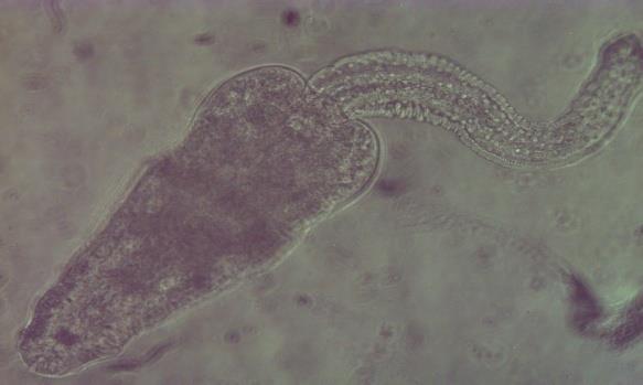
21 CHAPTER TWO LITERATURE REVIEW 2.1 The disease schistosomiasis Schistosomiasis, commonly known as bilharziasis, is a parasitic disease caused by blood flukes of the phylum Platyhelminthes, super family schistosomatoidae and belong to the genus Schistosoma (Gryseels et al., 2006; WHO 2013). A wide range of schistosome species under the genus Schistosoma parasitize both vertebrates and invertebrates. Five schistosome species of vertebrates have been identified as S. intercalatum, S. mekongi (Roca et al., 2002), S. mansoni, S. haematobium, and S. japonicum (Schmitt, 2006; Steinmann et al., 2006; Ayanda, 2009), with the latter three causing major pathology in man. Apart from S. haematobium which dwells in the venous plexus of the bladder and causes urogenital schistosomiasis, all the other four human schistosome parasites dwell in mesenteric vesicles, where they cause intestinal schistosomiasis (WHO, 2013). Schistosomes are atypical flukes; the only dioecious species among all blood flukes (Loker & Brant, 2006; Webster et al., 2013), with worms showing significant dimorphism (Brant & Loker, 2005). These parasites complete their life cycle using two hosts; an intermediate host and a definitive host, unlike the three-host system used by majority of the flukes (Loker & Brant, 2006). They invade their definitive hosts by penetrating their skin directly, before migrating through blood vessels to their final dwelling sites. 7
22 Male worms carry female worms in a specialized feature located on their ventral side, the gynaecophoric groove, where mating occurs. Once paired, female worms produce eggs throughout their lifetime (Gryseels et al., 2006), which they pass into the environment through either the urine (i.e S. haematobium) or stool (i.e S. mansoni). Schistosoma haematobium female worms produce about 100 eggs daily (Basch, 1991) while those of S. mansoni produce approximately 300 eggs (Loker, 1983; Bogitsh, 2005). Adult worms have a lifespan of between three and five years on the average (Gryseels et al., 2006). The disease and pathological conditions presented by schistosome parasites are classified as either urogenital or intestinal schistosomiasis and are mainly due to the presence of eggs in tissues (Brant & Loker, 2005; Barnabas et al., 2012). Urogenital schistosomiasis is primarily characterized by the presence of blood in the urine, a condition known as heamaturia (WHO, 2013), which may be accompanied by dysuria. In women, it may cause lesions in the cervix and vagina leading to vaginal bleeding, a risk factor for HIV transmission (Kjetland et al., 2006; Yirenya-Tawiah et al., 2011). In men, urogenital schistosomiasis can cause prostate cancer and damage to the seminal vesicles. Generally, chronic or advanced stages of the disease presents with bladder and ureteral fibrosis, hydronephrosis as well as cancer of the bladder (Parkin, 2008). Intestinal schistosomiasis is normally characterized by abdominal pains, diarrhoea and bloody stool. The liver and spleen enlarge, a condition known as hepatosplenomegaly in chronic stages of the disease (Parkin, 2008). 8
23 Generally, schistosomiasis causes aneamia, stunted growth, reduced learning and working ability as well as severe organ damage (King & Dangerfield-Cha, 2008). The disease infects persons of all ages who come into contact with freshwater infested with cercariae (Jordan & Webbe, 1993; Ross et al., 2002). Children are at a higher risk of infection compared to adults (Utzinger et al., 2009; Barnabas et al., 2012). Activities such as farming, fishing (WHO, 2002), playing and bathing along freshwater bodies increase human contact with the site of infection and play a role in disease transmission (Useh & Ejezie, 1999; WHO Expert Committee, 2002; Wagatsuma et al., 2003). High occurrence of snail vectors and intense human activities along freshwater bodies in endemic areas indicate active transmission of the disease (Yirenya-Tawiah et al., 2011). Nduka et al. (1995) and Musa et al. (2010) have reported children aged between 12 and 15 as showing the highest infection rates of urogenital schistosomiasis. Recent works by Deribe et al. (2011) on urogenital schistosomiasis and Barnabas et al. (2012) on both urogenital and intestinal schistosomiasis also reported children aged between 10 and 14 years as showing the highest infection rates. All these works confirmed boys have higher infection rates compared to girls. According to Jordan and Webbe (1993), schistosomiasis decreases with age, thus adults in endemic areas generally record very low infection rates. This condition may be associated with increased immunity against the disease, a possible result of continuous exposure to infection. 9
24 2.1.1 Life cycle of schistosome parasites Schistosoma spp. complete their life cycle utilizing two host species, intermediate hosts which are freshwater snails and definitive hosts which are humans and other animals. Following schistosome infections in the human hosts, eggs are expelled into the environment through urine in the case of S. haematobium infections and through stool during S. mansoni and S. japonicum infections (figure 1). Upon contact with water, the eggs hatch, releasing miracidia which swim and penetrate suitable vector snails in the water body. Snails of the genus Bulinus host miracidia from S. haematobium eggs while Biomphalaria snails harbour miracidia from S. mansoni eggs. Miracidia of S. japonicum eggs infect snails of the genus Oncomelania (Jordan et al., 1993; Waine & McManus, 1997). In the snail hosts, the miracidia develop into mother and daughter sporocysts and further develop into cercariae, the free-swimming stages. At 4 to 6 weeks post-infection, the snails shed cercariae into the water body in the presence of light and temperature range of 10 and 30 C. The free swimming cercariae then penetrate human skin which comes into contact with the cercariae infested water. Upon penetration, cercariae lose their forked tails and transform into schistosomulae, which are transported in blood vessels to the liver, where worms mature into adults and pair. Paired S. haematobium and S. mansoni worms migrate from the liver and finally dwell in the venous plexus of the urinary tract and/or the mesenteric venules of the intestines where they lay and deposit eggs (Jordan et al., 1993; Waine & McManus, 1997). 10

25 Figure 1: Life cycle of schistosome parasites Source: 11
26 2.2 Epidemiology of schistosomiasis Global epidemiology It is estimated that about 780 million people worldwide stand the risk of schistosomiasis disease (Hotez et al., 2006; Steinmann et al., 2006), of which more than 207 million people currently have the disease in 78 countries worldwide (Barnabas et al., 2012; WHO, 2013). This indicates the continuous spread of the disease worldwide; Schmitt (2006), Steinmann et al. (2006), Fenwick (2006 and 2009) and WHO (2010) earlier reported an estimated number of 200 million people with the disease in 74 countries. Of the over 200 million infected persons, 90% live in 46 African countries (Barnabas et al., 2012; WHO, 2013). Schistosoma haematobium which causes urogenital schistosomiasis is dominant in Africa and the Middle East (Schmitt, 2006) and has been documented in 53 countries (figure 2). According to Risikat and Ayoade (2012), about 90 million people living in these countries are positive for the disease. Intestinal schistosomiasis caused mainly by S. mansoni and S. japonicum is distributed in 54 countries (WHO, 1993b; Southgate, 1997; Urbani et al., 1997; Chitsulo et al., 2000). While S. mansoni is widely distributed across the globe, S. japonicum is confined to Asian countries. 12
 Centers for Disease Control and Prevention")
27 Figure 2: Global distribution of schistosomiasis Source: United States of America (USA) Centers for Disease Control and Prevention 13
28 2.2.2 Epidemiology of schistosomiasis in Ghana Schistosomiasis is most prevalent in many developing countries in Africa, including Ghana. Urogenital schistosomiasis, the most common form of the disease in Ghana, was first reported in 1895 while intestinal schistosomiasis was first reported in 1920 (CEGET- CNRS /OMS-WHO, 1987). Only two of the main parasites, S. haematobium and S. mansoni cause disease in Ghana (Bosompem et al., 2004). Epidemiological data on the two forms of the disease were first documented between 1952 and Before 1955 it was revealed that 20% of Ghana s total population suffered from urogenital schistosomiasis at some point in their lives (McCullough, 1954). The creation of the Volta lake in 1964 and 1981 at Akosombo and Kpong respectively (Zakhary, 1997; Aryeetey et al., 2000), the Weija lake in 1979 (Ampofo & Zuta, 1995) and other water reservoirs has led to the outbreak of the disease in the country, as the sudden reduction in the flow rate of these reservoirs, provided suitable breeding sites for the snail intermediate hosts to thrive (Nkegbe, 2010). Settlements along these lakes have since been described as endemic areas, with high infection rates recorded among the local populace (Ampofo & Zuta, 1995; Zakhary, 1997). Extensive epidemiological surveys and field researches have been carried out on the disease in Ghana, spanning over five decades. Works of McCullough, (1959); Obeng, (1997); Aryeetey et al. (2000); Bosompem etal. (2004); Aboagye and Edoh, (2009); Nkegbe, (2010); Yirenya Tawiah et al. (2011) and many others along several freshwater bodies in the country, notably the Volta and Weija Lakes, have contributed to the 14
29 knowledge on the disease. These studies have reported on the prevalence of both intestinal and urogenital schistosomiasis, snail vectors responsible for hosting parasites, as well as human activities that influence the distribution of the disease. Efficacy of drugs for treatment and measures for control of the disease have also been explored and documented. Report from the World Schistosomiasis Risk Chart (2012) indicates that, the disease caused by both parasites, is present in the country as a whole. However, the distribution is variable, with communities recording single infections of either S. haematobium or S. mansoni, or both. Lack of proper sanitation and the availability of conducive habitats that favour the survival of the vector snails contribute to the endemicity of the disease in many parts of the country. Generally, there are no data on the overall prevalence of schistosomiasis in Ghana. Prevalences recorded are based on the estimates of studies carried out in various parts of the country. Prevalence of S.haematobium infection has been found to range between 52% and 60% (Aryeetey et al., 2000; Nsowah-Nuamah et al., 2001; Aboagye & Edoh, 2009). On the other hand, prevalence of S. mansoni infection ranges between 10 and 49% (WHO, 2010), but this is likely to increase as this species is colonizing new areas which were initially noted for S. haematobium infections only (figures 3 and 4). 15
30 Figure 3: Distribution of S. haematobium in Ghana Source: WHO,

31 Figure 4: Distribution of S. mansoni in Ghana Source: WHO,
32 2.3 Morphology of schistosome parasites and eggs Adult worms Schistosoma parasites are cylindrical in shape. Adult male worms of S. mansoni and S. haematobium measure ~1cm by 1 mmin size. They possess a complex tegument, a blind digestive tract as well as oral and ventral suckers which they use for attachment to their definitive host. Coarse tubercles cover the entire tegument of S. mansoni male worms as opposed to the finer tubercles possessed by S. haematobium male worms. Tubercles are absent in S. japonicum male worms. Generally, male worms are shorter, stouter and possess a ventrally placed gynaecophoric canal (figure 5) in which they carry females (figure 6) for mating (Stewart, 2010). Conversely, female adult schistosome worms are generally longer and thinner than male worms (Danso-Appiah, 2009) (figure 6). S. mansoni females measure ~1.1cm by 0.2mm and S. haematobium females measure ~1cm by 1mm. Female worms also possess suckers; an oral sucker near the oesophageal gland and a ventral sucker near the genital pore, though these are not as pronounced as those found in male worms (Stewart, 2010). 18
")
33 Figure 5: Morphology of male schistosome worm Source: Figure 6: Male and female schistosome worms in copulation Source: Lewis (1998) 19
.")
34 2.3.2 The miracidium Miracidium is a non-feeding, sexually differentiated form of the parasite that hatches out of the egg upon contact with water. This stage of the parasite is infective to the vector snails. It is a free-swimming stage of the parasite that measures µm in length and µm in width (Stewart, 2010). As shown in figure 7, the body of a miracidium is covered with cilia for rapid movement in water. It possesses a terebratorium, located at its apex, which aids attachment to the snail host for effective penetration. The parasite also has a nervous system comprising of photoreceptors and chemoreceptors that help in locating and penetrating its snail host (Walker, 2011). Figure 7: Morphology of a miracidium Source: The cercaria Cercaria, the human infective stage of the schistosome parasite, develops from daughter sporocysts in the vector snail hosts. Akin to miracidium, it is a non-feeding and freeswimming stage of the parasite which is sexually differentiated. It measures ~325μm in 20
35 length and is characterized by a head region and a bifurcated tail region (figure 8) that propels it in water (Lewis, 1998). Cercaria is wholly covered in a single continuous layer by a carbohydrate-rich tegument which is made up of three layers of plasma membrane consisting of spines and sensory papillae. These help in the location of its host, man (Stewart, 2010). It also possesses an acetabulum that forms its ventral sucker, a preacetabular gland which contains enzymes such as proteases essential for efficient penetration of its host skin, a post-acetabular gland that secretes proteases and mucus for adherence to its host surface and an excretory system made of flame cells which help in osmoregulation (Walker, 2011). Figure 8: Morphology of a cercaria Source: 21

. Schistosoma mansoni eggs are also large, possessing lateral spines close to their posterior ends (figure 9b).")
. Most of the pathology associated with schistosomiasis are caused by the presence of eggs in tissues (Wynn et al.")
36 2.3.4 Human schistosome eggs Presence of schistosome eggs in urine and/or stool are used for diagnoses of the disease in man (Shane et al., 2011). Based on the presence and position of spines, eggs are classified as either S. haematobium, S. mansoni or S. japonicum. Schistosoma haematobium eggs are large, non-operculate, and have a transparent shell,with terminal spines located at their posterior end (figure 9a). They measure between μm in length (Kjetland et al., 2012). Schistosoma mansoni eggs are also large, possessing lateral spines close to their posterior ends (figure 9b). They measure between μm in length (Silva et al., 2002). Schistosoma japonicum eggs are smaller, measuring between μm in length (Centers for Disease Control and Prevention). These eggs also possess spines, although reduced (figure 9c). Most of the pathology associated with schistosomiasis are caused by the presence of eggs in tissues (Wynn et al., 2004; Danso- Appiah, 2009). a) S. haematobium egg b) S. mansoni egghttp:// c) S. japonicum egg Figure 9:Morphology of the three main schistosome eggs of public health importance 22
37 2.4 Vectors of schistosomiasis Vectors of schistosomiasis are freshwater snails belonging to various genera. Oncomelania spp. are the intermediate hosts of S. japonicum, while Biomphalaria and Bulinus spp. host S. mansoni and S. haematobium respectively (Madsen et al., 2008; Ayanda, 2009). They thrive and breed in stagnant freshwater bodies or slow sections of swift flowing water (Jones, 1993; Obeng, 1997; Yirenya-Tawiah et al., 2011), with aquatic vegetation at the fringes providing food, microhabitats, protection from strong water currents and cache for their eggs (Zakhary, 1997; Ofori, 1999). Clearing of aquatic vegetation may therefore help in controlling of vector populations at transmission sites (Yirenya-Tawiah et al., 2011). Vectors require favourable physiochemical conditions, including suitable ph and temperature to thrive well in the water bodies. Biomphalaria pfeifferi (figure 10) is responsible for the transmission of intestinal schistosomiasis in Ghana, while two species of the genus Bulinus: B. truncatus (figure 11) and B. globosus, have been identified as transmitting urogenital schistosomiasis, with the latter confined to small streams in Ghana (Chu et al., 1978). 23


38 Figure 10: Biomphalaria pfeifferi snails feeding on aquatic vegetation Figure 11: Bulinus truncatus snails feeding on aquatic vegetation 24
39 2.5 Mating interactions Mating interactions among schistosome parasites in concurrent infections have been studied. These interactions have resulted in the formation of homospecific and/or heterospecific pairs. Works by Southgate et al. (1995), Webster et al. (1999), Webster and Southgate (2003) have reported mating interactions between S. haematobium, and S. mattheei, S. mansoni or S. intercalatum respectively. These studies have shown that S. haematobium males have a greater heterospecific mate preference compared to males of the other species. This occurrence may be attributed to the presence of S. mansoni eggs in urine of some individuals as stated in a study by Cunin et al. (2003). The study also revealed, schistosome eggs eliminated through unusual routes may be due to high infections with that particular schistosome species; causing a spill-over of eggs. Similar to works done by Websteret al. (1999) in Senegal, there are areas in Ghana where S. haematobium and S. mansoni occur in sympatry, though the two parasites utilize vectors of different species. 2.6 Clinical signs and symptoms of schistosomiasis Clinical signs and symptoms of schistosomiasis may vary with the species of parasite, intensity and stage of infection. The disease can be manifested in three main forms; cercarial dermatitis (swimmers itch), acute schistosomiasis (katayama fever) and chronic schistosomiasis (Pearce & MacDonald, 2002). Cercarial dermatitis occurs mostly in naive individuals few minutes after cercariae penetrate the skin. This is usually 25
40 characterized by the formation of urticarial rash at the site of penetration which is able to persist for several days in the form of papules (Gryseels et al., 2006) Acute phase Acute schistosomiasis or katayama fever is generally an asymptomatic phase of the disease. It is a hypersensitive immune response against migrating schistosomulae (Gryseels et al., 2006) and progresses to the stage when female worms begin laying eggs (Stewart, 2010). Symptoms generally associated with this phase include fever, fatigue, myalgia (muscle pain), malaise, non-productive cough, eosinophilia infiltrates on thorax radiography (Gryseels et al., 2006; Caldas et al., 2008) and in severe cases bloody diarrhoea Chronic phase Individuals with chronic schistosomiasis, show symptoms months or years after initial exposure to cercariae. All chronic disease conditions of schistosomiasis are caused by the eggs trapped in tissues. Immune response by the hosts system against these eggs involve the release of cytokines, antibodies, and immune cells, including epithelial cells, macrophages and T cells, to mount an appropriate effector response (Wynn et al., 2004). This results in cellular inflammation of tissues leading to granuloma formation and subsequently fibrosis mostly in the liver and spleen (Stewart, 2010). This condition also leads to organ damage in the abdomen and hence ascites (accumulation of fluid in the abdomen) (Manzella et al., 2008). 26
41 2.7 Pathology associated with Schistosoma spp. infections Pathology of Schistosoma mansoni infection Individuals presenting with S. mansoni infections manifest the disease in two main forms: intestinal and hepatosplenic (Danso-Appiah, 2009). Eggs trapped in the intestines are engulfed by immune cells which cause inflammation resulting in granuloma formation. Some eggs are carried in the bloodstream to the liver where they get trapped in the liver tissues. Here again, immune cellular inflammatory response to the eggs leads to granuloma formation in the liver (figure 12). Over a long period of time, the granulomatous liver thickens and becomes fibrotic with an associated enlargement of the spleen resulting in a condition known as hepatosplenomegaly (figure 13) (Cheever, 1978; Cheever et al., 1978). As the liver continues to be inflamed due to immune reactions, liver cirrhosis sets in inhibiting blood flow and subsequently leading to portal hypertension. Once blood flow is obstructed, fluid accumulates in the vessels resulting in ascites and subsequent enlargement of the hepatic arteries forming varicose veins which eventually rapture causing anaemia as so much blood is lost. An infected person presenting with these symptoms may suffer from repeated variceal bleeding and haemorrhagic shock and may eventually die (Danso-Appiah, 2009). 27


42 Figure 12: Schistosome egg granuloma in human liver Source: Figure 13: Hepatosplenomegaly resulting from Schistosoma infection Source:
43 2.7.2 Pathology of Schistosoma haematobium infection Individuals with S. haematobium infections normally develop ulcerations in the walls of the bladder causing haematuria (bloody urine), dysuria (painful urination) and pollakisuria (frequent urination) as well as cystitis (inflammation of the bladder) over a long period of time (Saathoff et al., 2004). Eggs in the ureter may cause obstruction in the lower ends leading to conditions such as hydroureter and hydronephrosis (Badmos et al., 2009). In advanced stages of the disease, cancerous bladder develops and blood loss from the genitourinary tract results in anaemia in infected individuals. Infections with S. haematobium also cause genital disease in both men and women resulting in infertility (Yirenya-Tawiah et al., 2011) as eggs pass through and cause lesions with their spines in the spermatic ducts of men and the cervix as well as vagina of women. These lesions present in the reproductive organs of infected persons predispose them to acquiring and transmitting HIV during sexual intercourse (Kjetland et al., 2006; Yirenya-Tawiah et al., 2011). This ensures the spread of the virus and subsequently AIDS in an already schistosomiasis diseased population. 2.8 Granuloma formation in schistosomiasis infections The formation of granulomas in schistosomiasis infections is due to a delayed hypersensitivity reaction by the host immune response mechanism. It is a protective mechanism against severe schistosomiasis disease (Zuim et al., 2012) which results in the destruction of schistosome eggs (Warren & Domingo, 1970). A number of immune cells in granulomatous lesions play a role in the sequestration of toxins/antigens, released by 29
44 the trapped eggs. These include eosinophilic granulocytes, lymphocytes, fibroblasts and macrophages (Andrade & Azevedo, 1987; Lenzi et al., 1998; Baptista & Andrade, 2005). In liver tissues, infiltration of granulocytes and lymphocytes is a common feature of the egg induced granulomatous lesions. These lesions have been reported by Perrin and Philips (1989) to decrease in size as the disease progresses from an acute phase to a chronic phase. The length and size of granuloma lesions differ from one schistosome parasite to the other and are proportional to the persistence of eggs in tissue and the ability of host immune cells to destroy them (Lichtenberg, 1962, 1968). S. mansoni infections have been extensively studied using mice as animal models to ascertain the immune responses associated with the disease. Apart from mice showing a high compatibility with the parasite, their availability and low cost have added to their increase use in laboratory experiments (Farah et al., 2000) although the strain of mice influences disease progression to a large extent (Warren & Peters, 1967; Cheever et al., 2002). S. haematobium infections on the other hand, have been less studied in animal models due to their compatibility with limited animal models such as hamsters (Webster et al., 1999). According to McLaren and Smithers (1987), S. haematobium parasites rarely mature into adult worm stages when introduced into animal models. 30
45 2.9 Diagnosis and treatment of schistosomiasis Schistosomiasis can be diagnosed in four main ways including direct, indirect, immunological and molecular methods (Kahama et al., 1998) Direct and indirect methods of schistosomiasis diagnosis Diagnosis of schistosomiasis is based primarily on the examination of urine and stool samples for eggs of Schistosoma parasites using microscopy, a direct method (Gryseels et al., 2006). Schistosoma eggs and other intestinal helminthes in stool can be ascertained by microscopy using Kato-Katz technique, direct faecal smears and faecal concentration procedures. On the other hand, filtration techniques and sedimentation techniques are used to identify Schistosoma eggs in urine using microscopy. Although microscopy is inexpensive, straightforward (Feldmeier et al., 1998), and considered a standard method for diagnosing Schistosoma eggs (Gryseels et al., 2006) and other helminthes, it may not be useful particularly during the early stages of infection and when infection loads are low. Results obtained from microscopy, however, may not be a true representation of the egg/worm burden in an individual (Gryseels et al., 1996). Only urogenital schistosomiasis can be diagnosed using an indirect method, reagent strips. However, this method can also detect ulcerations caused by other infections other than urinary schistosomiasis, thus may not be effective for diagnosis. 31
46 2.9.2 Immunological and molecular diagnostic methods Immunologic diagnostics of schistosomiasis include serologic tests and immunoassays such as ELISA. These methods can measure levels of specific antibodies such as IgG and IgE. Antibodies are directed against soluble egg antigens or adult worm antigens in the serum of an individual. Since antibodies can be detected even after cure, this diagnostic tool is not able to differentiate an active infection from a passive one (Rabello et al., 1997). Circulating soluble egg antigens in the urine and blood can be tested using ELISA. This technique is highly sensitive and thus reliable as it gives a clear indication of egg load and pathology status of an individual. (Kahama et al., 1998; Stothard et al., 2009). Polymerase chain reaction, a molecular technique, is highly sensitive and can be specifically used in diagnosing schistosomiasis. It is however very expensive and thus its use is not widespread compared to microscopy. Molecular tests show positive results between one to two months after initial exposure to the parasite, and positive results may also indicate past exposure. Tissue biopsy is useful in identifying eggs that are lodged in tissues. In addition, ultrasound, chest X-rays, CT scans and MRI may be used to assess the damage of organs as inflicted by the presence of the parasites or their eggs. 32
47 2.9.3 Treatment Currently, praziquantel is the drug of choice for the treatment of schistosomiasis (Shekhar, 1991; Fallon & Doenhoff, 1994; Badmos et al., 2009). The drug is only effective in eliminating the adult worms, but not the eggs which cause most of the pathology associated with the disease. Other drugs such as oxamniquine and metrifonate are also useful in treating schistosomiasis (Shekhar, 1991; Fallon & Doenhoff, 1994). Currently, there are no vaccines for schistosomiasis and there is also the need to develop new drugs, particularly in areas where parasites have developed resistant to praziquantel. 33
48 CHAPTER THREE MATERIALS AND METHODS 3.1 Study area and study population The study was carried out in Tomefa, a coastal savannah vegetational zone, located along the Weija Lake, 17 km west of Accra in the Greater Accra region, with GPS coordinates N and W The nearest health facility is about 10km from the community whose inhabitants are predominantly Ewes and Ga-Adangbes numbering approximately 1,500 (Danso-Appiah et al., 2010). The major occupation of men in the community is fishing with the women mostly trading in fish among other wares. Tomefa has one school (Kindergarten to Junior high school) with a population of about 200 pupils who were recruited as subjects for the study. The pupils were aged between 0 and 26years. 3.2 Sample collection Informed consent was sought from community leaders, teachers, parents and adult pupils. Questionnaires were administered to the pupils to get information on their knowledge, practices and attitudes of schistosomiasis. Parasitological data were also collected to estimate the prevalence and intensity of the disease. 34
49 3.2.1 Urine sample collection and analysis The study participants were given 50ml labelled screw cap tubes to provide 15ml or more of urine. They were instructed on the appropriate sample collection procedures to follow to avoid mix-ups and contamination. Urine samples were then kept at 4 C in a cool box and transported to the Laboratory of the Parasitology Department of Noguchi Memorial Institute for Medical Research (NMIMR). Urine samples were later analyzed using the sedimentation and filtration methods and examined by microscopy to detect schistosome parasite eggs Sedimentation technique Each urine sample was homogenized and 10ml transferred into a new 50ml tube using a Pasteur pipette. The samples were then centrifuged at x1000 rpm for 5 minutes and the supernatant carefully decanted leaving a precipitate at the bottom of each tube. Each sample precipitate was transferred onto a slide with a Pasteur pipette and covered with a cover slip. The prepared slides were then examined for schistosome eggs using the x10 and x4 objectives of the Olympus CX21FS1 light microscope. Eggs observed were identified morphologically using the position of their spines to differentiate them into species. They were then counted and the numbers of each species recorded Filtration technique Urine samples were homogenized and for each sample, 10ml was drawn using a syringe. Each drawn sample was then filtered through a 25mm diameter Millipore filter 35
50 membrane of pore size 12µm secured in a swinnex support chamber to trap schistosome eggs. The membrane was then carefully removed from the chamber, using a pair of forceps, and placed on a microscope slide with the side on which eggs were trapped being in contact with the slide surface. The slides were then examined for schistosome eggs using the x10 and x4 objectives of the Olympus CX21FS1 light microscope. Eggs observed were identified morphologically using the position of their spines to differentiate them into species. They were then counted and the numbers of each species recorded Stool sample collection and analysis Participating pupils were each given a labelled plastic container to provide fresh stool sample. They were taken through appropriate procedures for sample collection to ensure the expected amount was received and contamination avoided. Collected stool samples were transported in a cool box to the laboratory (Parasitology Department, NMIMR) and stored at 4 C. Stool samples were later analyzed using the Kato-Katz and faecal concentration methods and examined microscopically to detect schistosome parasite eggs and eggs of other soil-transmitted helminths Kato-Katz technique Hydrophilic cellophane strips measuring 25 x 35mm 2 in size were soaked in 50% glycerol-malachite green solution for about 24 hours (hrs) before use. For each collected specimen, a gram of stool was transferred onto a pergamon paper using an applicator stick in a hood. A nylon mesh was then pressed on top of the sample and a flat-sided 36
51 wooden applicator stick used to scrape across the upper surface of the mesh to sieve the stool sample. The sieved sample was used to fill the hole of a 6mm diameter template placed on a clean microscope slide. This was then levelled with the applicator stick and the template removed carefully to leave a cylindrical sieved stool sample on the slide. The sample on the slide was covered with the treated cellophane strip, inverted, and the sample firmly pressed against the cellophane strip on a smooth surface to evenly spread out. The prepared slide was then placed in a slide holder for about 30minutes (mins) to clear, after which it was examined for eggs using the x10 and x4 objectives of the Olympus CX21FS1 light microscope. Observed eggs were identified, counted and recorded Faecal concentration technique One gram of stool was added to 10ml of 10% formalin solution in a centrifuge tube and stirred using an applicator stick to form a suspension. The suspension was then strained through two layers of gauze into another centrifuge tube. More 10% formalin solution was added to the strained suspension to bring the volume to 10ml. Three milliliters (3mm) of ethyl acetate was then added to the suspension in the tube and vigorously shaken for 10 seconds, after which it was centrifuged for 3 mins at 500g. Three layers of filtrate are formed in the tube; a layer of formalin at the bottom, a middle plug of faecal debris and a layer of the ethyl acetate on top. Using an applicator stick, the plug of faecal debris was carefully freed from the walls of the tube, after which the filtrate was decanted in a single movement. The remaining sediment was examined under an 37
52 Olympus CX21FS1 light microscope at x10 and x4 objectives. Observed eggs were identified, counted and recorded Snail sample collection and exposure to determine infectivity Schistosome vector snails were collected from the Weija Lake bordering the community with a scoop net into containers with water from the lake and transported to the laboratory (NMIMR). On arrival at the laboratory, the snails were separated, according to their species, into labelled aquaria containing distilled water and allowed 24hrs to adjust to laboratory conditions. Thereafter, snails were singly placed in each well of a 24-well plate containing distilled water. The plates were then placed under an artificial source of light for 2hrs to ensure maximum cercarial shedding by snails. After exposure, snails were observed under x10 and x4 objectives of a CK30-F200 inverted microscope. Snails that were found to have shed cercariae were noted in addition to type of cercariae shed. Snails that shed no cercariae were returned to their respective aquaria containing fresh distilled water and placed in a dark environment. Cercariae shedding snails were also transferred from the well plates to new aquaria labelled according to the snail species and type of cercariae shed. These snails were also put in a dark environment to minimize rate of cercariae shedding. Exposure was repeated for 4 weeks for non-shedding snails to allow enough time for snails with patent infection at field sampling time to begin cercariae shedding. Snails were observed for cercariae shedding in intervals of two days to isolate those that had begun shedding. 38
53 3.3 Hatching of miracidia and infection to laboratory snails Miracidium hatched from eggs in urine Remaining urine samples containing only S. haematobium eggs were pooled in a test tube and spun at x1000 rpm for 5 mins. The supernatant was decanted leaving pelleted eggs, which were re-suspended in normal saline and again spun at x1000rpm for 5 mins. Once again the supernatant was decanted leaving pelleted eggs. The eggs were re-suspended in distilled water and transferred gently unto the bottom of a conical flask containing distilled water. The conical flask was then covered with an aluminium foil up to the neck region leaving a portion for light penetration. More distilled water was then added to fill the conical flask up to the uncovered portion and left to stand on the laboratory bench at room temperature for at least 30 mins for egg hatching into miracidium Miracidium hatched from eggs in stool Stool from positive samples with only S. mansoni eggs were pooled and emulsified using normal saline in a beaker. This was then filtered using three different sieves of mesh sizes 150µm, 75µm and 45µm. Most of the debris in the stool was trapped by the sieves of mesh sizes 150µm and 75µm, with sieve of 45µm mesh size trapping mainly eggs. The contents of the sieve of mesh size 45µm was then washed with saline into a 50ml tube and spun at x1000rpm for 5 mins. Following, the supernatant was decanted leaving sedimented eggs which were re-suspended in normal saline and spun at x1000rpm for 5 mins. The supernatant was again decanted and the eggs re-suspended in distilled water which was then gently transferred unto the bottom of a conical flask containing distilled water. An aluminium foil was used to cover the conical flask up to the neck region 39
54 leaving a portion for light penetration. More distilled water was later added to fill the conical flask up to the uncovered portion and left to stand on the laboratory bench at room temperature for at least 30 mins for egg hatching into miracidium Miracidia infection to laboratory snails Five to six miracidia of S. mansoni and S. haematobium in solution were separately pipetted into 20 wells of two 24-well plates. Each plate contained miracidia of only one schistosome parasite species. Twenty (20) snails each of laboratory bred B. pfeifferi and B. globosus were singly placed in these wells containing their respective schistosome parasites. The snails were left in the miracidia solution for at least 3hrs to ensure maximum infection after which they were transferred to their respective aquaria. Snails used were three months old F2 generations obtained from uninfected wildtype species sampled from the study community. 3.4 Cercariae shedding by infected snails At four weeks post-parasite infection to B. pfeifferi and B. globosus snails respectively, the snails were transferred into a dark environment for 48hrs. The snails were then placed singly in wells of 24-well plates containing 1ml of distilled water. For at least 2hrs, the snails were exposed to an artificial source of light to induce cercarial shedding. 40
55 Exposed snails were then observed with the x4 and x10 objectives of the CK30-F200 inverted microscope to check for those that shed cercariae. Snails found not to have shed were returned to their respective aquaria. Cercariae counts were made for snails shedding cercariae after which they were placed in new aquaria according to their species. All snails were again placed in a dark environment for 48hrs and later observed for cercarial shedding. Cercariae were pooled into a test tube with the aid of a Pasteur pipette. About 20µl of cercariae solution was pipetted onto a slide and the cercariae counted. The counts were repeated three times to estimate the average number of cercariae in 20µl of cercariae solution. 3.5 Cercariae infection to laboratory mice Cercariae solution was pipetted into restrainer test tubes and distilled water added to about three-quarters the volume of each test tube. The tubes, each containing between human infective cercariae, were then placed in a restrainer rack and mice infected through their tails (figure 14). In all, 9 female ICR mice, (3 mice/ group) were each infected for an hour to ensure maximum infection. Group A mice were infected with S. mansoni cercariae only, whereas mice in group B were infected with only S. haematobium cercariae. The last group of mice, group C, were infected simultaneously with cercariae of S. mansoni and S. haematobium. 41

56 Source: Lewis (1998) Figure 14: An illustrative diagram and picture of cercariae infection to mice through their tails. 3.6 Perfusion of mice Infected mice were euthanized after 8 weeks and 12 weeks for S. mansoni and S. haematobium infections respectively. Mice infected simultaneously with S. mansoni and S. haematobium cercariae were also euthanized at 12 weeks post- parasite infection. Euthanized mice were dorsally placed onto a dissecting board and dissected to reveal the hepatic portal vein and mesenteric venous systems. Their livers were then cleaned by perfusing with citrate-saline buffer (prepared from 0.85% of sodium citrate and 0.9% of sodium chloride) and schistosome worms collected in the process (figure 15). Granulomatous livers and intestines of perfused mice were harvested, processed histologically and examined microscopically. 42

57 Source: Lewis (1998) Figure 15: A picture and an illustrative diagram of the perfusion of a mouse. 3.7 Schistosome eggs from perfused mice Cut portions of granulomatous liver and intestines of perfused mice were harvested and macerated in saline solution in an electric blender. The macerated tissue in solution was then sieved using three different sieves of mesh sizes 150µm, 75µm and 45µm. Most of the tissue was trapped by the sieve of mesh size 150µm, with sieves of 75µm and 45µm mesh sizes trapping mainly eggs. The contents of the sieves of mesh sizes 75µm and 45µm was then washed with saline into a 50ml tube and spun at x1000rpm for 5 mins. Following, the supernatant was decanted leaving sedimented eggs. The sedimented eggs were collected and hatched into miracidia using the process for hatching eggs from urine and stool. Miracidia obtained were then used to infect laboratory snails, following which infection to laboratory mice were made with cercariae obtained from the infected snails to maintain the life cycle in the laboratory. 43
58 3.8 Sampling and processing of mice liver and mesenteric vessels for histology Granulomatous liver and mesenteric tissues harvested from infected mice were fixed in 20ml of 10% buffered formalin. Fixed tissues were then taken through routine histological processes of dehydration, embedding, sectioning, de-waxing, rehydration, staining, dehydration and mounting. In dehydrating, tissues were passed through graded alcohol series of 50%, 70%, 95% and 100%. However before being embedded in paraffin wax, tissues were put in xylene, a solution that is easily miscible with wax to allow for proper embedding of tissue. Dehydrated tissues were then infiltrated with wax at 58 C after which they were put in labelled cassettes, filled with molten wax and allowed to cool to obtain a block of tissue embedded in wax. Tissue blocks were kept at -5 C in a refrigerator to further harden before sectioning with a Leica 2235 rotary microtome at a thickness of 5.0 µm. Sectioned tissue blocks were placed in a water bath of temperature 48 C to spread out and then picked with labelled microscope slides and allowed to dry in an oven. Each sectioned tissue on a slide was de-waxed by dipping in xylene and then rehydrated by passing through graded alcohol of 100%, 95%, 70% and 50%. Rehydrated sectioned tissues were then stained with haematoxylin and eosin (H&E) and dehydrated again in serial solutions of alcohol of 50%, 70%, 95%, 100% followed by 44
59 dipping in xylene. Stained tissue sections on slides were then prepared for microscopic examination by mounting with dibutylphalate in xylene, DPX. 3.9 Data analysis Parasite distribution according to sex and age of study participants were entered onto Microsoft Excel Spreadsheets and the prevalence and intensities of infection determined. All data were assessed for normality, following which non-normal data were normalized through transformations. ANOVA and unpaired t-tests were used to test for statistical significance. ANOVA was carried out to determine variations in mean intensities of parasite eggs among the various age groups of participants. A two sample unpaired t-test was also used to determine statistical significance between snail vectors shedding cercariae. All analyses were carried out at a set alpha ( ) level of Genstat Statistical Software (version 9.2) was used in all analyses except for questionnaire data. Data obtained from questionnaires administered were analyzed using Statistical Package for Social Science (SPSS) version Prevalence of infection The prevalence of both urogenital and intestinal schistosomiasisis is measured in percentage (%). Infection prevalence among age groups and sex was determined as the ratio of positive individuals to the total number of study participants and mathematically represented as: 45
60 Prevalence = Number of subjects testing positive x 100% Number of subjects investigated Intensity of infection The intensity of urogenital schistosomiasis was measured as the number of eggs in 10ml of urine (ep10ml). Positive urine samples were classified as light intensity (1 49ep10ml) or heavy intensity ( 50ep10ml). In the case of intestinal schistosomiasis, intensity was measured as total egg count in one gram of stool and classified as either light (1 99epg), moderate ( epg) or heavy (>400epg) infections. The mean egg loads were also calculated and expressed as: Mean egg = Sum of each individual epg Number of subjects being investigated 3.10 Definition of terms Single infections were defined as individuals with only one type of schistosome parasite egg present in either urine or stool. Concurrent infections were defined as individuals with both schistosome parasite eggs irrespective of route of elimination. 46
61 CHAPTER FOUR RESULTS 4.1 Demography and sample analysis of study participants As shown in table 1, a total of 202 pupils consisting of 102 males and 100 females from the Peace International School, Tomefa, who were aged between 0 and 26 years (yrs) were involved in the study. All the study participants gave urine samples whereas only 181 comprising of 91 males and 90 females provided stool samples (table 1). Following parasitological examination for the presence of schistosome eggs in the 202 stool samples, 163 tested positive for schistosome parasite eggs; this consisted 146 S. mansoni single infection and 17 concurrent infections of both S. mansoni and S. haematobium (S. mansoni+s. haematobium) (table 2). As shown in table 3, of the 202 children who presented urine samples, 135 were positive for schistosome eggs, comprising of 59 S. haematobium single infections, 3 S. mansoni single infections and 73 concurrent infections (S. mansoni+s. haematobium). Table 1: Demographic data of study participants. Urine samples (+ve individuals) Stool samples (+ve individuals) Age group (yrs) Male Female Male Female (3) 12 (2) 5 (5) 10 (7) (12) 17 (9) 18 (17) 15 (14) (7) 18 (10) 9 (9) 17 (16) (38) 45 (35) 48 (43) 40 (36) >15 19 (12) 8 (7) 11 (10) 8 (6) Total 102 (72) 100 (63) 91 (84) 90 (79) Overall total 202 (135) 181 (163) +ve = positive 47
62 Table 2: Infection status of intestinal schistosomiasis by age range of study participants. Age group (yrs) Number examined Infection status of positive individuals Sm single infections Sm+Sh infections > Total Sm = Schistosoma mansoni Sh = Schistosoma haematobium Table 3: Infection status of urogenital schistosomiasis by age range of study participants. Age group (yrs) Number examined Infection status of positive individuals Sh single infections (Sm only) Sm+Sh infections (1) (2) (0) (0) 38 > (0) 9 Total (3) 73 Sm = Schistosoma mansoni (Sm only) = individuals with only Sm eggs in urine Sh = Schistosoma haematobium 48
63 4.2 Prevalence of schistosomiasis Age distribution of intestinal schistosomiasis Prevalence of single S. mansoni infections, ranged between 59% and 91%, with the highest prevalence occurring in the (6 8) yrs age group. On the other hand, prevalence of both parasite species (S. mansoni+s. haematobium) at the same time in stool samples ranged between 2-21%, with the highest prevalence being recorded in the (0 5) yrs age group (figure 16). Overall prevalence of intestinal schistosomiasis among the pupils was 90.1% (163/181), and all age groups presented both concurrent and single infections in stool (table 2 and figure 16) Prevalence (%) Sm +Sh infections Sm infection only >15 Age group (years) Figure 16: Prevalence of intestinal schistosomiasis of single and concurrent infections by age range. 49
64 4.2.2 Age distribution of urogenital schistosomiasis Prevalence of S. haematobium single infections, ranged between 5-45%, with the highest prevalence occurring in the >15yrs age group. In the case of infections with both parasite species, the highest prevalence was recorded in the (9 11) yrs age group, with a range of between 23-49% across all age groups studied (figure 17). Generally, prevalence of urogenital schistosomiasis determined among the pupils was 66.8% (135/202), with all age groups presenting both S. mansoni+s. haematobium concurrent infections as well as S. haematobium single infections as shown in table 3 and figure Prevalence (%) Sm + Sh infections Sh infection only >15 Age group (years) Figure 17: Prevalence of urogenital schistosomiasis of single and concurrent infections by age range in study participants. 50
65 4.2.3 Sex distribution of intestinal schistosomiasis The prevalence of intestinal schistosomiasis based on the sex of study participants showed males had higher prevalence of infection (92.3%) compared to females (87.8%) among all the age groups (figure 18 and table 1). Whereas female infection prevalence was highest in the ages ranging from 6 15 yrs; male prevalence was generally high for all the age groups studied Prevalence (%) male female >15 Age group (years) Figure 18: Prevalence of intestinal schistosomiasis by sex across age groups in study participants. 51
66 4.2.4 Sex distribution of urogenital schistosomiasis Prevalence of urogenital schistosomiasis increased steadily from the lowest age group for both sexes, with peaks at the (9 11) yrs age group for males and >15yrs age group for females as shown in figure Prevalence (%) male female >15 Age range (years) Figure 19: Prevalence of urogenital schistosomiasis by sex across age groups in study participants. 52
67 4.3 Single and concurrent Schistosoma spp. infections in study participants As shown in table 4, majority of the study participants (131) showed concurrent infections relative to single infections (50). Of the 131 individuals with concurrent infections, 50 had S. mansoni eggs in stool and S. haematobium eggs in urine (type A concurrent infections), with the remaining showing both S. mansoni and S. haematobium eggs in either urine or stool, or both (type B concurrent infections). Table 5 further shows the distribution of parasites in individuals classified as type B concurrent infections. Among the 81 individuals, 63 had both S. mansoni and S. haematobium eggs in their urine only, with 6 individuals presenting both parasites in stool only. The remaining 12 individuals had both S. mansoni and S. haematobium eggs present in their urine as well as in the stool. Table 4: Single and concurrent Schistosoma spp. infections in study participants. Types of infections Number of individuals infected Total Single infections Concurrent infections Sm = S. mansoni Sm eggs only 43 Sh eggs only 7 Type A 50 Type B Total Sh = S. haematobium Single infections = individuals with only one type of parasite egg in stool/urine. Concurrent infections (type A) = individuals with both S. mansoni eggs in stool and S. haematobium eggs in urine. 53
68 Concurrent infections (type B) = individuals with both S. mansoni and S. haematobium eggs in urine and/or stool. Table 5: Categorization of type B concurrent infections in study participants. Category of type B concurrent infection Number of individuals Sm and Sh in urine 63 Sm and Sh in stool 6 Sh and Sm in urine and stool 12 Total 81 Sm = S. mansoni Sh = S. haematobium 4.4 Intensity of schistosomiasis infection Intestinal schistosomiasis intensities and mean egg loads As presented in table 6, among the 163 egg positive individuals, 131 individuals presented light infections, including all 12 individuals in the (0 5) yrs age group. The highest number of individuals with the infection intensity threshold described as moderate intensity of infection (30 positive individuals) belonged to the 6 8 and age groups. Again, only two individuals, each from the same age groups (6 8 and 12 15) yrs, had heavy intensity of infection. Although the calculated difference in the mean egg loads among all the age groups was not statistically significant (p=0.19), individuals in the (12 15) yrs age group had the highest mean egg load, with individuals in the (0 5) yrs age group presenting the lowest as shown in figure
69 Table 6: Intensity of intestinal schistosomiasis by age range of study participants. Age range (yrs) Light (1-99 epg) Infection Intensity Thresholds Moderate ( epg) Heavy ( 400 epg) > Total epg = eggs per gram Mean egg loads >15 Age group (years) Figure 20: Mean egg loads of intestinal schistosomiasis in different age groups of study subjects. 55
70 4.4.2 Urogenital schistosomiasis intensities and mean egg loads From table 7, study participants from all age groups had either light or heavy infections. Seventy (70) positive individuals presented light infections as against 65 with heavy infections. Of the heavy infections recorded, majority of those individuals were in the (6 8 and 12-15) yrs age group. For the calculated mean egg loads, individuals in the (6 8) yrs age group had the highest while the least was recorded in the >15 yrs age group (figure 21). The difference in the mean egg loads among all the age groups was however not statistically significant (p=0.59). Table 7: Intensity of urogenital schistosomiasis among the different age ranges of study participants. Infection intensity thresholds Age range (yrs) Light (1-49 ep10ml) Heavy ( 50 ep10ml) > Total ep10ml = eggs per 10ml 56
71 Mean egg loads >15 Age group (years) Figure 21: Mean egg loads of urogenital schistosomiasis. 57
72 4.5 Water contact activities of study participants and sewerage facilities available in study community Table 8 shows the various activities undertaken by male and female study participants at the side of the lake bordering the community. Fishing, fetching of water, washing and playing were the four main activities study participants engaged in along the lake. Generally, more males (102) engaged in fishing in the lake compared to females (7), with nearly an equal number of both sexes (95 males and 96 females) fetching water from the lake for domestic purposes, including cooking and bathing. All respondents of the questionnaire, 102 males and 100 females (table 1) stated the lake to be the main source of water supply to the community. Table 9 presents the various sewerage facilities utilised by the study population. It was evident in the study that there were inadequate toilet facilities in the community. Study participants used either of two main sewerage facilities- pit-latrines or nearby bushes mainly at the banks of the lake. Table 8: Water Contact Activities of Study Participants Water contact activities Males Females Total Fishing Fetching water Washing Playing Table 9: Sewerage facilities used by Study Participants Sewerage facility No. of individuals Flush toilet 0 Pit-latrine 133 Bush area
 and Ceratophyllum demersum (figure 23).")
73 4.6 Field snail vectors identification and collection Species of snail vectors sampled from the part of the lake used by the study community mainly for fishing and domestic activities were identified as B. pfeifferi and B. truncatus. Snail vectors were found mostly attached to aquatic vegetation, including Nymphea lotus (figure 22) and Ceratophyllum demersum (figure 23). Other aquatic vegetation found at the banks of the lake where snail vectors were sampled from included: Ipomea aquatica, Digitari insularis and Floscopa africana. Figure 22: Bulinus truncatus snails (indicated by arrows) attached to Nymphea lotus. 59
.")
74 Figure 23: Biomphalaria pfeifferi snails (indicated by arrows) attached to Ceratophyllum demersum. In table 10, a total of 1,676 snails consisting 780 B. pfeifferi and 896 B. truncatus were sampled from the lake. Although the numbers of B. truncatus snails were higher than that of B. pfeifferi snails, the difference was not statistically significant (p = 0.81). A hundred and fifty-six (156) B. pfeifferi out of the 780 collected and 85 out of the 896 B. truncatus snails were observed to be shedding at least one of three morphologically different cercariae parasites (figure 24a,b&c), of which 14 (8.9%) and 7 (8.2%) of the infected B. pfeifferi and B. truncatus snails respectively, shed cercariae infective to human only. The calculated means of snails of either species found to shed only human cercariae were equal (p = 0.24). Interestingly, one B. truncatus and one B. pfeifferi were each found to shed both human and one other cercariae not known to be infective to man (figure 24a&b). 60

 Unknown")
 Unknown cercariae")


75 a) Human infective cercariae shed by both B. truncatus and B. pfeifferi snails b) Unknown cercariae type 1 shed by both B. truncatus and B. pfeifferi snails c) Unknown cercariae type 2 shed by both B. truncatus and B. pfeifferi snails Figure 24: Three morphologically distinct cercariae types shed by both B. truncatus and B. pfeifferi snails 61
76 Table 10: Infection status of snails collected from the field. Number of snails shedding cercariae Number Snail vector species examined Human Other Human+other Total only Bulinus truncatus Biomphalaria pfeifferi Total Infections to laboratory-bred snail vectors As shown in table 11, of a total of 57 second filial generation (F2) snails consisting of 32 B. pfeifferi and 25 B. truncatus that were infected with human cercariae, eight (25%) of the B. pfeifferi snails shed cercariae whereas none of the B. truncatus snails shed cercariae after 4 weeks. Table 11: Infection status of laboratory-bred snails at four weeks post-infection. Snail vector species Snail numbers Infected Alive at exposure Shedding at exposure Biomphalaria pfeifferi Bulinus truncatus Total
77 4.8 Infection status of laboratory mice infected with human Schistosoma cercariae Table 12 shows the infection status of 3 groups of mice. Group A mice (S. mansoni infected) gave a total of 127 schistosome worms, comprising 45 single and 41 paired worms. Fifteen worms consisting 11 single and 2 paired ones were obtained from group B mice (S. haematobium infected). Of a total 235 worms collected from group C mice (S. mansoni+s. haematobium infected), 90 were paired and 55 were single. Table 12: Schistosome worms recovered from laboratory mice infected with human infective schistosome cercariae Infection group Mouse number Single worms collected Paired worms collected Group A Total Total Group B Total Group C Total
78 4.9 Processing and staining of infected mice tissues (liver and intestines) Upon dissection of infected mice, the intestines were found enlarged and the livers hardened with clear granulomatous lesions. Further processing of the tissues histologically, revealed granuloma formations around schistosome eggs trapped in the tissues. As shown in figures 25a&b, 26a&b, 27a&b, 28a&b, 29a&b and 30a&b granuloma lesions formed around eggs varied for the three mice groups. In comparison with tissues of mice infected with only S. mansoni (group A) and S. haematobium (group B) cercariae, tissues of mice infected with cercariae of both species (group C) showed gross pathology revealing an up-regulation of inflammatory cells (mainly eosinophils, basophils, mast cells and neutrophils) in the entire tissue. The granulomatous lesions around mixed (S. mansoni and S. haematobium) parasite eggs were also larger in size due to the high intensity of infiltration of inflammatory cells. Whereas granuloma formation was large in livers and intestines of group A mice which were presented with S. mansoni eggs, quite smaller granulomas were found in those of group B mice which presented no schistosome eggs but only a worm in the liver. 64


 and b) show x40 and x100 magnifications respectively of")
79 a) b) Figure 25: Liver of mice infected with cercariae of only S. mansoni parasites. Panels a) and b) show respectively, x40 and x100 magnifications of sectioned mice liver stained with haematoxylin and eosin. Arrows indicate size of granuloma formations surrounding S. mansoni eggs in mice liver. a) b) Figure 26: Intestines of mice infected with cercariae of only S. mansoni parasites. Panels a) and b) show x40 and x100 magnifications respectively of sectioned mice intestines stained with haematoxylin and eosin. Arrows indicate granuloma formations surrounding S. mansoni eggs. 65


 b Figure 28: Intestines of mice infected with cercariae of only S.")

80 a) b) Figure 27: Liver of mice infected with cercariae of only S. haematobium parasites. As shown in panels a) and b) of x40 and x100 magnifications respectively, sectioned liver tissues were stained with haematoxylin and eosin. Arrows indicate granulocytes surrounding a worm. a) b Figure 28: Intestines of mice infected with cercariae of only S. haematobium parasites. Panels a) and b) show x40 and x100 magnifications respectively of sectioned mice intestines stained with haematoxylin and eosin. Tissues were obtained from mice infected with cercariae of only S. haematobium parasites. Arrows indicate aggregations of granulocytes. 66


 b) Figure 30: Intestines of mice infected with cercariae of both S.")
 and b) show x40 and x100 magnifications respectively of sectioned mice")
81 a) b) Figure 29: Liver of mice infected with cercariae of both S. mansoni and S. haematobium parasites. Panels a) and b) show sectioned liver viewed under x4 and x10 objective lens of a light microscope. Sectioned liver tissues were stained with haematoxylin and eosin. Arrows show intense infiltration of tissue with granulocytes as well as trapped schistosome eggs. a) b) Figure 30: Intestines of mice infected with cercariae of both S. mansoni and S. haematobium parasites. Panels a) and b) show x40 and x100 magnifications respectively of sectioned mice intestines stained with haematoxylin and eosin. Arrows show intense infiltration of tissue with granulocytes as well as trapped schistosome eggs. 67
Schistosomiasis. World Health Day 2014 SMALL BITE: Fact sheet. Key facts
 Fact sheet Key facts is an acute and chronic disease caused by parasitic worms. At least 249 million people required preventive treatment for schistosomiasis in 2012. The number of people reported to have
Helminths: Schistosoma mansoni. Schistosoma japonicum 10/14/2009. Trematoda - non-segmented flat worms
 Helminths: Trematoda - non-segmented flat worms The schistosomes: Schistosoma mansoni Schistosoma haematobium Schistosoma japonicum Schistosoma mekongi Aquatic freshwater snails are the intermediate hosts
10/14/2009. Helminths: Trematoda - non-segmented flat worms. The schistosomes: Schistosoma mansoni Schistosoma haematobium. Schistosoma mekongi
 Helminths: Trematoda - non-segmented flat worms The schistosomes: Schistosoma mansoni Schistosoma haematobium Schistosoma japonicum Schistosoma mekongi 1 Japan is schistosome-free as of 1976 2 Aquatic
ESCMID Online Lecture Library. by author
 Schistosoma epidemiology & diagnosis José Manuel Correia da Costa INSA National Institute of Health Dr. Ricardo Jorge Brindley PJ & Hotez PJ (2013) Break Out: Urogenital Schistosomiasis and Schistosoma
PARASITOLOGY CASE HISTORY 11 (HISTOLOGY) (Lynne S. Garcia)
 (Lynne S. Garcia)") PARASITOLOGY CASE HISTORY 11 (HISTOLOGY) (Lynne S. Garcia) A 37-year-old man from the United Arab Emirates was admitted to the hospital for complaints of upper and lower abdominal pain. The greater omentum
PARASITOLOGY CASE HISTORY 11 (HISTOLOGY) (Lynne S. Garcia) A 37-year-old man from the United Arab Emirates was admitted to the hospital for complaints of upper and lower abdominal pain. The greater omentum
Schistosomiasis / Bilharzia
 Schistosomiasis / Bilharzia An update for registered healthcare practitioners April 2017 Aims of resource:- To raise awareness of Schistosomiasis/Bilharzia infection with registered healthcare practitioners
Schistosomiasis. Epidemiology. Global epidemiology
 Schistosomiasis Introduction Epidemiology Risk for Travellers Transmission Signs and Symptoms Treatment Prevention References Reading List Introduction Schistosomiasis, or bilharzia, is caused by worms
General Characters of Trematodes
 Parasitology Department General Characters of Trematodes By Hala Elwakil, MD Intended Learning Outcomes By the end of this lecture, the student will be able to know: 1. General morphology of trematodes
WRAIR- GEIS 'Operational Clinical Infectious Disease' Course
 Schistosomiasis WRAIR- GEIS 'Operational Clinical Infectious Disease' Course The opinions or assertions contained herein are the private views of the author, and are not to be construed as official, or
BIO Parasitology Spring 2009
 BIO 475 - Parasitology Spring 2009 Stephen M. Shuster Northern Arizona University http://www4.nau.edu/isopod Lecture 13 Important Orders a. Echinostomatiformes b. Strigeiformes c. Opisthorchiformes d.
[black box of sprorocyst/rediae]
![[black box of sprorocyst/rediae]](/thumbs/88/114695814.jpg "[black box of sprorocyst/rediae]") Digeneans: All life cycles involve a mollusc and a vertebrate All life cycles are similar but different All life cycles are a variation of one theme. Adult-egg-miracidium-[black box of sprorocyst/rediae]
Digeneans: All life cycles involve a mollusc and a vertebrate All life cycles are similar but different All life cycles are a variation of one theme. Adult-egg-miracidium-[black box of sprorocyst/rediae]
Acknowledgements. Schistosomiasis Epidemiology
 Pulmonary Arterial Hypertension and Schistosomiasis Rubin M. Tuder, M. D. Brian Graham, M.D. Program in Translational Lung Program Division of Pulmonary Sciences and Critical Care Medicine, Department
V. Subclass Digenea (Chapters 15-18, BLY ) A. Background 1. Flukes 2. Digenea refers to having an alternation of hosts in the life cycle B.
 A. Background 1. Flukes 2. Digenea refers to having an alternation of hosts in the life cycle B.") V. Subclass Digenea (Chapters 15-18, BLY 459 2009) A. Background 1. Flukes 2. Digenea refers to having an alternation of hosts in the life cycle B. Characteristics 1. Cuplike suckers without hooks a..
V. Subclass Digenea (Chapters 15-18, BLY 459 2009) A. Background 1. Flukes 2. Digenea refers to having an alternation of hosts in the life cycle B. Characteristics 1. Cuplike suckers without hooks a..
Schistosomiasis: Challenges and Opportunities
 Schistosomiasis: Challenges and Opportunities Charles H. King MD Center for Global Health and Diseases Case Western Reserve University Cleveland, Ohio and Schistosomiasis Consortium for Research and Elimination,
General Description. Cause. Transmission Mode. Epidemiology of Schistosomiasis
 General Description Schistosomiasis, also known as bilharziasis, is a human disease caused by a zoonotic parasite that infects a wide range of animals, including cattle, dogs, pigs, and rodents. It most
Filariasis. Nematoda Systemic round worms
 Filariasis Nematoda Systemic round worms Filaria -a roundworm -Adult filaria live in body cavities, lymphatics, and subcutaneous tissues -embryos (microfilaria) live in blood or dermis -all require an
One of largest flukes: 30 x 13 mm
 One of largest flukes: 30 x 13 mm The adult parasites reside in the intra-hepatic bile ducts, produce eggs, and the eggs are passed in the host's feces. After passing through the first intermediate host
Hussein et al., World J. Biol. Med. Science Volume 2 (3), 74-80, 2015
, 74-80, 2015") Indexed, Abstracted and Cited: ISRA Journal Impact Factor, International Impact Factor Services (IIFS), Directory of Research Journals Indexing (DRJI), International Institute of Organized Research and
Indexed, Abstracted and Cited: ISRA Journal Impact Factor, International Impact Factor Services (IIFS), Directory of Research Journals Indexing (DRJI), International Institute of Organized Research and
INTERNATIONAL JOURNAL OF WOMEN'S HEALTH AND REPRODUCTION SCIENCES
 Letter to the Editor INTERNATIONAL JOURNAL OF WOMEN'S HEALTH AND REPRODUCTION SCIENCES http://www.ijwhr.net doi: 10.15296/ijwhr.2014.17 Pathogenic Effects of Genital Schistosomiasis on Men and Women Health
Department of Pure and Applied Zoology, Federal University of Agriculture, Abeokuta, Nigeria
 DEVELOPING NEW BEHAVIOURAL CHANGE INTERVENTIONS FOR SCHISTOSOMIASIS CONTROL Department of Pure and Applied Zoology, Federal University of Agriculture, Abeokuta, Nigeria SCHISTOSOMIASIS Disease of poverty
ORIGINAL ARTICLE AFRICAN JOURNAL OF CLINICAL AND EXPERIMENTAL MICROBIOLOGY SEPTEMBER 2011 ISBN X VOL 12(3) AJCEM/201092/21133 COPYRIGHT 2011
 AJCEM/201092/21133 COPYRIGHT 2011") ORIGINAL ARTICLE AFRICAN JOURNAL OF CLINICAL AND EXPERIMENTAL MICROBIOLOGY SEPTEMBER 2011 ISBN 1595-689X VOL 12(3) AJCEM/201092/21133 -http://wwwajolinfo/journals/ajcem COPYRIGHT 2011 doi: 104314/ajcemv12i35
ORIGINAL ARTICLE AFRICAN JOURNAL OF CLINICAL AND EXPERIMENTAL MICROBIOLOGY SEPTEMBER 2011 ISBN 1595-689X VOL 12(3) AJCEM/201092/21133 -http://wwwajolinfo/journals/ajcem COPYRIGHT 2011 doi: 104314/ajcemv12i35
Heterophyiasis Heterophyes heterophyes. Samar N. El-Beshbishi Prof. of Medical Parasitology Mansoura Faculty of Medicine
 Heterophyiasis Heterophyes heterophyes Samar N. El-Beshbishi Prof. of Medical Parasitology Mansoura Faculty of Medicine 2 Objectives 1. Overview on heterophyiasis. 2. Geographical distribution. 3. Morphology
The prevalence of schistosomiasis among school children in Zagazig district, Sharkyia Governorate, Egypt
 Middle East Journal of Applied Sciences Volume : 05 Issue : 04 Oct.-Dec. 2015 Pages: 1267-1272 The prevalence of schistosomiasis among school children in Zagazig district, Sharkyia Governorate, Egypt 1
Investigation of Intestinal Schistosomiasis among School Children in Nteje Town, Oyi Local Government Area of Anambra State
 Jan 2016 Vol 3, No.1 Investigation of Intestinal Schistosomiasis among School Children in Nteje Town, Oyi Local Government Area of Anambra State Anyaegbunam, Lucy C Department of Biological Sciences,Chukwuemeka
NEWSLETTER. ACTION AGAINST WORMS FROM ANIMATION TO REALITY IN THIS ISSUE: NOVEMBER 2008 ISSUE 12
 ACTION AGAINST WORMS NOVEMBER 2008 ISSUE 12 IN THIS ISSUE: A child taking praziquantel tablets during a deworming campaign in Madagascar, 2008. From animation to reality Prevalence of schistosomiasis Action
Chapter 12 Part 2. The Worms Platyhelminthes, Nematoda & Annelida
 Chapter 12 Part 2 The Worms Platyhelminthes, Nematoda & Annelida Phylum: Platyhelminthes Examples: Flatworms, Planaria sp., tapeworms and blood flukes Acoelomate, Invertebrate, Simplest critter w/ bilateral
The impact of global warming and snail susceptibility to schistosomiasis
 The impact of global warming and snail susceptibility to schistosomiasis Matty Knight The George Washington University, Washington DC University of the District of Columbia, Washington DC Life cycle of
Spatially explicit modeling of schistosomiasis transmission dynamics: applications to Senegal
 Spatially explicit modeling of schistosomiasis transmission dynamics: applications to Senegal Lorenzo Mari Dipartimento di Elettronica, Informazione e Bioingegneria Politecnico di Milano mailto: lorenzo.mari@polimi.it
Chapter 30 Nonvertebrate Chordates, Fishes, and Amphibians Name
 Chapter 30 Nonvertebrate Chordates, Fishes, and Amphibians Name Lab Dissecting a Perch Background Information Fish are the largest group of vertebrates found in fresh and salt water. In fact, over 25,000
MODULE. Schistosomiasis. Degree Program For the Ethiopian Health Center Team. Haramaya University
 MODULE \ Schistosomiasis Degree Program For the Ethiopian Health Center Team Laikemariam Kassa, Anteneh Omer, Wutet Tafesse, Tadele Taye, Fekadu Kebebew, Abdi Beker Haramaya University In collaboration
Killingly Public Schools
 Grade 11 Draft: Jan. 2005 Killingly Public Schools Aquaculture/Natural Resources III Tilapia Production CONTENT STANDARD 11 AQ III 1: The students will understand the origin of Tilapia culture, the worldwide
A Public Health Project in Methods of Schistosomiasis haematobium Control in Adasawase, Ghana
 Project Number: JXR-2010 A Public Health Project in Methods of Schistosomiasis haematobium Control in Adasawase, Ghana A Major Qualifying Project submitted to the Faculty of WORCESTER POLYTECHNIC INSTITUTE
Introduction. Case study 4 - Koi herpes virus. Major impact on commercial food carp production. History. KHV and other species
 Introduction Case study 4 - Koi herpes virus Dr. David Huchzermeyer Sterkspruit Veterinary Clinic Lydenburg Koi Herpesvirus is a recently emerged viral disease of carp (Cyprinus carpio) in all of its varieties
The Increased Risk of Schistosomiasis Caused by High Frequency of Rainfall and Open-Defecation Habit in Indonesia
 International Proceedings of Chemical, Biological and Environmental Engineering, V0l. 93 (2016) DOI: 10.7763/IPCBEE. 2016. V93. 2 The Increased Risk of Schistosomiasis Caused by High Frequency of Rainfall
Test for Transgenerational Immune Priming in the Snail, Biomphalaria glabrata. by Sonal Anand A THESIS. submitted to. Oregon State University
 Test for Transgenerational Immune Priming in the Snail, Biomphalaria glabrata by Sonal Anand A THESIS submitted to Oregon State University University Honors College in partial fulfillment of the requirements
Schistosomiasis in travellers and migrants
 Travel Medicine and Infectious Disease (2011) 9, 6e24 available at www.sciencedirect.com journal homepage: www.elsevierhealth.com/journals/tmid REVIEW Schistosomiasis in travellers and migrants Jan Clerinx
New Equimax Elevation.
 New Equimax Elevation. Gets worming right, New Equimax Elevation offers 3-way action for lifelong protection. Controlling worms in foals and young horses is important. The immature immune systems of foals
THE ROLE OF JAKARA DAM IN THE TRANSMISSION OF SCHISTOSOMIASIS
 Bayero Journal of Pure and Applied Sciences, 2(1): 58-63 Received: February, 2009 Accepted: April, 2009 THE ROLE OF JAKARA DAM IN THE TRANSMISSION OF SCHISTOSOMIASIS 1 Duwa, M.R.* and 2 Oyeyi, T.I 1 School
Fish Dissection. Background
 Fish Dissection The Fish Dissection program at Hatfield Marine Science Center is a 50-minute hands-on program for 4th through 12th grade students. Students will work in small groups as they examine a variety
PRODUCT INFORMATION TESTOVIRON DEPOT. (testosterone enanthate)
") PRODUCT INFORMATION TESTOVIRON DEPOT (testosterone enanthate) NAME OF THE MEDICINE Testosterone enanthate is designated chemically as 17 beta-heptanoyloxy-4-androstene-3- one. The empirical formula of
PRODUCT INFORMATION TESTOVIRON DEPOT (testosterone enanthate) NAME OF THE MEDICINE Testosterone enanthate is designated chemically as 17 beta-heptanoyloxy-4-androstene-3- one. The empirical formula of
Assay of Some Biological Attributes Of Schistosome Snail Hosts and Schistosomiasis Transmission in Taraba North Senatorial District Nigeria
 IOSR Journal of Pharmacy and Biological Sciences (IOSR-JPBS) e-issn: 2278-3008, p-issn:2319-7676. Volume 9, Issue 4 Ver. II (Jul -Aug. 2014), PP 112-117 Assay of Some Biological Attributes Of Schistosome
Health Education as Intervention in Promotion of Knowledge, Attitudes and Practices among School Pupils as Regards Schistosomiasis
 EUROPEAN ACADEMIC RESEARCH Vol. II, Issue 9/ December 2014 ISSN 2286-4822 www.euacademic.org Impact Factor: 3.1 (UIF) DRJI Value: 5.9 (B+) Health Education as Intervention in Promotion of Knowledge, Attitudes
The current status of Schistosoma mansoni among biomphalaria snails in Kabaka s lake, Kampala Uganda
 Research Journal of Agriculture and Environmental Management Vol. 6(2), pp. 031-035, March, 2017 Available online at http://www.apexjournal.org ISSN 2315-8719 2017 Apex Journal International Full Length
HYBRIDIZATION OF SCHISTOSOMA MANSONI AND SCHISTOSOMA JAPONICUM IN MICE
 HYBRIDIZATION OF S. MANSONI AND S. JAPONICUM IN MICE HYBRIDIZATION OF SCHISTOSOMA MANSONI AND SCHISTOSOMA JAPONICUM IN MICE PC Fan and LH Lin Institute and Department of Parasitology, National Yangming
The Truth About Snails!
 I want to know! The Truth About Snails! Based on the research of Dr. Bajope Baluku, CRSN-Lwiro, DRC Learn about: The importance of snails in the environment Which snails carry disease, and how you can
EPIDEMIOLOGICAL SURVEY OF URINARY SCHISTOSOMIASIS AMONG PRIMARY SCHOOL CHILDREN IN MICHIKA, ADAMAWA STATE, NORTH-EASTERN NIGERIA
 IJCRR Vol 05 issue 05 Section: Healthcare Category: Research Received on: 22/12/12 Revised on:14/01/13 Accepted on: 22/02/13 EPIDEMIOLOGICAL SURVEY OF URINARY SCHISTOSOMIASIS AMONG PRIMARY SCHOOL CHILDREN
M0BCore Safety Profile. Pharmaceutical form(s)/strength: 5 mg SE/H/PSUR/0002/006 Date of FAR:
/strength: 5 mg SE/H/PSUR/0002/006 Date of FAR:") M0BCore Safety Profile Active substance: Finasteride Pharmaceutical form(s)/strength: 5 mg P-RMS: SE/H/PSUR/0002/006 Date of FAR: 16.05.2014 4.3 Contraindications Finasteride is not indicated for use in
M0BCore Safety Profile Active substance: Finasteride Pharmaceutical form(s)/strength: 5 mg P-RMS: SE/H/PSUR/0002/006 Date of FAR: 16.05.2014 4.3 Contraindications Finasteride is not indicated for use in
Worm Essential Questions
 1. What are the four major phyla of marine worms? (in order from least to most complex) Worm Essential Questions Marine Biology Platyhelminthes - flatworms Nemertea ribbon worms or proboscis worms Nematoda
Pr oject Summar y. Principal Investigators: Walter Cook, Elizabeth Williams, Fred Lindzey, and Ron Grogan. University of Wyoming
 Pr oject Summar y Chronic wasting disease in white-tailed deer in Wyoming: Interaction with livestock, movement patterns, and evaluation of ante-mortem diagnostic tests Principal Investigators: Walter
Department of Biology Work Sheet Respiratory system
 Department of Biology Work Sheet Respiratory system 1. Name the following : i. A muscular sheet separating the thoracic and abdominal cavities. ii. A respiratory tube supported by cartilaginous rings.
ANNUAL REPORT JOHN I. BRUCE, PH.D. AUGUST 25, Supported. Center for Tropical Diseases University of Lowell Lowell, Massachusetts 01854
 _Report No. 04-4267/3 0 NTESTING OF COMPOUNDS FOR EFFICACY AGAINST SCHISTOSOMIASIS AD ANNUAL REPORT JOHN I. BRUCE, PH.D. AUGUST 25, 1988 D T 1O ifelecten NV0818 D Supported by U.S. ARMY MEDICAL RESEARCH
Review Article Indian Schistosomes: A Need for Further Investigations
 Parasitology Research Volume 2011, Article ID 250868, 4 pages doi:10.1155/2011/250868 Review Article Indian Schistosomes: A Need for Further Investigations M. C. Agrawal 1 and V. G. Rao 2 1 Department
Comparison of the reproductive ability of varroa mites in worker and drone brood of Africanized Honey Bees
 Comparison of the reproductive ability of varroa mites in worker and drone brood of Africanized Honey Bees Rafael A. Calderón F. Tropical Beekeeping Research Center (CINAT) Universidad Nacional, Costa
Lung Volumes and Capacities
 Lung Volumes and Capacities Normally the volume of air entering the lungs during a single inspiration is approximately equal to the volume leaving on the subsequent expiration and is called the tidal volume.
Fish Dissection Background
 Fish Dissection Background Introduction Living things are similar to and different from each other. For example, when we look at the inside of a fish, we learn that the organ systems of fish are similar
Creative brief for Radio Skits for Uganda Schistosomiasis Prevention Campaign
 Creative brief for Radio Skits for Uganda Schistosomiasis Prevention Campaign Priority Audiences: 1) Adults who are at high risk of Schistosomiasis. These include parents of children who are also at risk.
Elements for a public summary
 VI.2 VI.2.1 Elements for a public summary Overview of disease epidemiology Prostate gland enlargement is a common condition as men get older. Also called benign prostatic hyperplasia (BPH) and prostatic
Gynaecomastia. Benign breast conditions information provided by Breast Cancer Care
 Gynaecomastia This booklet tells you about gynaecomastia. It explains what gynaecomastia is, what causes it, how it s diagnosed and what will happen if it needs to be treated or followed up. Benign breast
Introduction. Major impact on commercial food carp production. Worldwide distribution. History. Spring viraemia of carp (SVC)
") Introduction KOI HERPESVIRUS: USING BIOSECURITY AND SURVEILLANCE TO PRODUCE DISEASE FREE FISH David Huchzermeyer Sterkspruit Veterinary Clinic Lydenburg Koi herpesvirus is a recently emerged viral disease
Introduction KOI HERPESVIRUS: USING BIOSECURITY AND SURVEILLANCE TO PRODUCE DISEASE FREE FISH David Huchzermeyer Sterkspruit Veterinary Clinic Lydenburg Koi herpesvirus is a recently emerged viral disease
Risk of spread after introduction of the virus in to wild boar population
 Available tools for the prevention and control of diseases in wildlife, with focus on ASF in wild boar in infected country: hunting biosecurity and wild boar carcass disposal Marius Masiulis FAO international
Unit 19.2: Fish. Vocabulary fish spawning swim bladder
 Unit 19.2: Fish Lesson Objectives Describe structure and function in fish. Explain how fish reproduce and develop. Give an overview of the five living classes of fish. Summarize the evolution of fish.
Frog Dissection. External Observation
 Frog Dissection External Observation Use the diagram below to locate and identify the external features of the head. Find the mouth, external nares, tympani (ear drum), eyes, and nictitating membranes
Marine Fishes. Chapter 8
 Marine Fishes Chapter 8 Fish Gills The construction of the gill is the same in all fish gill arch supports the entire structure, gill rakers are on the forward surface of the gill arch and gill filaments
A 2-Dimensional Model for the Transmission Dynamics of Schistosomiasis in the Human-Snail Hosts
 merican Journal of pplied Sciences 3 (5): 1846-185, 6 ISSN 1546-939 6 Science Publications -Dimensional Model for the Transmission Dynamics of Schistosomiasis in the Human-Snail Hosts G. Besigye-Bafaki
Brian Talpos Brandon Elliott
 Investigation of a Topical Ointment to Prevent against the Penetration of Trichobilharzia ocellata Cercariae Causing Schistosome Cercarial Dermatitis Brian Talpos Brandon Elliott Introduction Schistosome
Male pattern baldness is the most common type of balding among males. It affects roughly, 50% of men by the age of 50,
 Dihydrotestosterone (DHT) Male pattern baldness is the most common type of balding among males. It affects roughly, 30% of men by the age of 30, 50% of men by the age of 50, 57% of men by the age of 60.
Student Information Document
 Applied Science and Technology 557-306 Secondary Cycle Two, Year One June 2009 Doctors Unlimited A Summer in Colombia Theory Examination Student Information Document Secondary Cycle Two 557-306 Student
Phylum Platyhelminthes. You will need: five colours of pencil crayon or pen (preferably blue, green, red, orange and purple)
") Phylum Platyhelminthes You will need: five colours of pencil crayon or pen (preferably blue, green, red, orange and purple) Phylum Platyhelminthes bilaterally symmetrical have all 3 embryonic germ layers:
Phylum Platyhelminthes You will need: five colours of pencil crayon or pen (preferably blue, green, red, orange and purple) Phylum Platyhelminthes bilaterally symmetrical have all 3 embryonic germ layers:
Natural Hair Transplant Medical Center, Inc Dove Street, Suite #250, Newport Beach, CA Phone
 Natural Hair Transplant Medical Center, Inc. 1000 Dove Street, Suite #250, Newport Beach, CA 92660 Phone-949-622-6969 Finasteride (PROPECIA ) Acknowledgement Finasteride is an oral medication, manufactured
Pharmacologyonline 3: (2009) Newsletter Dighe et al. A REVIEW ON: SNAIL FEVER
 Newsletter Dighe et al. A REVIEW ON: SNAIL FEVER") A REVIEW ON: SNAIL FEVER Nachiket S Dighe 1,Shashikant R Pattan 1,Sanjay B Bhawar 2, Deepak Thakur 1, Vinayak M Gaware 1,Mangesh B Hole 1 and Ravindra B Laware 3 1- Department Of Medicinal Chemistry,Pravara
A REVIEW ON: SNAIL FEVER Nachiket S Dighe 1,Shashikant R Pattan 1,Sanjay B Bhawar 2, Deepak Thakur 1, Vinayak M Gaware 1,Mangesh B Hole 1 and Ravindra B Laware 3 1- Department Of Medicinal Chemistry,Pravara
The Sustainability of Atlantic Salmon (Salmo salar L.) in South West England
 in South West England") The Sustainability of Atlantic Salmon (Salmo salar L.) in South West England Submitted by Sarah-Louise Counter to the University of Exeter as a thesis for the degree of Doctor of Philosophy in Biological
The Sustainability of Atlantic Salmon (Salmo salar L.) in South West England Submitted by Sarah-Louise Counter to the University of Exeter as a thesis for the degree of Doctor of Philosophy in Biological
Studies on Urinary Schistosomiasis in Selected Villages around Gusau Dam Site, Zamfara State, Nigeria
 Available online at http://www.ajol.info/index.php/njbas/index Nigerian Journal of Basic and Applied Science (September, 2012), 20(3): 189-194 ISSN 0794-5698 Studies on Urinary Schistosomiasis in Selected
1001 West Broadway, Vancouver, BC V6H 4B1. Topical Finasteride
 1001 West Broadway, Vancouver, BC V6H 4B1 Topical Finasteride 1 Topical finasteride is a solution containing the drug finasteride typically sold under the brand names Propecia and Proscar. The Finasteride
Atypical Appendicitis: Schistosomal Infection Causing Perforated Appendicitis.
 ISPUB.COM The Internet Journal of Surgery Volume 28 Number 2 Atypical Appendicitis: Schistosomal Infection Causing Perforated Appendicitis. V Gundlapalli, M Shah, A Baskara, J Fobia Citation V Gundlapalli,
GSK Medicine: Study Number: Title: Rationale: Phase: Study Period: Study Design: Centres: Indication: Treatment: Objectives:
 The study listed may include approved and non-approved uses, formulations or treatment regimens. The results reported in any single study may not reflect the overall results obtained on studies of a product.
Trematode Parasites of the Opossum, Didelphis virginiana, from Florida
 Proc. Helminthol. Soc. Wash. 46(2), 1979, pp. 207-212 Trematode Parasites of the Opossum, Didelphis virginiana, from Florida G. PREMVATI AND THOMAS D. BAiR1 Department of Zoology, University of Lucknow,
Frog Dissection. PreLab: 1. Where do frogs get their energy? Draw a simple food chain to illustrate.
 Name Date Frog Dissection Class # PreLab: Amphibian Reading As members of the class Amphibia, frogs may live some of their adult lives on land, but they must return to water to reproduce. Eggs are laid
AQUATIC ANIMAL BIOSECURITY: CHALLENGES AND SOLUTIONS. THE CASE OF KOI HERPESVIRUS
 AQUATIC ANIMAL BIOSECURITY: CHALLENGES AND SOLUTIONS. THE CASE OF KOI HERPESVIRUS David Huchzermeyer and Philippa Colly Sterkspruit Veterinary Clinic Introduction Koi herpesvirus is a recently emerged
MS/^^SM RE. Hydraulics Research. WaUingford FOR COW'W^1' ^sio-tbie^siii
 MS/^^SM 2 4 5. 10 83 RE Hydraulics Research WaUingford FOR COW'W^1' I x ^sio-tbie^siii A review of the literature on schistosomiasis and its control N H G Deacon Tech Note OD/TN3 September 1983 TABLE OF
West Coast Rock Lobster. Description of sector. History of the fishery: Catch history
 West Coast Rock Lobster Description of sector History of the fishery: The commercial harvesting of West Coast rock lobster commenced in the late 1800s, and peaked in the early 1950s, yielding an annual
A Swimmer s Itch Control Program for Higgins Lake
 A Swimmer s Itch Control Program for Higgins Lake Annual Report for Maintenance Year 1 December 4, 2018 by Curtis L. Blankespoor, Ph.D. and Randall De Jong, Ph.D. * This report was written for the Higgins
FISH ANATOMY DIAGRAM AND QUESTIONS
 Name Block FISH ANATOMY DIAGRAM AND QUESTIONS External: 1. What percentage of fish are bony fish? 2. What is the operculum s function? 3. The nostrils are used for, not. 4. Which fins keeps the fish level
Phylum: Porifera (sponges)
") Phylum: Porifera (sponges) (8,761 known species) General Description: Simplest animals, multicellular No organs or body systems Skeleton composed of spongin (soft) and spicules (hard) Symmetry: Asymmetrical
Phylum: Porifera (sponges) (8,761 known species) General Description: Simplest animals, multicellular No organs or body systems Skeleton composed of spongin (soft) and spicules (hard) Symmetry: Asymmetrical
Summer Newsletter DO YOU SHOW IN USHJA HUNTER, JUMPER, OR EQUITATION SHOWS?
 DO YOU SHOW IN USHJA HUNTER, JUMPER, OR EQUITATION SHOWS? Beginning December 1, 2017, all horses competing at USEF recognized horse shows must have a microchip to accumulate points. With the start of the
GENETIC ANALYSIS OF PRAZIQUANTEL RESISTANCE IN SCHISTOSOMA MANSONI
 GENETIC ANALYSIS OF PRAZIQUANTEL RESISTANCE IN SCHISTOSOMA MANSONI You-Sheng Liang 1,2, Jian-Rong Dai 1, Yin-Chang Zhu 1, Gerald C Coles 2, Michael J Doenhoff 3 1 Jiangsu Institute of Parasitic Diseases,
Bivalved molluscs filter feeders
 Class Bivalvia Bivalved molluscs have two shells (valves). Mussels, clams, oysters, scallops, shipworms. Mostly sessile filter feeders. No head or radula. Class Bivalvia Part of the mantle is modified
*e. mail:
 The Real Threat of European Schistosomiasis JérômeBoissier, SébastienGrech-Angelini, Bonnie L. Webster*, Jean-François Allienne, Tine Huyse, Santiago Mas-Coma, Eve Toulza, Hélène Barré-Cardi, David Rollinson,
Self-declaration by New Zealand of its status of freedom from Equine Viral Arteritis
 Self-declaration by New Zealand of its status of freedom from Equine Viral Arteritis Self-declaration submitted to the OIE on xxxx2014, by Dr Matthew Stone, Chief Veterinary Officer, Ministry for Primary
Animal Systems: The Respiratory System
 Animal Systems: The Respiratory System Tissues, Organs, and Systems of Living Things Cells, Cell Division, and Animal Systems and Plant Systems Cell Specialization Human Systems The Digestive The Circulatory
Title: May 31 2:42 PM (1 of 23) Phylum Mollusca
 Phylum Mollusca") Title: May 31 2:42 PM (1 of 23) Phylum Mollusca Title: May 31 3:25 PM (2 of 23) often referred to as mollusks second largest phylum has 7 classes only looking at 4 bilateral symmetry, true body cavity
Title: May 31 2:42 PM (1 of 23) Phylum Mollusca Title: May 31 3:25 PM (2 of 23) often referred to as mollusks second largest phylum has 7 classes only looking at 4 bilateral symmetry, true body cavity
Respiration. Chapter 33
 Respiration Chapter 33 Learning Objectives: Understand the basis of gas exchange and factors that influence diffusion of gases in and out of tissues Compare and contrast different respiratory systems among
Colic Fact Sheet One hell of a belly ache
 Colic Fact Sheet One hell of a belly ache No other word strikes fear in the hearts and minds of horse owners more than the word Colic - it can affect any horse at any time for a multitude of reason. Sadly,
SEA GRANT PROGRESS REPORT
 SEA GRANT PROGRESS REPORT Project Title: The relationship between seasonal migrations of berried female lobster Homarus americanus, egg development and larval survival. Principal Investigator: Winsor H.
The Effect of a Seven Week Exercise Program on Golf Swing Performance and Musculoskeletal Screening Scores
 The Effect of a Seven Week Exercise Program on Golf Swing Performance and Musculoskeletal Screening Scores 2017 Mico Hannes Olivier Bachelor of Sport Science Faculty of Health Sciences and Medicine Bond
Internal Anatomy of Fish
 Internal Anatomy of Fish The Systems of a Fish Skeletal System Muscular System Respiratory System Digestive System Circulatory System Nervous System Reproductive System Special Organs Skeletal System
Advanced Animal Science TEKS/LINKS Student Objectives One Credit
 First Six Weeks Career/Safety/Work Habits AAS 1(A) The student will identify career development and entrepreneurship opportunities in the field of animal systems. AAS 1(B) The student will apply competencies
Dogfish Shark Dissection
 Dogfish Shark Dissection Name Date Period Fun Facts: Materials: The teeth of sharks are modified scales embedded in the skin of its mouth Sharks have pits on their face used to detect electric fields Sharks
What if we are wrong about the lionfish invasion? Craig Layman North Carolina State University
 What if we are wrong about the lionfish invasion? Craig Layman North Carolina State University Lionfish prove to be tireless terminator-like invasive species Godzilla Lionfish Threatening Cayman Paradise
ACUTE TEMPERATURE TOLERANCE OF JUVENILE CHINOOK SALMON FROM THE MOKELUMNE RIVER
 ACUTE TEMPERATURE TOLERANCE OF JUVENILE CHINOOK SALMON FROM THE MOKELUMNE RIVER Charles H. Hanson, Ph.D. Hanson Environmental, Inc. SUMMARY A series of static acute tests were performed to determine the
Dead Perch Parts. ACADEMIC STANDARDS: 4 th Grade B. Know that living things are made up of parts that have specific functions.
 Dead Perch Parts Fish Anatomy Adapted from: An original Creek Connections activity created from the Fish Anatomy model. Grade Level: Intermediate or advanced Duration: 30 minutes Setting: classroom Summary: