Surface Ultrastructure of Larvae and Adults of Gnathostoma binucleatum obtained in Mexico
|
|
|
- Catherine Cummings
- 5 years ago
- Views:
Transcription
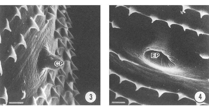
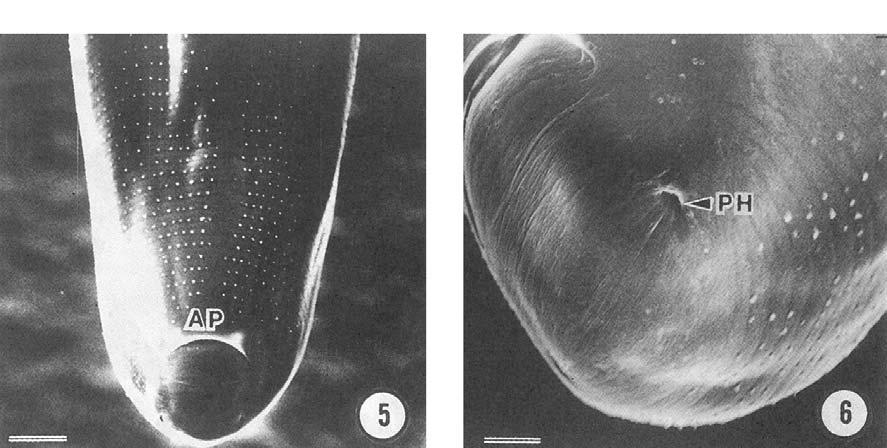
1 Surface Ultrastructure of Larvae and Adults of Gnathostoma binucleatum obtained in Mexico Masataka Koga 1), Hiroshige Akahane 2), Rafael Lamothe-Argumedo 3), David Osorio-Sarabia 3), Luis García-Prieto3), Juan Manuel Martinez-Cruz 4), Sylvia paz Diaz Camacho 5) 1) Department oí Parasitology, Faculty oí Medicine, Kyushu. University, Fukuoka , ]apan 2) Department oí Parasitology, School oí Medicine, Fukuoka. University, Fukuoka , ]apan 3) Laboratorio de Helmintologia, Departament de Zoología. Instituto de Biologia, Universidad Nacional Autonoma de Mexico D. F., Mexico 4) Pedro Garcia No. 918 Tierra Blanca, Veracruz, Mexico 5) Facaltad de Ciencias Quimico-Biologicas, Universidad. Autonoma de Sinaloa, Culiacan, Sinaloa, Mexico Abstract: We examined the morphology of gnathostome larvae obtained in Temazcal, Sinaloa and Tepic Mexico, mainly using scanning electron microscopy (SEM). The larvae from Temazcal further experimentally infected two dogs and 8-9 months later. We found adults of those larvae in the stomachs of them. They are al so examined the surface morphology by SEM. Upon the larvae obtained from the muscles oí fishes and pelicans, the average body length was mm. The head had four transverse rows of hooklets, and average number of each row was 39.3, 43.9, 46.6 and 49.8 respectively. The bodies were wholly covered with minute cuticular spines along their transverse striations. The average number of striations varied from The cervical papillae were situated between the 13th-17th transverse striations. An excretory pore also located ventrally between the 24th-28th transverse striations. On the other hand, the morphology of the adult worms was examined by SEM. The adults demonstrated tridentate cuticular spines in the anterior forefront regions. The shape of the cuticular spines changed to di- and monodentate forms in the anterior one-third of the body. Very minute monodentate spines covered the posterior two-thirds of the body. The ventral surface of the male tail had 4 pairs of caudal papillae and 3 pairs of small papillae. The spines in this area were short. We found no pits on the eggshell surface of our specimens. The three previously reported indigenous to Latin America, G.turgidum, G.procyonis, and G.americanum, have many pits on their eggshells. The present species was more similar to G.binucleatum (G.spinigerum). Introduction Gnathostomiasis is an important parasitic zoonosis, which is mainly endemic in Asia so faro In such countries as ]apan, Thailand and Vietnam, people often eat raw fresh water fish. For this reason, this food-borne disease was thought to be limited to Southeast Asian countries for the past 50 years. In 1970, however, a case of human gnathostomiasis was first reported in Mexico (Pelaez and Perez-Reyes, 1970). This patient was neither a traveler nor an immigrant from Southeast Asia. After that, the number of reported gnathostomiasis patients increased drastically and up to the present time, more than 1,000 cases have been diagnosed in Mexico. The endemic area in Mexico includes six states, which are roughly divided into three regions including the Pacific coast (Culiacan), Atlantic coast areas (Tampico) and adjacent regions (Veracruz) of north American country (Ogata et al. 1998). Previously, Lamothe-Argumedo et al. (1989) and Armeyda-Artigas (1991) examined the morphology of Gnathostoma larvae from fish in Oaxaca-Veracruz and later Akahane et al. (1994) al so examined the morphology of the larvae collected from Pelicans in the same are a by light microscopy. We herein report the morphology of Gnathostoma specimens which are considered to be the same species reported from human in Mexico. Larvae from fish and pelicans, and adults recovered from dogs which had infected experimentally with the larvae were examined mainly using scanning electron microscopy. Materials and Methods We collected 3 pelicans from the Presidente Miguel Aleman Reservoir in Temazcal and examined their muscles to collect gnathostome larvae. The muscles were removed, chopped into small pieces, and the pieces cut into thin slices. The slices were then placed between 2 glass plates (10 by 10cm), pressed by hands, and examined under a dissecting microscope. The inspected muscle remnants were next digested in artificial gastric juice (0.2g pepsin in 0.7ml HCl/100ml distilled water) for 3 hours at 37 C to harvest any larvae that had been overlooked. We eventually found a 1
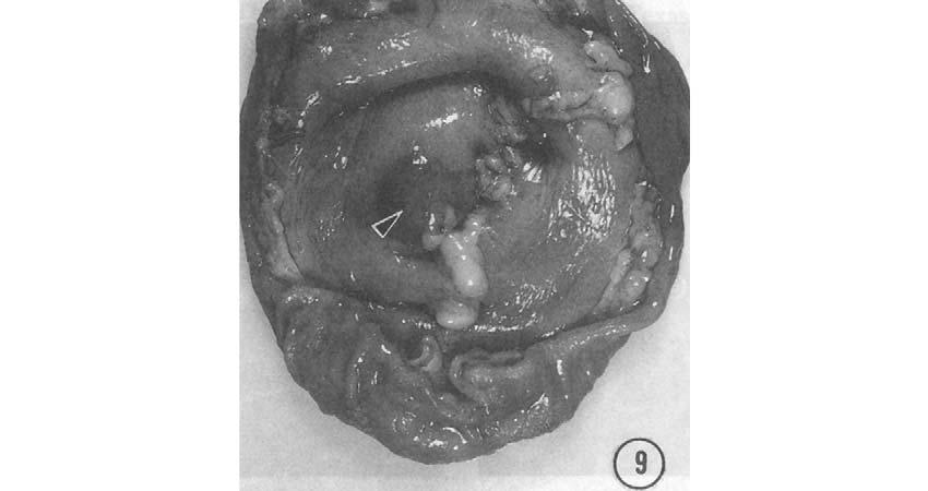
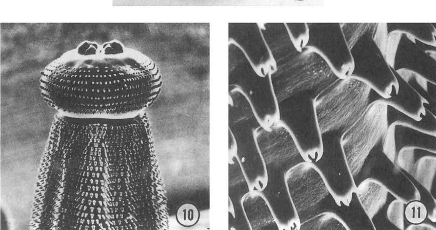
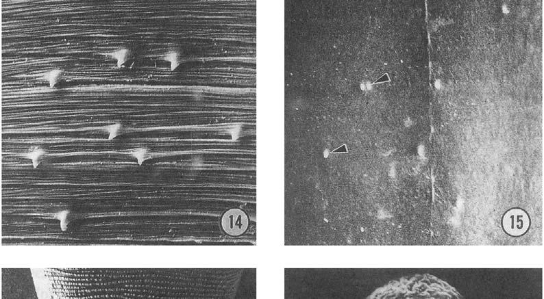
2 total of 570 lavae. We also examined a natural second intermediate fish host to collect the larvae, especially in Culiacan and Tepic. Two dogs were each infected experimentally with 20 larvae obtained from pelicans, and thereafter, they were maintained at an animal center. To assess egg shedding, fecal examinations were performed once a month starting at the fifth month postinfection. Gnathostoma eggs were first observed in the feces 8 and 9 months after infection. The dogs were then anesthetized by sodium barbital and killed by bleeding from the cervical arteries. The peritonea were then gently opened, and the stomachs were removed and opened by cutting along the lesser curvature. A single hard nodule was evident in the mucous membrane in each stomach. In the nodules from each dogs, 4 (1, 3 ) and 9 (6,3 ) adult worms were obtained. The eggs were moved from the uteri of the gravid female worms for further examination. First the larvae were processed for morphological observations by both light and scanning electron microscopy. Paraffin section specimens were prepared by the conventional methods and stained H E. For the SEM specimen preparations, ten viable larvae from Temazcal and Tepic, and three from Culiacan were washed in distilled water and stored in a refrigerator until the worms died naturally and stretched out completely. They were then fixed in 10% formalin for 7 days. Next, the larvae were washed in running tap water overnight to remove the fixative, and then were transferred to distilled water. The specimens were rinsed twice in Millonig's phosphate buffer and postfixed overnight in 1 % OS04 in the same buffer, as a result bodies became rigid. AlI specimens were then carefully and gradually dehydrated in an ascending series of ethanol, since such specimens often shrink or have surface wrinkles due to rapid dehydration. They were then transferred into amyle acetate, and COz critical-point dried with a Hitachi HCP2 dryer. The specimens were sputter coated with gold and examined with a leol lsm-u3 SEM operated at 15KV. Viable male and female worms and eggs were processed same procedures for preparing SEM specimens. Results As many as 570 larvae were thus finally obtained from three Pelicans in Temazcal.Almost same number of larvae were obtained from about 200 fish in Tepic. On the other hand, only three larvae were found in five egrets in Culiacan. The average body length (lo larvae) of these Mexican larvae was 4.67 mm measured in a relaxed state after nutural death in cold distilled water. The head had four transverse rows of hooklets (Fig. 1), the average number (10 larvae) of each rows was 39.3, 43.9, 46.6 and 49.8 respectively. The typical hooks on the head bulb had sharp tapering points composed of hard keratin that emerged from an oblongate chitinous base (Fig. 2). The body were entirely covered with minute cuticular spines along their transverse striations. The average number (lo larvae) of striations varied from 227 to 275. A pair of cervical papillae was laterally situated between the 13th-17th transverse striations (Fig. 3). In most specimens, the papillae were located between 14th-15th striations. An excretory pore was observed ventrally and the location was between 24th-28th transverse striations (Fig. 4). On the ventral surface of terminal end, a wide anal opening (AP) was visible and transverse striations were spaced at this openings (Fig. 5). The extremity of the larva had a pair of phasmidial pores (PH) which were late rally located (Fig. 6). The morphological features of intestinal cells were of multiple nuclei in the larvae from Temazcal (Fig. 7). The larvae from Sinaloa also had 2-7 nuclei in each intestinal cell (Fig. 8). The either dogs infected the mexican larvae collected in Temazcal had adult worms in their stomach nodules (Fig. 9). The body length of them were cm in female and cm in maleo The adult worm specimens had a hemispherical head bulb armed with 8-9 transverse rows of cephalic hooks (Fig. 10). The body spines irnmediately behind the cephalic bulb were tridentate (Fig.l1) and not multidigitated. One pair of cervical papillae was located laterally near the twentieth transverse striations and had a mammiform shape (Fig. 12). The shapes of the spines around these papillae were mixed, with 2-3 denticles, and only rare unidentate spines. A domelike excretory pore was situated ventrally a little behind the cervical papillae and was covered only by single spines (Fig. 13). These unidentate spines gradually decreased in size posteriorly along the body (Fig. 14), and most posterior spines were minute (Fig. 15). At the posterior half of the body, very minute spines were sparsely evident as less as those in the Thai specimens of G.spinigerum. On the ventral side of the tail of the mail, single-toothed unidentate spines were densely distributed only over the entire extremity. Four pairs of caudal papillae and a few small papillae, which bore no spines were also seen on this side (Fig. 16). The fertilized uterine eggs of this Gnathostoma species were oval (66!: 2.92 by 40!: 3. 1J.1.IIl) and had operculum on one end. The eggshell surface was plain and without pits (Fig.17). This plain, nonpitted appearance is characteristic of this species and is major feature differentiating from G.spinigerum. Discussion Lamothe-Argumedo et al. (1989) described their larval Gnathostoma specimens obtained from Temazcal as Gnathostoma sp. alone. However, based on our observations, his specimens seemed to be the same as those reported by Almeyda-Artigas (1991). Because both specimens of larvae were from fish and waterfowls in the same endemic area of human gnathostomiasis and the descriptions of larval morphology were quite similar. We thus identify this specimen as G.binucleatum (Almeyda-Artigas, 1991) in Table 1. Lamothe-Argumedo et al. (1989) had previously observed larvae 2
3 from Oaxace, Temazcal, Mexico by SEM. We think that their SEM observations were somewhat insufficient, especially regarding the location of excretory pares and number of transverse striations on larval bodies. We thus re-examined the Temazcal specimens using SEM and discovered some new findings. Furthermore, we also examined the surface structures of the specimens from Sinaloa, Culiacan and Tepic, Nayarit. Previously, five specimens from Sinaloa were observed by Carnacho et al. (1998) using SEM. They mentioned the number of hooklets of four rows on the head buld as 39, 42, 44 and 49 respectively. Furthermore, they recognized one pair of cervical papillae located between the 13th- 15th striation of the cuticular spines on a single larva. The number of transverse striations on the body was more than 200. There were no descriptions regarding the location of the excretory pore. The situations of cervical papillae, excretory pore and the number of transverse striations are very important for the identification of Gnathostoma species at larval stage. As shown in Table 1, the number of transverse striations is even more than 200 in G.spinigerum. However, the number is less than 200 in most G.d%resi. On the other hand, the cervical papillae and excretory pore of G.hispidum were situated more anteriorly than those of above two species. In the present study, we compared the larvae from three districts, Temazcal, Sinaloa and Tepic, and found that those morphologies quite resemble that of G.spinigerum in Thailand. In adult specimens, the morphology is also very similar to that of G. spinigerum in Thailand (Koga et al. 1991). The most noticeable difference between G. spinigerum and the present Mexican species is the surface of the eggshell. The eggshells of G.spinigerum and other gnathostome species have many pits on the surface, and the shape of these pits is species-specific (Koga, 1996), while the eggshell of the Mexican species are not pitted. In North-Central, South America, 3 species of Gnathostoma have been recorded: G. procyonis (Chandler, 1942) from a raccoon in Texas, G.turgidum (Stossich, 1902, quoted in travassos, 1925) from an opossum, and G.americanum (Travassos, 1925) from Felis tigrina, the latter two in Brazil. The adults of these 3 species have multidigitate (4-5 teeth) spines of their anterior region immediately behind the head bulb, and have dense spines on the posteriorhalf of their body surfaces, except for G.turgidum. HQwever, G.turgidum has bipolar plugged eggs. In contrast, our specimens had very minute spines sparsely distributed over the posterior half of their bodies, and these spines were recognizable only by SEM examination. The eggs have unipolar plug. Therefore our new specimens are easily distinguishable from previously reported species in America. Consequently, a new species gives rise to occur the mexican gnathostomiasis. 3
4 References 1) Akahane, H., R. Lamothe-Argumedo, ]. M. Martinez-Cruz, D. Osorio-Sarabia, and L. Garcia-Priento. (1994): A morphological observation oí the advanced third-stage larvae oí Mexican Gnathostoma. ]apanese ]ournal oí Parasitology, 43, ) Almeyda-Artigas, R.]. (1991): Hallazgo de Gnathostoma binuc/etum n.sp. (NEMATODA: SPIRURIDA) en íelinos silvestres y el papel de peces dulceacuicolas y oligohalinos como vectores de la gnathostmiasis humana en la cuenca baja del rio Papaloapan, OaxacaVeracruz, Mexico. Anales del instituto de Ciencias del Mar y Limnologia, Universidad Nacional Autonoma de Mexico, 18, ) Ash, 1. R. (1962): Development oí Gnathostoma procyonis Chandler, 1942, in the first and second intermediate hosts. ]ournal oí Parasitology, 48, ) Camacho, S. P. D., Zazueta-Ramos, E. Ponce- Torrecillas, 1. Osuna-Ramires, R. CastroVelazquez, A. Elores- Gaxiola, ]. Baquera-Heredia, K. Willms, H. Akahane, K.Ogata, and Y. Nawa. (1998): Clinical Maniíestations and immunodiagnosis oí gnathostomiasis in Culiacan, Mexico. American ]ournal oí Tropical Medicine and Hygiene, 59, ) Chandler, A. C. (1942): The helminths oí raccoons in east Texas. ]ournal oí Parasitology, 28, ) Koga, M. and Y. Ishii. (1987): Surface morphology oí the advanced third-stage larvae of Gnathostoma doloresi - An electron microscopic study-. ]apanese ]ournal oí Parasitology, 36, ) Koga, M., ].Ishibashi, Y.Ishii, and T.Nishimura. (1988): Scanning electron microscopic comparisons among the early and advanced third-stage larvae oí Gnathostoma hispidum and the Gnathostoma larvae obtained from loaches. ]apanese ]ournal oí Parasitology, 37, ) Koga, M., H. Akahane and Y. Ishii. (1991): Surface ultrastructure oí adults and eggs oí Gnathostoma spinigerum (Nema toda : Gnathostomatidae). Transactions oí the American Microscopical Society, 110, ) Koga, M., H. Akahane, and Y. Ishii and S. Kojima. (1994): External morphology oí the advanced third-stage larvae oí Gnathostoma spinigerum: A scanning electron microscopy. ]apanese ]ournal oí Parasitology, 43, ) Koga, M. (1996): Comparative surface ultrastructure oí adults and eggs oí Gnathostoma obtained in ]apan. Southeast Asian ]ournal oí Tropical Medicine and Public Health, 27, ) Lamothe-Argumedo, R., R.1. Medina- Vences, S. Lopez- ]imenez, and 1. Garcia-Prieto. (1989): Hallazgo de la íorma iníectiva de Gnathostoma sp., en peces de Temazcal, Oaxaca, Mexico, Anales del Institutio de Biologia de la Universidad Nacional Autonoma de Mexico., Series-Zoologia, 60, ) Ogata, K., Y. Nawa, H. Akahane, S. P. Camacho, R. Lamothe-Argumedo and A. Cruz Reyes. (1998): Gnathostomiasis in Mexico. American ]ournal oí Tropical Medicine and Hygiene, 58, ) Pelaez, D. and R. Perez-Reyes. (1970): Gnathostomiasis humana en America. Revista Latino-Americana de Microbiologia, 12, ) Travassos, L. (1925): Contrbuicoes para o conhecimento da íauna helmintologica brasileira. 18 Sobre as especies brasileiras do genero Gnathostoma Owen, Sciencia Medica, 3,

5 5

6 6


7 7
8 Figure regends Figs.1-6 and Scanning electron micrographs. Fig.1. Lateral view oí the head bulb oí Gnathostoma binucleatum from Temazcal. The arrowhead indicates the cervical papilla. Fig.2. An enlarged riew oí the hooklets. The base oí each hooklets has an oblongate shape. Sharp keratin hooks armed posteriorly. Fig.3. A mammiform oí the cervical papillae protruding írom the tegumento Fig.4. An oval shaped opening oí the excretory pore which opened ventrally. Fig.5. The terminal end oí a larva. The anal opening is clearly visible on the ventral surface of the larve, which has a crescent shape. Fig.6. A terminal extremity oí a larva. A phasmidial pore can be laterally seen. Figs.7-8. Cross sections oí the larval intestines. Fig. 7. A cross section oí the Temazcal larva. Multiple nuclei were evident in one cell. Fig. 8. A cross section oí the Sinaloan larva. Arrowheads indicate the cells bearing 5 nuclei each. Fig. 9. Parasitic nodule (arrowhead) in the stomach wall of a dog iníected with the Mexican larvae. Fig.10. Lateral view oí the head buld of adult Ganthostoma binucleatum. Fig.11. Short, stumpy tridentate spines lying immediating behind the head bulbo Fig. 12. Mammiform cervical papilla located between the eighteenth and twentieth transverse striations, with spines of trididentates on the body surface. Fig. 13. Dome-shaped excretory pore. Around this pore, all spines are unidentate with acute tips. Fig. 14. Smaller unidentate spines located at middle part oí the body. Fig.15. Very minute spines (arrowheads) are sparsely distributed around the posterior half of the body. Fig.16. Ventral surface of the tail of the maleo Dome-shaped caudal papillae and some small papillae without spines are evident. Fig.17. An egg from the uterus oí a gravid female of Gnathostoma binucleatum bearing an operculum on one end, showing smooth surface. 8
GNATHOSTOMIASIS IN THAILAND: A SURVEY ON INTERMEDIATE HOSTS OF GNATHOSTOMA SPP. WITH SPECIAL REFERENCE TO A NEW TYPE OF LARVAE FOUND IN FLUTA ALBA
 GNATHOSTOMIASIS IN THAILAND: A SURVEY ON INTERMEDIATE HOSTS OF GNATHOSTOMA SPP. WITH SPECIAL REFERENCE TO A NEW TYPE OF LARVAE FOUND IN FLUTA ALBA P Setasuban', Supapom Nuamtanong', V Rojanakittikoon',
GNATHOSTOMA INFECTION IN NAKHON NAYOK AND PRACHIN BURI, CENTRAL THAILAND
 GNATHOSTOMA INFECTION IN NAKHON NAYOK AND PRACHIN BURI, CENTRAL THAILAND Wichit Rojekittikhun 1, Tossapon Chaiyasith 1, Supaporn Nuamtanong 1, Somchit Pubampen 1, Wanna Maipanich 1 and Rungsunn Tungtrongchitr
Perch Dissection Lab
 Name: Block: Due Date: Perch Dissection Lab Background The fish in the class Osteichthyes have bony skeletons. There are three groups of the bony fish: ray-finned, lobe-finned, and the lungfish. The perch
Trematode Parasites of the Opossum, Didelphis virginiana, from Florida
 Proc. Helminthol. Soc. Wash. 46(2), 1979, pp. 207-212 Trematode Parasites of the Opossum, Didelphis virginiana, from Florida G. PREMVATI AND THOMAS D. BAiR1 Department of Zoology, University of Lucknow,
General Characters of Trematodes
 Parasitology Department General Characters of Trematodes By Hala Elwakil, MD Intended Learning Outcomes By the end of this lecture, the student will be able to know: 1. General morphology of trematodes
Perch Dissection Lab
 Perch Dissection Lab Introduction: The fish in the class Osteichthyes have bony skeletons. There are three groups of the bony fish - -- ray-finned fish, lobe-finned fish, and the lung fish. The perch is
A new species of Spirophilometra (Nematoda: Philometridae) from the yellowfin snook Centropomus robalito (Osteichthyes) in southern Mexico
 from the yellowfin snook Centropomus robalito (Osteichthyes) in southern Mexico") FOLIA PARASITOLOGICA 54: 215 219, 2007 A new species of Spirophilometra (Nematoda: Philometridae) from the yellowfin snook Centropomus robalito (Osteichthyes) in southern Mexico František Moravec 1, Ana
FOLIA PARASITOLOGICA 54: 215 219, 2007 A new species of Spirophilometra (Nematoda: Philometridae) from the yellowfin snook Centropomus robalito (Osteichthyes) in southern Mexico František Moravec 1, Ana
Schwenkiella orietanlis Singh and Agarwal, 1997 (PLATE-XXVII-XXVIII)
") Schwenkiella orietanlis Singh and Agarwal, 1997 (PLATE-XXVII-XXVIII) Host : Periplaneta americana Locality : Meerut Location : Intestine No. of hosts examined : 250 No. of hosts found infected : 200 Female
Schwenkiella orietanlis Singh and Agarwal, 1997 (PLATE-XXVII-XXVIII) Host : Periplaneta americana Locality : Meerut Location : Intestine No. of hosts examined : 250 No. of hosts found infected : 200 Female
Gulf Research Reports
 Gulf Research Reports Volume 8 Issue 2 January 1989 A Redescription of Oncholaimoides elongatus Hopper, 1961 (Nematoda: Enoplida) with Descriptions of the Other Two Members of the Genus Edwin J. Keppner
Chapter 30 Nonvertebrate Chordates, Fishes, and Amphibians Name
 Chapter 30 Nonvertebrate Chordates, Fishes, and Amphibians Name Lab Dissecting a Perch Background Information Fish are the largest group of vertebrates found in fresh and salt water. In fact, over 25,000
TREMATODE INFECTION RATES OF FISH FROM A WASTEWATER TREATMENT FACTORY POLISHING POND AND A CANAL IN PHUKET, THAILAND
 TREMATODE INFECTION RATES OF FISH FROM A WASTEWATER TREATMENT FACTORY POLISHING POND AND A CANAL IN PHUKET, THAILAND D Krailas, T Janecharat, S Ukong,W Junhom, S Klamkhlai, N Notesiri and P Ratanathai
Microbrotula randalli Cohen and Wourms, Samoa and Vanuatu at 30 to 38 m near reef-sand interface. Rare.
 click for previous page Ophidiiform Fishes of the World 107 Diagnosis and description: Body completely covered with small imbricate scales; head partly naked; snout depressed; eyes small, more than 6 times
Brasacanthus sphoeroides gen. n., sp. n. (Acanthoceph ala, Echi norhynch idae) from a coastal marine fish of Parana State, Brazil 1
 from a coastal marine fish of Parana State, Brazil 1") Brasacanthus sphoeroides gen. n., sp. n. (Acanthoceph ala, Echi norhynch idae) from a coastal marine fish of Parana State, Brazil 1 Vernon E. Thatcher 2 ABSTRACT. Brasacanlizus sphoeroides gen. n.. sp.
Brasacanthus sphoeroides gen. n., sp. n. (Acanthoceph ala, Echi norhynch idae) from a coastal marine fish of Parana State, Brazil 1 Vernon E. Thatcher 2 ABSTRACT. Brasacanlizus sphoeroides gen. n.. sp.
New oxyuroid nematodes of the genera Ichthyouris and Spinoxyuris from South American freshwater fishes
 FOLIA PARASITOLOGICA 48: 311-320, 2001 New oxyuroid nematodes of the genera Ichthyouris and Spinoxyuris from South American freshwater fishes František Moravec 1 and Vernon E. Thatcher 2 1 Institute of
NATIONAL BIORESOURCE DEVELOPMENT BOARD Dept. of Biotechnology Government of India, New Delhi
 NATIONAL BIORESOURCE DEVELOPMENT BOARD Dept. of Biotechnology Government of India, New Delhi MARINE BIORESOURCES FORMS DATA ENTRY: Form- 1(general) For office use: Fauna: Flora Microorganisms General Category:
Heterophyiasis Heterophyes heterophyes. Samar N. El-Beshbishi Prof. of Medical Parasitology Mansoura Faculty of Medicine
 Heterophyiasis Heterophyes heterophyes Samar N. El-Beshbishi Prof. of Medical Parasitology Mansoura Faculty of Medicine 2 Objectives 1. Overview on heterophyiasis. 2. Geographical distribution. 3. Morphology
Development and Identification of Three Species of Thai Ricefish, Oryzias, in the Mekong Basin
 Tropical Natural History 12(1): 75-88, April 2012 2012 by Chulalongkorn University Development and Identification of Three Species of Thai Ricefish, Oryzias, in the Mekong Basin APICHART TERMVIDCHAKORN
O'opu Prints and Dissections
 O'opu Prints and Dissections Tina Alcain Konawaena High School HCPS III Science Standards Addressed: SC.BS.4.6 Grade Level: 9 th -12th Project Time Span: 2-3 class periods To The Teacher: This lesson is
Helminths: Schistosoma mansoni. Schistosoma japonicum 10/14/2009. Trematoda - non-segmented flat worms
 Helminths: Trematoda - non-segmented flat worms The schistosomes: Schistosoma mansoni Schistosoma haematobium Schistosoma japonicum Schistosoma mekongi Aquatic freshwater snails are the intermediate hosts
Lumbricus terrestris - preserved specimens for dissection
 Lumbricus terrestris - preserved specimens for dissection External Anatomy: Prostomium (observe under dissecting microscope for external sensory organs), peristomium, clitellum, setae (dissecting microscope),
10/14/2009. Helminths: Trematoda - non-segmented flat worms. The schistosomes: Schistosoma mansoni Schistosoma haematobium. Schistosoma mekongi
 Helminths: Trematoda - non-segmented flat worms The schistosomes: Schistosoma mansoni Schistosoma haematobium Schistosoma japonicum Schistosoma mekongi 1 Japan is schistosome-free as of 1976 2 Aquatic
FAO SPECIES IDENTIFICATION SHEETS FISTULARIIDAE. Cornetfishes, flutemouths
 click for previous page FIST 1982 FAO SPECIES IDENTIFICATION SHEETS FISHING AREA 51 (W. Indian Ocean) FISTULARIIDAE Cornetfishes, flutemouths Body elongate and depressed. Mouth small, at end of a long
-8- spinous. nape caudal fin. body depth. pectoral fin. anus. total length Fig. 4
 click for previous page -8-1.3 Illustrated Glossary of Technical Terms and Measurements External Morphology and Measurements spinous dorsal fin soft nape caudal fin interorbital body depth snout lateral
F?arQsitps. of South Afri«;.an.frpshwater fish... V.
 Onderstepoort Journal of Veterinary Research, 6: 139-145(1993) F?arQsitps. of South Afri«;.an.frpshwater fish... V. De~criRtion of two new s~ecies.9f the genus Spinitectus Fourment,.1 883 (Nematoda: Cystidicolidae)
Fish. Water Dwelling Animals
 Fish Water Dwelling Animals Class Agnatha (Jawless fish) They are believed to be the most primitive and oldest vertebrates. Lamprey and hagfish are the only 2 living members of this class and are placed
Exercise 18B Class Chondrichthyes Cartilaginous Fishes
 AP Biology Chapter 24 Exercise #18: Chordates: Fish Cartilaginous Fishes Lab Guide Exercise 18B Class Chondrichthyes Cartilaginous Fishes This group contains about 970 species that are characterized by
Notes on Varroa destructor (Acari: Varroidae) parasitic on honeybees in New Zealand
 parasitic on honeybees in New Zealand") Systematic & Applied Acarology Special Publications (2000) 5, 9-14 Notes on Varroa destructor (Acari: Varroidae) parasitic on honeybees in New Zealand ZHI-QIANG ZHANG Landcare Research, Private Bag 92170,
Systematic & Applied Acarology Special Publications (2000) 5, 9-14 Notes on Varroa destructor (Acari: Varroidae) parasitic on honeybees in New Zealand ZHI-QIANG ZHANG Landcare Research, Private Bag 92170,
THE PREVALENCE OF CESTODE INFECTION IN A FRESHWATER CATFISH, SPERATA SARWARI INTRODUCTION
 Punjab Univ. J. Zool., Vol. 21 (1-2), pp. 41-47, 2006 THE PREVALENCE OF CESTODE INFECTION IN A FRESHWATER CATFISH, SPERATA SARWARI HAFIZ ABDULLAH SHAKIR, ABDUL MAJID KHAN AND MUHAMMAD ABID Department of
Dogfish Shark Dissection Introduction 1. What are two reasons why spiny dogfish are used for study in laboratories?
 Dogfish Shark Dissection Introduction 1. What are two reasons why spiny dogfish are used for study in laboratories? 2. Someone who studies fish is called an. 3. Sharks and fish belong to the Phylum a.
WHAT TO DO WITH ILL OR DEAD FROGS
 WHAT TO DO WITH ILL OR DEAD FROGS Lee Berger and Rick Speare School of Public Health, Tropical Medicine & Rehabilitation Sciences, James Cook University, Townsville, Qld, 4811 CONTENTS 1. Introduction...
* A New Species of Cichlid Fish From Lake Malawi. Pseudotropheus tursiops, \(I75 Tropical Fish Hobbyist a'l (3) : 8 L-? 0. ,$ IOU.
 : 8 L-? 0. ,$ IOU.") ,$ IOU. \(I75 Tropical Fish Hobbyist a'l (3) : 8 L-? 0. * 2.37 Pseudotropheus tursiops, A New Species of Cichlid Fish From Lake Malawi by Warren E. Burgess and Dr. Herbert R. Axelrod Among the cichlid
,$ IOU. \(I75 Tropical Fish Hobbyist a'l (3) : 8 L-? 0. * 2.37 Pseudotropheus tursiops, A New Species of Cichlid Fish From Lake Malawi by Warren E. Burgess and Dr. Herbert R. Axelrod Among the cichlid
Cylicostephanus asymetricus
 Cylicostephanus asymetricus 50 µm 50 µm 00 µm 00 µm Figure 9a. dorsal gutter Figure 9b. arms and hands Figure 9c Figure 9d This species is uncommon in Kentucky. It is almost identical to Cylicostephanus
Redescription of Rhabdochona anguillae (Nematoda: Rhabdochonidae), a parasite of eel, Anguilla anguilla, in Europe
, a parasite of eel, Anguilla anguilla, in Europe") FOLIA PARASITOLOGICA 45: 233-238, 1998 Saraiva and Moravec: Redescription of Rhabdochona anguillae Redescription of Rhabdochona anguillae (Nematoda: Rhabdochonidae), a parasite of eel, Anguilla anguilla,
FOLIA PARASITOLOGICA 45: 233-238, 1998 Saraiva and Moravec: Redescription of Rhabdochona anguillae Redescription of Rhabdochona anguillae (Nematoda: Rhabdochonidae), a parasite of eel, Anguilla anguilla,
Philometroides africanus sp. n. (Nematoda: Philometridae), a new tissue parasite of the African pike Hepsetus odoe (Pisces) in Botswana
, a new tissue parasite of the African pike Hepsetus odoe (Pisces) in Botswana") FOLIA PARASITOLOGICA 48: 127-131, 2001 Philometroides africanus sp. n. (Nematoda: Philometridae), a new tissue parasite of the African pike Hepsetus odoe (Pisces) in Botswana František Moravec 1 and Jo
FOLIA PARASITOLOGICA 48: 127-131, 2001 Philometroides africanus sp. n. (Nematoda: Philometridae), a new tissue parasite of the African pike Hepsetus odoe (Pisces) in Botswana František Moravec 1 and Jo
Development of Procamallanus saccobranchi (Nematoda: Camallanidae), a parasite of a freshwater fish in India
, a parasite of a freshwater fish in India") FOLIA PARASITOLOGICA 47: 216-226, 2000 Development of Procamallanus saccobranchi (Nematoda: Camallanidae), a parasite of a freshwater fish in India Nimai C. De and Rabindra N. Maity Helminthology Laboratory,
FOLIA PARASITOLOGICA 47: 216-226, 2000 Development of Procamallanus saccobranchi (Nematoda: Camallanidae), a parasite of a freshwater fish in India Nimai C. De and Rabindra N. Maity Helminthology Laboratory,
CALLES A, VINCX M (submitted)
") 112871 Gonionchus ecuadoriensis sp. n. Paper prepared as CALLES A, VINCX M (submitted) Description of Gonionchus ecuadoriensis sp. n., (Nematoda: Xyalidae) a dominant species on Ecuadorian sandy beaches.
112871 Gonionchus ecuadoriensis sp. n. Paper prepared as CALLES A, VINCX M (submitted) Description of Gonionchus ecuadoriensis sp. n., (Nematoda: Xyalidae) a dominant species on Ecuadorian sandy beaches.
First Report of Clavinema mariae (Nematoda: Philometridae) in Cultured Rockfish, Sebastes schlegeli, in Cheonsuman (Bay), the Republic of Korea
 in Cultured Rockfish, Sebastes schlegeli, in Cheonsuman (Bay), the Republic of Korea") ISSN (Print) 0023-4001 ISSN (Online) 1738-0006 BRIEF COMMUNICATION Korean J Parasitol Vol. 55, No. 2: 219-224, April 2017 https://doi.org/10.3347/kjp.2017.55.2.219 First Report of (Nematoda: Philometridae)
ISSN (Print) 0023-4001 ISSN (Online) 1738-0006 BRIEF COMMUNICATION Korean J Parasitol Vol. 55, No. 2: 219-224, April 2017 https://doi.org/10.3347/kjp.2017.55.2.219 First Report of (Nematoda: Philometridae)
FICHES D IDENTIFICATION DU PLANCTON
 FICHES D IDENTIFICATION DU PLANCTON Edited by G.A. ROBINSON Institute for Marine Environmental Research Prospect Place, The Hoe, Plymouth PLl 3DH, England FICHE NO. 176 GADIDAE Ciliata Couch, 1832 by NECLA
ROSINA C. KRECEK Department of Parasitology, Faculty of Veterinary Science, University of Pretoria, Private Bag X04, Onderstepoort 0110, South Africa
 Proc. Helminthol. Soc. Wash. 56(2), 1989, pp. 183191 Habronema malani sp. n. and Habronema tomasi sp. n. (Nematoda: Habronematidae) from the BurchelPs Zebras and Hartmann's Mountain Zebras in Southern
ON FOUR NEW SPECIES OF AVIAN NEMATODES FROM ORISSA, INDIA
 Rec. zool. Surv. India, 85(4) : 467-480, 1989 ON FOUR NEW SPECIES OF AVIAN NEMATODES FROM ORISSA, INDIA By N. MAJUMDAR AND S. R. DEY SARKAR Zoological Survey of India, Calcutta-700053 INTRODUCTION In the
PREVALENCE OF PARASITIC INFECTIONS OF FARMED TILAPIA (Oreochromis niloticus) AND CATFISH (Clarias gariepinus) IN NYERI COUNTY, KENYA
 AND CATFISH (Clarias gariepinus) IN NYERI COUNTY, KENYA") PREVALENCE OF PARASITIC INFECTIONS OF FARMED TILAPIA (Oreochromis niloticus) AND CATFISH (Clarias gariepinus) IN NYERI COUNTY, KENYA KVA ANNUAL SCIENTIFIC CONFERENCE 27TH-29 TH APRIL, 2016 PRESENTER: Dr
PREVALENCE OF PARASITIC INFECTIONS OF FARMED TILAPIA (Oreochromis niloticus) AND CATFISH (Clarias gariepinus) IN NYERI COUNTY, KENYA KVA ANNUAL SCIENTIFIC CONFERENCE 27TH-29 TH APRIL, 2016 PRESENTER: Dr
Slide 1. Slide 1. Next. 5:30:08 AM
 Slide 1 Slide 1 http://www3.utep.edu/leb/mosquito/larvslide1.htm10/27/2004 5:30:08 AM Slide 1 Slide 2 Recognition that the specimens are mosquito larvae is a prerequisite to identification of the genera.
Ree. zool. Surv. India, 91 (3-4) : , 1992
 : , 1992") Ree. zool. Surv. India, 91 (3-4) : 319-323, 1992 RECORDS OF TWO CERCARIAE, HETEROPHYES SP. AND HAPLORCHIS SP. FROM THE SNAIL, MELANO/DES TUBERCULATA (MULLER) FROM TAMIL NADU. N. VEERAPPAN AND H. N. ACHUTHAN
Ree. zool. Surv. India, 91 (3-4) : 319-323, 1992 RECORDS OF TWO CERCARIAE, HETEROPHYES SP. AND HAPLORCHIS SP. FROM THE SNAIL, MELANO/DES TUBERCULATA (MULLER) FROM TAMIL NADU. N. VEERAPPAN AND H. N. ACHUTHAN
Chapter 12 Part 2. The Worms Platyhelminthes, Nematoda & Annelida
 Chapter 12 Part 2 The Worms Platyhelminthes, Nematoda & Annelida Phylum: Platyhelminthes Examples: Flatworms, Planaria sp., tapeworms and blood flukes Acoelomate, Invertebrate, Simplest critter w/ bilateral
TWO NEW SPECIES OF ARGULUS MULLER (CRUSTACEA: BRANCHIURA) FROM RIVER CAUVERY WITH A KEY TO INDIAN SPECIES
 FROM RIVER CAUVERY WITH A KEY TO INDIAN SPECIES") TWO NEW SPECIES OF ARGULUS MULLER (CRUSTACEA: BRANCHIURA) FROM RIVER CAUVERY WITH A KEY TO INDIAN SPECIES M. M. THOMAS AND M. DEVARAJ Central Marine Fisheries Research Institute Regional Centre, Mandapam
TWO NEW SPECIES OF ARGULUS MULLER (CRUSTACEA: BRANCHIURA) FROM RIVER CAUVERY WITH A KEY TO INDIAN SPECIES M. M. THOMAS AND M. DEVARAJ Central Marine Fisheries Research Institute Regional Centre, Mandapam
LECTURE 6 - OUTLINE. Evolution & Classification - Part II. Agnatha (cont.) Gnathostomata
 Gnathostomata") LECTURE 6 - OUTLINE Evolution & Classification - Part II Agnatha (cont.) 6. Myxini 7. Cephalaspidomorphi Gnathostomata 1. Phylogenetic relationships 2. Placodermi 3. Acanthodii BIOL 4340 Lecture 6-1 Class
LECTURE 6 - OUTLINE Evolution & Classification - Part II Agnatha (cont.) 6. Myxini 7. Cephalaspidomorphi Gnathostomata 1. Phylogenetic relationships 2. Placodermi 3. Acanthodii BIOL 4340 Lecture 6-1 Class
TWO NEW SPECIES OF COPEPODS JAPANESE FISHES. Author(s) Yamaguti, Satyu; Yamasu, Terufumi.
 Yamaguti, Satyu; Yamasu, Terufumi.") Title TWO NEW SPECIES OF COPEPODS JAPANESE FISHES PARASIT Author(s) Yamaguti, Satyu; Yamasu, Terufumi Citation PUBLICATIONS OF THE SETO MARINE BIO LABORATORY (1960), 8(1): 137-140 Issue Date 1960-05-30
Title TWO NEW SPECIES OF COPEPODS JAPANESE FISHES PARASIT Author(s) Yamaguti, Satyu; Yamasu, Terufumi Citation PUBLICATIONS OF THE SETO MARINE BIO LABORATORY (1960), 8(1): 137-140 Issue Date 1960-05-30
Institute of Parasitology, Biology Centre of the Academy of Sciences of the Czech Republic, Branišovská 31, České Budějovice, Czech Republic;
 Folia Parasitologica 56[4]: 305 312, 2009 ISSN 0015-5683 (print), ISSN 1803-6465 (online) Institute of Parasitology, Biology Centre ASCR http://www.paru.cas.cz/folia/ Redescription of Spinitectus tabascoensis
Chapter 12 Marine Fishes
 Chapter 12 Marine Fishes Marine Protochordates Phylum: Chordata (nerve cord) Subphylum: Protochordata first chordates/primitive Primitive species of marine vertebrates Do not have advanced features (backbone)
CAPILLARIASIS PHILIPPINENSIS: A FISH-BORNE PARASITIC ZOONOSIS*
 CAPILLARIASIS PHILIPPINENSIS: A FISH-BORNE PARASITIC ZOONOSIS* John H Cross' and Virginia Basaca-Sevilla US Naval Medical Research Unit No. 2; Department of Health, Manila, Philippines. Abstract. Fish
BIO Parasitology Spring 2009
 BIO 475 - Parasitology Spring 2009 Stephen M. Shuster Northern Arizona University http://www4.nau.edu/isopod Lecture 13 Important Orders a. Echinostomatiformes b. Strigeiformes c. Opisthorchiformes d.
Contribution to the morphology of the third-instar larvae of Laccophilus poecilus KLUG (Coleoptera: Dytiscidae)
") Genus Vol. 15(1): 31-36 Wroc³aw, 30 III 2004 Contribution to the morphology of the third-instar larvae of Laccophilus poecilus KLUG (Coleoptera: Dytiscidae) EUGENIUSZ BIESIADKA and IWONA KA KAŹMIERSKA
Genus Vol. 15(1): 31-36 Wroc³aw, 30 III 2004 Contribution to the morphology of the third-instar larvae of Laccophilus poecilus KLUG (Coleoptera: Dytiscidae) EUGENIUSZ BIESIADKA and IWONA KA KAŹMIERSKA
Fish Dissection. Background
 Fish Dissection The Fish Dissection program at Hatfield Marine Science Center is a 50-minute hands-on program for 4th through 12th grade students. Students will work in small groups as they examine a variety
Chapter 10. Part 1: Cartilaginous Fishes
 Chapter 10 Part 1: Cartilaginous Fishes Objectives Understand how hagfishes and lampreys differ from all other fishes. Describe how sharks, skates, and rays are related. Differentiate between cartilaginous
Description of the Immature Stages of Galindomyia leei Stone and Barreto, 1969l. Abdiel J. Adames* and Pedro Galindo*
 132 Description of the Immature Stages of Galindomyia leei Stone and Barreto, 1969l Abdiel J. Adames* and Pedro Galindo* The genus and species Galindomyia Zeei, a member of the tribe Culicini, was described
Crayfish Dissection. Materials:gloves, preserved crayfish, paper towel, dissecting pan, scissors, forceps, dissecting. Background: LME-305
 Living Science LME-305 Crayfish Dissection Materials:gloves, preserved crayfish, paper towel, dissecting pan, scissors, forceps, dissecting needle, dissecting pins, and pen or pencil Background: Like all
ON SOME NEMATODES OF BIRDS FROM INDIA PART II. SPIRURIDAE AND HEDRURIDAE
 RES. BULL. MEGURO PARASIT. MUS. No.5, 1971 21 ON SOME NEMATODES OF BIRDS FROM INDIA PART II. SPIRURIDAE AND HEDRURIDAE Durdana S. ]AIRAJPURI AND Ather H. SIDDIQI (Section of Parasitology, Department of
a. Kingdom: b. Phylum: c. Class: d. Order: e. Family: f. Genus: g. Species:
 Pre-lab Discussion: The earthworm belongs to a group of animals called annelids (segmented worms). The body of an annelid is usually divided internally and externally into well-defined segments, which
By Charles Hawks Sponsored By The Angelfish Society
 By Charles Hawks Sponsored By The Angelfish Society About this presentation In July, 2011, The Angelfish Society held its second annual Angelfish Show at the American Cichlid Association convention in
Broodstock and Hatchery Management
 Broodstock and Hatchery Management Ryan L. Lane, PhD Fisheries & Illinois Aquaculture Center Aquaculture in U.S. Demand for lean, mild-flavored products Striped bass Morone saxatilis Depletion of wild
Handbook for the identification of yellowfin and bigeye tunas in fresh, but less than ideal condition
 0 WCPFC SC1 FT IP 1 1st Meeting of the Scientific Committee of the Western and Central Pacific Fisheries Commission WCPFC SC1 Noumea, New Caledonia 8 19 August 2005 Handbook for the identification of yellowfin
NOTES ON EGGS, LARV^ AND JUVENILES OF FISHES FROM INDIAN WATERS
 NOTES ON EGGS, LARV^ AND JUVENILES OF FISHES FROM INDIAN WATERS V. Euthynnus affinis (Cantor)* BY S. JONES {Central Marine Fisheries Research Station) LARVAL stages of Katsuwonus pelamis (Linnaeus) and
LIBRARY. Class\ V"^ A *Ii:T_
 LIBRARY Class\ V"^ A *Ii:T_ ^ Publications OP FIELD MUSEUM OF NATURAL HISTORY ZOOLOGICAL SERIES Volume X Chicago, U. S. A. 1909-1923 7/,3 ^Issued September 18, 19 12. 69 NEW SPECIES OF FISHES FROM
NATIONAL BIORESOURCE DEVELOPMENT BOARD Dept. of Biotechnology Government of India, New Delhi
 NATIONAL BIORESOURCE DEVELOPMENT BOARD Dept. of Biotechnology Government of India, New Delhi MARINE BIORESOURCES FORMS DATA ENTRY: Form- 1(general) For office use: Fauna: Flora Microorganisms General Category:
Department of Biology, Lafayette College, Easton, PA 18042
 Journal of the Pennsylvania cademy of Science 86(1): 61-68, 2012 LIGHT ND SNNING ELETRON MIROSOPI OSERVTIONS OF THE LRVL STGES ND DULT OF SPHERIDIOTREM GLOULUS (TREMTOD: PSILOSTOMIDE) 1 Jane E. Huffman
A New Species of Glossodoris (Mollusca: Nudibranchia), of the Glossodoris atromarginata Color Group, from Indonesia 1
, of the Glossodoris atromarginata Color Group, from Indonesia 1") A New Species of Glossodoris (Mollusca: Nudibranchia), of the Glossodoris atromarginata Color Group, from Indonesia 1 A ngel Valdés 2,3 and Mary Jane Adams 2 Abstract: A new species, Glossodoris tibboeli
A New Species of Glossodoris (Mollusca: Nudibranchia), of the Glossodoris atromarginata Color Group, from Indonesia 1 A ngel Valdés 2,3 and Mary Jane Adams 2 Abstract: A new species, Glossodoris tibboeli
Mehratreraa polynemusinis, n.sp.
 TREMATODES FROM INDIAN MARINE FISHES Part III. Description of a New Species of the Genus Mehratrr (Family Monodhelminthidae Dollfus, 1937) BY B. S. CHAUHAN (College of Science, Nagpur, India) Received
Gynaecotyla squatarolae (Digenea: Microphallidae) from rats experimentally infected with metacercariae from the shore crab, Macrophthalmus dilatatus
 from rats experimentally infected with metacercariae from the shore crab, Macrophthalmus dilatatus") Korean Journal of Parasitology Vol. 45, No. 3: 199-204, September 2007 DOI: 10.3347/kjp.2007.45.3.199 Gynaecotyla squatarolae (Digenea: Microphallidae) from rats experimentally infected with metacercariae
Korean Journal of Parasitology Vol. 45, No. 3: 199-204, September 2007 DOI: 10.3347/kjp.2007.45.3.199 Gynaecotyla squatarolae (Digenea: Microphallidae) from rats experimentally infected with metacercariae
On a New Nematode, Procamallanus inuelleri n. sp. from the Stomach of a Freshwater Fi h, Heteropneustes fossilis1
 204 PROCEEDINGS OF THE [VOL. 33, No. 2 On a New Nematode, Procamallanus inuelleri n. sp. from the Stomach of a Freshwater Fi h, Heteropneustes fossilis1 VlNOD AGRAWAL2 INTRODUCTION Baylis (1923) created
František Moravec 1 / +, Guillermo Salgado-Maldonado 2, Juan M Caspeta-Mandujano 3
 52 Mem Inst Oswaldo Cruz, Rio de Janeiro, Vol. 105(1): 52-56, February 2010 Spinitectus osorioi (Nematoda: Cystidicolidae) in the Mexican endemic fish Atherinella alvarezi (Atherinopsidae) from the Atlantic
Frog Dissection. External Observation
 Frog Dissection External Observation Use the diagram below to locate and identify the external features of the head. Find the mouth, external nares, tympani (ear drum), eyes, and nictitating membranes
Gen Bio 2 Lab #10: Chondrichthyes and Osteichthyes
 Name: Date Gen Bio 2 Lab #10: Chondrichthyes and Osteichthyes Pre-Lab Reading: pages 687-690 Pre-Lab Vocabulary: 1) Ampullae of Lorenzini 2) Claspers 3) Lateral line 4) Ovoviviparous 5) Squalene 6) Viviparous
Raillietnemakritscheri sp. n. (Nematoda: Cosmocercidae) from Cichlasoma spp. (Pisces) from Mexico
 from Cichlasoma spp. (Pisces) from Mexico") Ann. Naturhist. Mus. Wien 94/95 B 35-40 Wien, 1993 Raillietnemakritscheri sp. n. (Nematoda: Cosmocercidae) from Cichlasoma spp. (Pisces) from Mexico By F. MORAVEC 1 ), G. SALGADO MALDONADO 2 ) and R. PINEDA
Ann. Naturhist. Mus. Wien 94/95 B 35-40 Wien, 1993 Raillietnemakritscheri sp. n. (Nematoda: Cosmocercidae) from Cichlasoma spp. (Pisces) from Mexico By F. MORAVEC 1 ), G. SALGADO MALDONADO 2 ) and R. PINEDA
SHAHNAZ BANO. Chemotherapy Division, C.D.R.I., Lucknow, V.P. (With 2 text-figures) I-DESCRIPTION. Lutztrema singbi sp. n.
 I-DESCRIPTION. Lutztrema singbi sp. n.") STUDIES ON HELMINTH PARASITES OF CROWS. TWO NEW SPECIES OF THE GENUS LUTZTREMA TRAVASSOS FROM CORVUS SPLENDENS (VIEILLOT) AND A DISCUSSION ON THE GENUS LUTZTREMA By SHAHNAZ BANO Chemotherapy Division,
STUDIES ON HELMINTH PARASITES OF CROWS. TWO NEW SPECIES OF THE GENUS LUTZTREMA TRAVASSOS FROM CORVUS SPLENDENS (VIEILLOT) AND A DISCUSSION ON THE GENUS LUTZTREMA By SHAHNAZ BANO Chemotherapy Division,
HELMINTH FAUNA OF THE PRUSSIAN CARP, CARASSIUS GIBELIO (BLOCH, 1782), FROM THE SREBARNA BIOSPHERE RESERVE
, FROM THE SREBARNA BIOSPHERE RESERVE") Trakia Journal of Sciences, Vol. 3, No. 6, pp 36-40, 2005 Copyright 2005 Trakia University Available online at: http://www.uni-sz.bg ISSN 1312-1723 Original Contribution HELMINTH FAUNA OF THE PRUSSIAN
Trakia Journal of Sciences, Vol. 3, No. 6, pp 36-40, 2005 Copyright 2005 Trakia University Available online at: http://www.uni-sz.bg ISSN 1312-1723 Original Contribution HELMINTH FAUNA OF THE PRUSSIAN
Cylicocyclus spp. Walls of the Buccal Capsule:
 Figure 8a Cylicocyclus ashworthi Figure 9a Cylicocyclus auriculatus Figure 0a Cylicocyclus brevicapsulatus Figure a Cylicocyclus elongatus Figure 3a Cylicocyclus insigne Figure 4a Cylicocyclus radiatus
SOME ADDITIONAL NOTES ON EXTERNAL FEATURES AND ON THE JAW MUSCLES OF ORTHRAGORISCUS MOLA (L.)
") SOME ADDITIONAL NOTES ON EXTERNAL FEATURES AND ON THE JAW MUSCLES OF ORTHRAGORISCUS MOLA (L.) by J. M. VAN ROON In 1939 J. M. van Roon and J. J. ter Pelkwijk published some data on two specimens of Orthragoriscus
SOME ADDITIONAL NOTES ON EXTERNAL FEATURES AND ON THE JAW MUSCLES OF ORTHRAGORISCUS MOLA (L.) by J. M. VAN ROON In 1939 J. M. van Roon and J. J. ter Pelkwijk published some data on two specimens of Orthragoriscus
- 7 - DESCRIPTION OF SPECIES
 I - 7 - DESCRIPTION OF SPECIES./' Anguilla bicolor McClelland ' Level-finned eel (Figs.i & 2) Length of head 6-8 times in length of body; Diameter of eye 8-10 times, Inter-orbital length 2-2.5 times, Gape
SPINITECTUS OSORIOI N. SP. (NEMATODA: CYSTIDICOLIDAE) FROM CHIROSTOMA SPP. (OSTEICHTHYES: ATHERINIDAE) IN LAKE PÁTZCUARO, MICHOACÁN, MÉXICO
 FROM CHIROSTOMA SPP. (OSTEICHTHYES: ATHERINIDAE) IN LAKE PÁTZCUARO, MICHOACÁN, MÉXICO") J. Parasitol., 87(3), 2001, p. 648 655 American Society of Parasitologists 2001 SPINITECTUS OSORIOI N. SP. (NEMATODA: CYSTIDICOLIDAE) FROM CHIROSTOMA SPP. (OSTEICHTHYES: ATHERINIDAE) IN LAKE PÁTZCUARO,
J. Parasitol., 87(3), 2001, p. 648 655 American Society of Parasitologists 2001 SPINITECTUS OSORIOI N. SP. (NEMATODA: CYSTIDICOLIDAE) FROM CHIROSTOMA SPP. (OSTEICHTHYES: ATHERINIDAE) IN LAKE PÁTZCUARO,
Czech Republic 2 Institute of Biology, National Autonomous University of Mexico, AP, , 04510, México, DF, Mexico
 Systematic Parasitology 45: 53 59, 2000. 2000 Kluwer Academic Publishers. Printed in the Netherlands. 53 Records of the bird capillariid nematode Ornithocapillaria appendiculata (Freitas, 1933) n. comb.
Chapter 3: General Characteristics
 Chapter 3: General Characteristics Chapter 3 of the Bettas4all Standard describes the general characteristics that all show betta should possess regardless of their fin and/or color variety. General remark:
NATIONAL BIORESOURCE DEVELOPMENT BOARD Dept. of Biotechnology Government of India, New Delhi
 NATIONAL BIORESOURCE DEVELOPMENT BOARD Dept. of Biotechnology Government of India, New Delhi For office use: MARINE BIORESOURCES FORMS DATA ENTRY: Form- 1(general ) (please answer only relevant fields;
What is an insect? An insect is an invertebrate (an animal with no spine) that has three pairs of legs and three
 that has three pairs of legs and three") What is an insect? An insect is an invertebrate (an animal with no spine) that has three pairs of legs and three Small minnow mayfly body divisions. The mouth, antennae, and eyes are located on the head;
What is an insect? An insect is an invertebrate (an animal with no spine) that has three pairs of legs and three Small minnow mayfly body divisions. The mouth, antennae, and eyes are located on the head;
Age and growth of the young swordfish Xiphias gladius L. in Taiwan waters using otolith. Chi-Lu Sun, Hsiao-Ling Lin, an Su-Zan Yeh
 SCTB15 Working Paper BBRG-8 Age and growth of the young swordfish Xiphias gladius L. in Taiwan waters using otolith Chi-Lu Sun, Hsiao-Ling Lin, an Su-Zan Yeh Institute of Oceanography National Taiwan University
FAO SPECIES IDENTIFICATION SHEETS ACROPOMATIDAE. (= "Percichthyidae") Glow-bellies and splitfins
 Glow-bellies and splitfins") click for previous page ACRO 1983 FAO SPECIES IDENTIFICATION SHEETS FISHING AREA 51 (W. Indian Ocean) ACROPOMATIDAE (= "Percichthyidae") Glow-bellies and splitfins Body oblong, more or less compressed.
click for previous page ACRO 1983 FAO SPECIES IDENTIFICATION SHEETS FISHING AREA 51 (W. Indian Ocean) ACROPOMATIDAE (= "Percichthyidae") Glow-bellies and splitfins Body oblong, more or less compressed.
FAO SPECIES IDENTIFICATION SHEETS CAESIONIDAE. Fusiliers
 click for previous page CAES FAO SPECIES IDENTIFICATION SHEETS FISHING AREA 51 (W. Indian Ocean) CAESIONIDAE Fusiliers Lutjanoid fishes, moderately deep-bodied to slender and fusiform, laterally compressed.
Frog Dissection. PreLab: 1. Where do frogs get their energy? Draw a simple food chain to illustrate.
 Name Date Frog Dissection Class # PreLab: Amphibian Reading As members of the class Amphibia, frogs may live some of their adult lives on land, but they must return to water to reproduce. Eggs are laid
Dogfish Shark Dissection
 Dogfish Shark Dissection Name Date Period Fun Facts: Materials: The teeth of sharks are modified scales embedded in the skin of its mouth Sharks have pits on their face used to detect electric fields Sharks
PHYLUM CHORDATA: Subphylum vertebrata
 PHYLUM CHORDATA: Subphylum vertebrata There are three basic characteristics that distinguish Phylum Chordata from all other animal phyla: The presence of a flexible, rod-like, internal supporting structure
Thresher Shark Alopias vulpinus
 Thresher Shark Alopias vulpinus 30 Upper tooth Lower tooth Diagnosis The only Alaska species of shark with a distinctly elongate upper caudal-fin lobe. Dentition Teeth small, somewhat triangular, without
A NEW SPECIES OF MONOGENEAN PARASITE GENUS, BYCHOWSKYELLA ACHMEROW, 1952 FROM THE FISHES OF CLARIAS Anuradha Pandey*, Nirupma Agrawal
 A NEW SPECIES OF MONOGENEAN PARASITE GENUS, BYCHOWSKYELLA ACHMEROW, 1952 FROM THE FISHES OF CLARIAS Anuradha Pandey*, Nirupma Agrawal *Corresponding Author Address: Department of Zoology, University of
Comparison of the reproductive ability of varroa mites in worker and drone brood of Africanized Honey Bees
 Comparison of the reproductive ability of varroa mites in worker and drone brood of Africanized Honey Bees Rafael A. Calderón F. Tropical Beekeeping Research Center (CINAT) Universidad Nacional, Costa
Intestinal damage in locally occurring game fish infected with the acanthocephalan, Leptorhynchoides thecatus
 Intestinal damage in locally occurring game fish infected with the acanthocephalan, Leptorhynchoides thecatus F. B. Reyda 1, C. Lange 2, J. Sheehan 2, U. Habal 2, D. Willsey 2, L. Laraque 2, & M. O Rourke
[Type the abstract of the document here. The abstract is typically a short summary of the contents of the document. Type the abstract of the document
 [Type the abstract of the document here. The abstract is typically a short summary of the contents of the document. Type the abstract of the document here. The abstract is typically a short summary of
Shark Lab Key. dorsal surface. click on picture for ventral surface
 Shark Lab Key Study this basic information about the spiny dogfish shark. Print this Shark Lab Report Guide. Pre-Lab Research Study this website. It provides several useful videos of large shark dissections.
A NEW SPECIES MYLONCHULUS WASIMI (MONONCHIDA : NEMATODA) FROM SOUTH 24-PARGANAS, W.B., INDIA
 FROM SOUTH 24-PARGANAS, W.B., INDIA") Rec. zool. Surv. India : l08(part-1) : 25-31, 2008 A NEW SPECIES MYLONCHULUS WASIMI (MONONCHIDA : NEMATODA) FROM SOUTH 24-PARGANAS, W.B., INDIA TIASI JANA 1 *, AMALENDU CHAITERJEE 1 AND BUDDHADEB MANNA2
Rec. zool. Surv. India : l08(part-1) : 25-31, 2008 A NEW SPECIES MYLONCHULUS WASIMI (MONONCHIDA : NEMATODA) FROM SOUTH 24-PARGANAS, W.B., INDIA TIASI JANA 1 *, AMALENDU CHAITERJEE 1 AND BUDDHADEB MANNA2
Larval Bucephalids (Trematoda: Digenea) Parasitizing Bivalve Molluscs in the Galveston Bay Area, Texas
 Parasitizing Bivalve Molluscs in the Galveston Bay Area, Texas") J. Helminthol. Soc. Wash. 57(1), 1990, pp. 5-11 Larval Bucephalids (Trematoda: Digenea) Parasitizing Bivalve Molluscs in the Galveston Bay Area, Texas WILLIAM J. WARDLE Department of Marine Biology, Texas
J. Helminthol. Soc. Wash. 57(1), 1990, pp. 5-11 Larval Bucephalids (Trematoda: Digenea) Parasitizing Bivalve Molluscs in the Galveston Bay Area, Texas WILLIAM J. WARDLE Department of Marine Biology, Texas
Info Counting chamber (haemacytometer)
") Info Counting chamber (haemacytometer) 1. What is a counting chamber and what is it used for? A counting chamber is a precision measuring instrument made of special optical glass. It is used to count cells
Info Counting chamber (haemacytometer) 1. What is a counting chamber and what is it used for? A counting chamber is a precision measuring instrument made of special optical glass. It is used to count cells
FAO SPECIES IDENTIFICATION SHEETS MUGILOIDIDAE. (Parapercidae of some authors) Sandsmelts, sandperches, grubfishes
 Sandsmelts, sandperches, grubfishes") click for previous page MUGILO 1983 FAO SPECIES IDENTIFICATION SHEETS FISHING AREA 51 (W. Indian Ocean) MUGILOIDIDAE (Parapercidae of some authors) Sandsmelts, sandperches, grubfishes Body elongate, subcylindrical,
click for previous page MUGILO 1983 FAO SPECIES IDENTIFICATION SHEETS FISHING AREA 51 (W. Indian Ocean) MUGILOIDIDAE (Parapercidae of some authors) Sandsmelts, sandperches, grubfishes Body elongate, subcylindrical,
Title. Author(s)Yamada, Seitaro; Fukumoto, Shin-ichiro. CitationJapanese Journal of Vetrinary Research, 59(2&3): 101. Issue Date DOI.
Yamada, Seitaro; Fukumoto, Shin-ichiro. CitationJapanese Journal of Vetrinary Research, 59(2&3): 101. Issue Date DOI.") Title Isolation of sporocyst broodsacs of the Genus Leucoc host, Succunea lauta, in Japan Author(s)Yamada, Seitaro; Fukumoto, Shin-ichiro CitationJapanese Journal of Vetrinary Research, 59(2&3): 101 Issue
Title Isolation of sporocyst broodsacs of the Genus Leucoc host, Succunea lauta, in Japan Author(s)Yamada, Seitaro; Fukumoto, Shin-ichiro CitationJapanese Journal of Vetrinary Research, 59(2&3): 101 Issue