Anatomy and Histology of the digestive system of the carnivorous fish, the brown-spotted grouper, Epinephelus chlorostigma
|
|
|
- Anthony Osborne
- 5 years ago
- Views:
Transcription
1 Anatomy and Histology of the digestive system of the carnivorous fish, the brown-spotted grouper, Epinephelus chlorostigma(pisces; Serranidae) from the Red Sea Adel A. Hassan 1,2 1 Department of Zoology, Faculty of Science, Ain Shams University, Cairo, ARE. 2 Faculty of Science and Arts, MuhaielAseer,Tihama Branch, King Khalid University, KSA. e_adelali@yahoo.com Abstract: The present investigation aims to give a detailed anatomical and histological studies on the digestive system of a carnivorous serranid fish, Epinephelus chlorostigma, in correlation with its feeding habits. The anatomical studies revealed that the mouth opening is large leading to a large bucco-pharyngeal cavity containing teeth on dentary, premaxillary, vomer, palatine and superior and inferior pharyngeal bones. The pharynx opens into a short muscular oesophagus which ends with the stomach. The stomach is divided into three regions: cardiac, pyloric and caecal portions. The pyloric portion terminates as a narrow, highly muscular valve, the pyloric sphincter. The anterior intestine originates just posterior to the pyloric sphincter, giving rise to thirty six blind tubes, the pyloric caeca. The intestine narrows posteriorly at the ileo-rectal valve that demarcates it from the rectum. The rectum is terminated at the anus. The liver is a brownish compact gland, which is not completely divided into lobes. The bile is stored within a greenish gall bladder and then dispersed to the duodenum through a long and stout bile duct. The juxtahepatic pancreas is scattered throughout the mesenteries and viscera of the peritoneal cavity. The exocrine pancreas surrounds the hepatic portal veins. The endocrine pancreas is formed of a large principle and several smaller islets of Langerhans. The histological examination of the digestive system revealed that the mucosa of the buccal cavity, pharynx, tongue and anterior oesophagus is composed of stratified epithelium with numerous large oval and circular goblet cells and taste buds. Posteriorly, the stratified epithelium of the oesophageal mucosa gradually changes to a columnar type, which will proceed throughout the rest of the alimentary canal.the submucosa has two layers, a stratum compactum of dense connective tissue and an underlying areolar connective tissue. The oesophageal muscularis is formed of a striated circular muscle layer, which become two layers in the rest of the alimentary canal: an inner circular and an outer longitudinal unstriated muscle layers. The serosa is formed of a connective tissue band covered by simple squamous epithelium. The hepatic parenchyma is formed of hepatocytes in a diffuse or radial patterns. The exocrine pancreas is located within mesenteric fat and surrounds the portal veins. The islets of Langerhans of the endocrine pancreas contain several hundreds of endocrine cells surrounded by a fibrous connective tissue layer. [Adel A. Hassan.Anatomy and Histology of the digestive system of the carnivorous fish, the brownspotted grouper, Epinephelus chlorostigma (Pisces; Serranidae) from the Red Sea] Life Sci J 2013;10(2): ] (ISSN: ) Key Words: Epinephelus chlorostigma, Anatomy, Histology, Alimentary Canal, Glands. 1. Introduction The study of the digestive system of the teleost fishes is of special interest where it shows a great diversity in form and structure. There is usually a direct correlation between the structure of the digestive system and the feeding habits of the fish. Although extensive studies had been carried out on the digestive system of teleost fishes in general, yet a very little work had been done on that of the serranid fishes. From the pioneer rare workers who dealt with the gross and microscopic anatomy of the digestive system of some serranids are Dawes (1929) on the plaice, Blake (1930) on the black sea bass and Groman (1982) on the stripped bass. The digestive system is one of the most studied aspects of fish anatomy and histology. Worthmentioning are the studies of Green (1912) on king salmon; Blake (1936) on sea robin;rogick (1931) on minnow; Curry (1939) on common carp; Al-Husaini (1946, 1947 and 1949) on bottom and plankton feeders; Islam (1951) on freshwater teleosts; Kapoor (1953) on a siluroid fish; Weinreb and Bilstad (1955) on rainbow trout; Hale (1965) on guppy and threespine stickleback; Bucke (1971) on northern pike; Chao (1973) on oncunner; Wassersung and Johnson (1976) on a lepisauroid fishes; Thurmond (1979) on American eel; Sis et al. (1979) on channel catfish; Walker et al. (1981) on fathead minnow; Martin and Balber ( 1984) on Ambassidae; Cataldiet al. (1987) on sea bream; Grauet al. (1992) on amberjack; Gargiuloet al. (1998) on on a hybrid teleost; Albrecht et al. (2001) on two neotropical omnivorous fishes; Park and Kim (2001) on mud loach; Petrinecet al. (2005) on northern pike;suicmez and Ulus (2005) on Orthorias;Carasonet al. (2006) on a sparid; Ameret al. (2008) on two Nile fishes;khojastehet al. (2009) on rainbow trout; Ikpegbuet al. (2012) on African catfish; and Khojasteh(2012) on teleost fishes. 2149
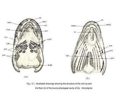
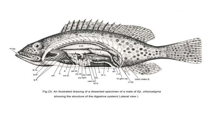
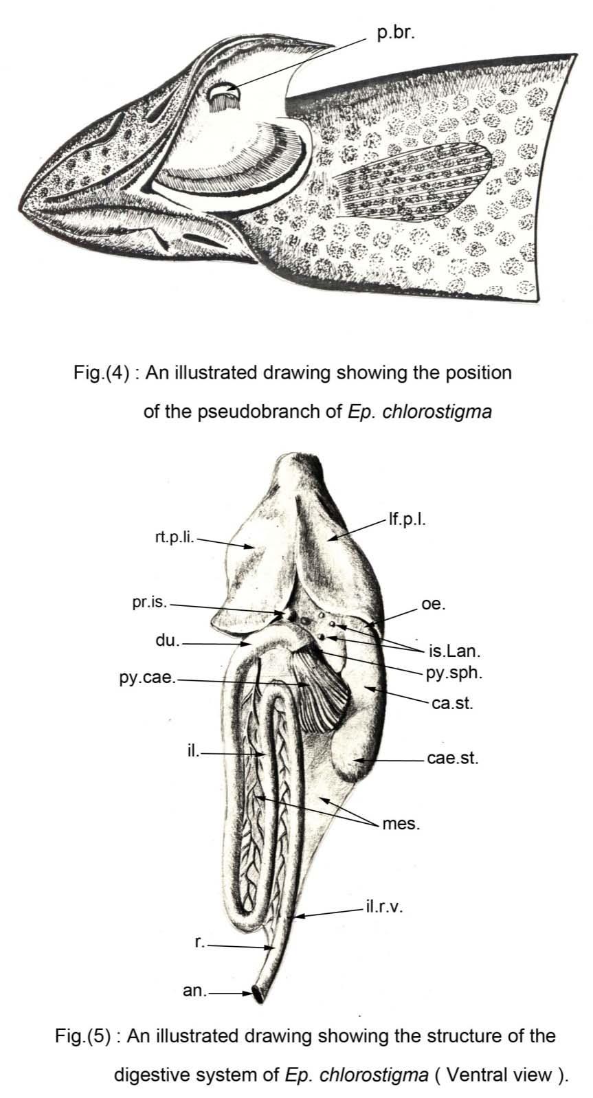
2 2. Material and Methods Specimens of the brown-spotted grouper, Epinephelus chlorostigma, were collected alive from the coral reefs in the neighbourhood of the Marine Biological Station, Ghardaqa, Red Sea. These specimens were carefully dissected to expose the digestive system and study its anatomical features. For histological purposes, small pieces of the different regions of the digestive system were fixed in aqueous Bouin's fluid for 24 hours. After fixation and washing for several times in 70% ethyl alcohol, dehydration was carried out in ascending grades of ethyl alcohol. These materials were then cleared in terpineol and embedded in molten paraplast. Sections of 5-7 µm thick were cut, mounted on glass slides and stained with Harris' haematoxylin and counterstained with eosin. Some sections were stained with Mallory's triple stain. The sections were examined by a light microscope and measurements were carried out using the eyepiece micrometer, calibrated by the stage micrometer. Photomicrographs were made as required. 3. Results and Discussion The brown-spotted grouper is a carnivorous fish feeding on fishes and different invertebrates. Its alimentary canal reflects this carnivory: large mouth, buccal cavity and pharynx; short expandable oesophagus; large distensible stomach; and relatively short intestine. These results agree with those of Grau et al. (1992), Petrinec et al. (2005) and Garasson et al. (2006). The digestive system has two functional units: an alimentary canal and digestive glands. Anatomy of the Alimentary Canal a- The mouth and the bucco-pharyngeal cavity: The mouth opening of the brown-spotted grouper is large in size, terminal in position and slightly oblique. It is protractile and bounded by upper and lower jaws. The lower jaw is slightly projecting in front of the upper jaw. The maxillae are broad and exposed posteriorly. They lie posterior to the premaxillae. The upper and the lower jaws are provided with lips (Fig. 1). These results agree with those of Kapoor (1953). The jaws are provided with small villiform teeth. On the upper jaw, several rows of maxillary teeth are arranged on the inner sides of the premaxillae (Fig. 2a); The teeth of the outer row are slightly longer than the others. The teeth are curved posteriorly. A pair of large canines occurs at the anterior end of each premaxilla. The maxillae are toothless. On the lower jaw, several rows of mandibular teeth are arranged on the sides of the dentaries (Fig. 2b); the outer row is slightly longer and strongly curved posteriorly. There is also a pair of large canines at the anterior end of each dentary. These results agree with those of Kapoor (1953), Woolcott (1957) and Groman (1982). The mouth opening leads into a spacious buccal cavity. The mucous membrane lining the roof of the buccal cavity is thrown into longitudinal folds which are not represented on the floor of the buccal cavity (Fig. 2a). The maxillary and the mandibular oral valves are narrow, thin semilunar membranes (Figs 2a,b). These result agrees with that of Kapoor (1953). On the roof of the buccal cavity, there are three patches of small villiform teeth: a crescentic patch of vomerine teeth on the vomers and two large oval patches of palatine teeth on the palatines(fig. 2a). The floor of the buccal cavity is flat with shallow transverse folds which are deeper at the posterior end (Fig. 2b). The tongue is small, elongate, free anteriorly and attached posteriorly to the floor of the buccal cavity (Fig. 2b). It lacks teeth, a result which contradicts with that of Groman (1982). The buccal cavity leads into a narrower pharyngeal cavity (Figs 2a,b). Four pairs of gills are arranged on the sides of the pharynx. Each gill consists of a long curved gill arch, two rows of gill filaments as well as two rows of toothed gill rakers (Fig. 3). The outer row of gill rakers of the first anterior gill arch is longer than the inner row of the same arch and those on the other arches (Fig. 3). These gill rakers form a screen that directs food particles toward the esophagus and protects the gill filaments. These results agree with those of Groman (1982). There is usually a well developed pseudobranch in the roof of each branchial chamber (Fig. 4). Five small patches of villiform teeth, the superior pharyngeal teeth, are arranged on each side of the roof of the pharynx (Fig. 2a). Two large triangular patches of inferior pharyngeal teeth also occur on the sides of the floor of the pharynx (Fig. 2b). They lie against those occurring on the roof of the pharynx. The teeth of the buccal cavity and those of the pharynx are homodont villiform teeth. They are pointed at the apex and broad at the base, being embedded in depressions and bent downwards and backwards. These results agree with those of Kapoor (1953). Since the present species is a carnivorous fish, therefore, its teeth are not used for mastication of food, but they are specialized for preventing the escape of the preys out of the buccal cavity. This conclusion was verified by the occurrence of complete preys inside the stomach. b- The oesophagus: Posteriorly, the pharynx of the brownspotted grouper narrows gradually to attain a cylindrical form, merging into a short, stout and muscular oesophagus (Fig. 3). This extends posteriorly over the lobes of the liver and below the air bladder. The oesophagus opens directly into the stomach without any external demarcation. 2150
3 Although the air bladder has no digestive function, but it is always associated with the digestive system. The air bladder is a large, cylindrical and single-chambered sac of the physoclistous type. This result agrees with that of Harder (1975) and Groman (1982). This hydrostatic organ fills most of the dorsal peritoneal cavity extending the length of the trunk kidney. This result agrees with that of Dobbin (1941) and Groman (1982). c- The stomach: The oesophagus leads into a large sac-like cylindrical stomach (Fig. 3). The stomach of the brown-spotted grouper is of the caecal type. It extends on the ventral side of the gas bladder. It can be divided into three definite portions: The proximal cardiac portion, the distal blind fundus or caecal portion and the mesial pyloric portion (Fig. 3). This result agrees with that of Harder (1975). In addition to digestion, the stomach seems to serve in this case for the storage of food. It is highly distensible so that the stomach of a small fish can accommodate a large prey. d- The intestine: It follows the pyloric portion of stomach, being separated from it by a pyloric sphincter (Figs. 3,5). The intestine of the brown-spotted grouper extends forward for a short distance where it curves strongly backward towards the cardiac portion of stomach (Fig. 5). On reaching a short distance behind the middle region of the body cavity, the intestine curves forward again ventral to the caecal portion of stomach making a U-shaped loop. Opposite to the pyloric portion of stomach, the intestine curves strongly backward again. This free limb slightly dilates just posterior to the bottom of the U-shaped loop and continues backward. At the point of dilation there is a slight constriction marking the presence of an internal ileo-rectal valve. These results agree with those of Groman (1982). The part of the intestine lying posterior to this valve represents the rectum. The rectum opens to the exterior by the anus which is one of three external openings on the postero-ventral surface of the body, just anterior to the anal fin (fig. 3). From anterior to posterior, these openings are the anus, the genital and the urinary openings. This result agrees with that of Groman (1982). In the mean time, it is very difficult to differentiate externally between the other two regions of the intestine, namely the duodenum and the ileum, due to the lack of any external clear demarcation. However, the first part of the of the intestine following the pyloric sphincter (the duodenum) is slightly wider than that of the other portions of the intestine (Fig. 5). At the anterior end of the duodenum where it turns backward, Just posterior to the pyloric sphincter, there are about thirty to thirty six blind tubes, the pyloric caeca (Figs 3,5). These are arranged in two groups of a circular pattern. The function of these pyloric caeca is poorly understood, but they may secrete some intestinal enzymes. It is also considered likely that they are important in neutralizing the acidity of the chyme before it reaches the intestine, where the environment is alkaline. Anatomy of the Digestive Glands a- The liver: The liver of the brown-spotted grouper is a brownish compact gland situated centrally within the peritoneal cavity, ventral to the esophagus as well as to the cardiac and pyloric portions of stomach (Figs 3,5). It is not completely divided into lobes. A shallow depression is present on the antero-mesial part of the ventral aspect of the liver giving the impression of a bilobed structure (Fig. 5). The gall bladder is a greenish oval structure situated in the right side of the liver (Fig. 3). This result agrees with that of Harder (1975). Bile produced in the liver can be stored within the gall bladder and then dispersed to the duodenum through a bile duct. This duct is long and stout and extends over the first part of intestine, the duodenum (Fig. 3). These results agree with those of Fange and Grove (1979). b- The pancreas: The pancreas of the brown-spotted grouper is represented by whitish areas scattered throughout the mesenteries and viscera of the peritoneal cavity, being more concentrated in the gastero- and duodeno-hepatic omenta (Fig. 5). This result agrees with that of Khalilov (1966). Small exocrine pancreatic patches almost always surround the hepatic portal veins as well as their branches which extend through the intestinal mesenteries. This result agrees with that of Groman (1982). The pancreatic tissue is usually concealed in large areas of adipose tissue found in the body cavity. The endocrine pancreas occurs as a large principle and several smaller islets of Langerhans scattered in the mesenteries adjacent to the neck of the gall bladder (Fig. 5). This endocrine pancreas is surrounded by exocrine pancreatic tissue. These results agree with those of Brinn (1973) and Groman (1982). The islets of Langerhans are generally whitish in colour and are usually encapsulated since they possess definite shape within the pancreatic tissue. There are no external signs to indicate that the pancreatic tissue invades the liver, a case referred to as the juxtahepatic pancreas. Histology of the Alimentary Canal a- The bucco-pharyngeal cavity: The mucosa of the roof of the buccal cavity and pharynx of the brown-spotted grouper is composed of a layer of stratified squamous epithelium,wich is thicker in the pharynx (Fig. 7) 2151
4 than that in the buccal cavity (Fig. 6). Numerous large goblet cells of circular and oval shapes are present in the distal epithelium (Figs. 6,7). These goblet cells secrete mucus to assist in swallowing food. These results agree with those of Kapoor (1953). Taste buds are sparsely present at the distal end of the epithelial folds. This result agrees with those of Walker et al. (1981). A thick lamina propria underlies the buccal and pharyngeal stratified squamous epithelium, which is composed of dense fibrous connective tissue lacking vascularization and cell diversity (Figs. 6,7). This result agrees with that of Groman (1982). The tongue mucosa is formed also of stratified squamous epithelium, containing goblet cells and taste buds (Figs. 8,9). The goblet cells are large circular and ovoid in shape. Superficially, the taste buds are numerous and bulb-shaped papillae (Fig. 9). This result agrees with that of Walker et al. (1981). The tongue mucosa is supported by a dense lamina propria that encircles a central core of loose areolar connective tissue and a hyalinizedcentral lingual cartilage (Fig. 8). These results come in agreement with those of Groman (1982). b- The oesophagus: The oesophagus of the brown-spotted grouper comes after the pharynx, which is a muscular tube that leads to the stomach. Its wall consists of four layers;the mucosa, the submucosa, the muscularis and the serosa. The anterior oesophageal mucosa is formed of stratified squamous epithelium that rests on a thick basement membrane (Fig.10). No lamina propria is present as it is replaced by the stratum compactum of the submucosa. Taste buds occur in the distal portion of the primary folds of the anterior esophagus, whereas the large circular and oval goblet cells are situated centrally, on the sides and at the bases of the folds (Fig. 10). The goblet cells secrete mucus that keeps the food lubricated and helps it to move along the tube. These results agree with those of Groman (1982). Posteriorly, in the transitional area between the oesophagus and the stomach, the posterior oesophagus, the mucosal folds lenghthen and gradually the stratified epithelium changes into columnar epithelium with numerous circular goblet cells in between (Fig. 12). These results agree with those of Flood et al. (1975) and Groman (1982). At the posterior end of the oesophagus the mucosa extends posteriorly to form an oesophagealstomach junction (Fig. 13).No muscularis mucosa exists in the oesophagus. The oesophageal submucosa has two layers, a stratum compactum of dense connective tissue that supports the mucosa and an underlying zone of loose areolar connective tissue (Fig. 11). Interfasicular connective tissue of the submucosa surrounds bundles of longitudinal striated submucosal muscles which occur in triangularshaped cords that terminates in the anterior part of the cardiac stomach. This result agrees with that of Groman (1982). The oesophageal external muscularis is formed of a single thick circular striated muscle layer (Fig. 11). The serosa is present only where the oesophagus extends into the peritoneal cavity. It consists of an underlying fibrous connective tissue layer covered by simple squamous epithelium (Fig. 11). c- The stomach: The stomach of the brown-spotted grouper has two histologically distinct areas: the cardiocaecal stomach region and the pyloric stomach region (Fig. 13). The gastric mucosa shows primary and secondary longitudinal folding of their mucous membranes (Fig. 14). The mucosal surface of the stomach is made up of simple columnar epithelium with basal nuclei forming a secretory sheath which has no goblet cells (Fig. 15). This sheath secretes mucus to protect the stomach against self digestion. The secretory sheath is followed by branched tubular gastric glands which secrete the gastric juice into the lumen of the stomach through gastric pits (Fig. 15). The mucosal epithelium of the cardiac portion of stomach stomach is continuous with that of the caecal and pyloric portions (Fig. 13). The complexity of the mucosal folds diminishes posteriorly toward the pyloric stomach, which contains no gastric glands and has a thin lamina propria (Fig. 16). This result agrees with that of Kapoor (1953). There is a prominent constriction, called the pyloric sphincter, between the pyloric portion of stomach and the intestine which may serve to control the food passage into the duodenum (Figs 16,17). This result agrees with that of Weinreb and Bilstad(1955), Martin and Balber (1984), Albrecht et al. (2001), Mai et al. (2005) and Amer et al. (2008). There is no muscularis mucosa. The submucosa of the stomach has two zones, an inner stratum compactum of dense collagenous connective tissue and an outer loose connective tissue (Fig. 14). The muscularis of the stomach begins in the cardiac portion where the striated muscles of the esophagus changes into smooth muscles. It consists of two layers, a thick inner circular and a thin outer longitudinal smooth muscle layers (Fig. 14). This bi-layer arrangement of the muscularis is retained throughout the remainder of the of the alimentary canal. The serosa of the stomach is a continuation of that covering the oesophagus with a thin fibrous connective tissue layer covered by simple squamous epithelium. d- The intestine: 2152
5 The intestine of the brown-spotted grouper can be distinguished histologically into three regions: the duodenum, the ileum and the rectum which are more or less similar in structure. Generally, the intestinal wall is constructed of the main four layers: the mucosa, submucosa, muscularis and serosa. The lymphocytes are numerous and scattered in the mucosal epithelium and the lamina propria of the whole intestine, which may play a role in protecting the fish from pathogenic organisms. This result comes in agreement with that of Osman and Caceci (1991)and Park and Kim (2001). The anterior intestine and its appendages, the pyloric caeca, originate immediately posterior to the pyloric sphincter (Fig. 17). They differ histologically from the stomach by having decreased amounts of submucosal and muscular tissues and longer mucosal folds (Fig 18). This result agrees with that of Groman (1982). The pyloric caeecal mucosa is built up of columnar epithelium with goblet cells. The lamina propria of the pyloric caeca extend throughout the complex folds of the mucosa (Fig. 18). The submucosa is very thin vascularized loose connective tissue (Fig. 18). The muscularis of the pyloric caeca is formed of an inner thick circular and an outer thin longitudinal smooth muscle layers (Fig. 18). The serosa is formed of simple squamous epithelium. This is absent in areas of connection with pyloric stomach, where it is replaced by loose connective tissue. The mucosa of the three regions of the intestine is built up of two types of cells: the enterocytes and the goblet cells, and contain a thin vascular lamina propria (Figs 19,20and 21). This result agrees with that of Cataldi et al. (1987) and Gargiulo et al. (1998). The enterocytes of the mucosa has the typical structure of the absorptive cells. They are tall columnar cells with prominent brush borders and numerous goblet cells in between (Fig. 22). These results agree with that of Olsen et al. (1999). There are variations in the morphology of the mucosal folds in the different parts of the intestine. The mucosa of the duodenum is thrown up into long and highly branched villi (Fig. 19), while that of the ileum is simple unbranched villi (Fig. 20). This result agrees with that of Amer et al. (2008). Posteriorly, the mucosal folds of the ileum become shorter toward the ileorectal valve (Fig. 23). The rectum has shorter primary and few secondary villi with numerous goblet cells between the enterocytes (Fig. 21). These goblet cells secrete mucus which may serve as a lubrication aid to defecation. This result agrees with that of Martin and Balber (1984) and Amer et al. (2008). The ileo-rectal valve (or the intestinal valve) is the morphologic separation between the ileum and the rectum. It consists of a circular tissue flap with a central opening that is directed posteriorly within the rectal lumen(fig. 24). The mucosal and submucosal layers of the valve are continuous with those of the ileum; however, only the circular layer of the muscularis is present in the valve (Fig. 25). These results agree with those of Groman (1982). The intestinal submucosa is formed of thin dense connective tissue (Figs 19, 20 and 21). There is no muscularis mucosa. The intestinal muscularis is built up of two layers: a thick inner circular and a thin outer longitudinal smooth muscle layers. These two layers remain intact throughout the intestine, but their relative proportions change between the anterior intestine and the rectal sections. In the rectum, the circular muscle layer increases in thickness (Fig. 21). This result agrees with that of Grau et al. (1998), but contradicts with that of Gargiulo et al. (1998). The serosa of the anterior intestine and rectum is composed of a layer of a simple squamous epithelium. Histology of the Digestive Glands a- The liver: The hepatic parenchyma of the brownspotted grouper contained hepatocytes. The hepatocytes are polyhedral in shape and each contained a basophilic central nucleus (Figs 26,27). The hepatocytes are arranged in a diffuse or radial patterns (Fig 26). These results agree with those of Raskovie et al. (2011), Rocca et al. (1994) and Iqpegbu et al. (2012). Some hepatocytes surround the central veins in a radial pattern, while others surround the hepatic portal venules and the hepatic arterioles in a diffuse pattern (Figs 26,27). The bile is connected in the bile canaliculi found between the adjacent hepatocytes (Fig.26). These canaliculi are lined with a low simple cuboidal epithelium and surrounded by a thin layer of of fibrous connective tissue (Fig. 27). The canaliculi are connected to larger hepatic ductules (Figs 26,27), which join together to form the hepatic duct that emerges out of the liver. Each ductule is lined with simple cuboidal to short columnar epithelium which is surrounded by a supporting fibrous connective tissue sheath (Fig. 27). These results agree with those of Kapoor (1953). b- The pancreas: The pancreas consists mainly of two components: an exocrine and an endocrine pancreas. The brown-spotted grouper has a diffuse pancreas that can be found scattered within the mesenteries and viscera in the peritoneal cavity (Fig.28). The exocrine pancreas is located within the mesenteric adipose tissue and surrounds the branches of the hepatic portal veins (Figs. 29,30). This result agrees with that of Groman(1982). It is made up of large polyhedral cells attached by their basal ends to the portal veins (Figs 29,30). Each 2153
6 cell has a rounded nucleus, situated centrally to basally on one side (eccenteric nucleus), surrounded by a cytoplasm which is granular in nature (Fig. 30). Zymogen granules are located in the apical ends of these cells. This result agrees with that of Leeson and Leeson (1970). The pancreatic juice is collected in the interlobular excretory ducts, which are lined with columnar epithelium surrounded by connective tissue (Fig. 28). This result agree with that of Fiore et al. (1973). These interlobular ducts empty their contents into the main pancreatic duct. The endocrine pancreas occurs as a principle and several smaller islets of Langerhans scatterred in the intestinal mesenteries (Fig. 28). This result agrees with that of Groman(1982). The islets of Langerhans usually contain several hundreds of endocrine cells surrounded by a fibrous connective tissue layer containing blood capillaries (Fig. 31). References Al-Hussaini, A.H. (1946): The anatomy and histology of the alimentary tract of the bottomfeeder Mulloides auriflamma(forsk). J. Morph., 78 : Al-Hussaini, A.H. (1947): The anatomy and histology of the alimentary tract of the plankton- feeder Atherina forskali (Rupp.). J. Morph., 80: Al-Hussaini, A.H. (1949): On the functional morphology of the alimentary tract of some fish in relation to differences in their feeding habits: anatomy and histology. Quart. J. Micr. Sci., 90 (2): Albrecht, M.P.; M.F.N. Ferreira and E.P. Caramaschi (2001): Anatomical features and histology of the digestive tract of two related neotropical omnivorous fishes (Characiformes; Anostomidae). J. Fish Biol., 58: Amer, F.I.; S.A.A. Naguib and F.A. Abdel Ghafar (2008): Comparative study on the intestine of Schilbe mystus and Labeo niloticus in correlation with their feeding habits. Egypt. J. Aquat. Biol. And Fish., 12 (4): Blake, I.H. (1930): Studies on the comparative histology of the digestive tube of certain teleost fishes. A predaceous fish, the sea bass, Centropristes striatus. J. Morph., 50: Blake, I.H. (1936): Studies on the comparative histology of the digestive tube of certain teleost fishes. A bottom feeding fish, the sea robin, prionotus carolinus. J. Morph., 60: Brinn, J.E. (1973): The pancreatic islets of bony fishes. Amer. Zoolog., 13: Bucke, D. (1971): Anatomy and histology of the alimentary tract of the carnivorous fish, the pike, esoxluicius. J. Fish Biol., 3: Carrasson, M.; A. Grau; L.R.Dopazo and S. Crespo (2006): A histological, histochemical and ultrastructural study of the digestive tract of Dentex dentex (Pisces; Sparidae). Hist. Histopathol., 21 (6): Cataldi, E.; S. Cataudella; G. Monaco; A. Rossi and L. Tancioni (1987): A study of the histology and morphology of the digestive tract of the sea bream, Sparus aurata. J. Fish Biol., 30: Chao, L.N. (1973): Digestive system and feeding habits of the cunner,tautogolabris adspersus, a stomachless fish. Fish. Bull., 71: Curry, E. (1939): The histology of the digestive tube of the carp, Cyprinus carpio communis. J. Morph., 65: Dawes, B. (1929): The histology of the alimentary tract of the plaice, pleuronectes platessa. Quart. J. Micr. Sci., 12: Dobbin, C.N. (1941): A comparative study of the gross anatomy of the air bladder of ten families of fishes of New York and other eastern states. J. Morph., 68: Fange, R. and D. Grove (1979): Digestion. Pp in W.S. Hoar, D.J. Randall J.R. Brett, editors.fish Phys., vol. 8. Academic press, N.Y., N.Y.,USA. Fiore, M.S.H., R.E. Mancini and E.D.L. Roberts (1973): New atlas of histology. Lea and Febiger, Phila., Pen., USA. Flood, M.T.; R.F. Nigrelli and J.F. Gennaro (1975): Some aspects of the ultrastructure of the stabchendrusenzellen, a peculiar cell associated with the endothelium of the bulbus arteriosus and with other fish tissues. J. Fish Biol., 7: Gargiulo, A.M.; P. Ceccarelli; C. Dall'Aglio and V. Pedini (1998): Histology and ultrastructure of the gut of the tilapia (Tilapia spp.), a hybrid teleost. Anat. Histol. Embryol., 27: Grau, A.; S. Crespo; M.C. Sarasquete and M.L.G. De Canales (1992): The digestive tract of the amberjack Serioladumerilli; Risso: a light and scanning electron microscope study. J. Fish Biol., 41: Greene, C.W. (1912): Anatomy and histology of the alimentary tract of the king salmon. Bull. US Bureau.Fish., 32: Groman, D.B. (1982): Histology of the striped bass. Monog.3, ISSN , Bet., Mary., USA. Hale, P.A. (1965): The morphology and histology of the digestive system of two freshwater teleosts, Poecilia reticulate and Gasterosteu saculeatus. J. Zool. (London), 146: Harder, W. (1975): Anatomy of fishes. E. SchweizerbartscheVerlagsbuchhandlung (Nagel V. Obermiller), Stutt, W. Germ. Ikpegbu, E.; U.C. Nelebedum; O. Nnadozie and I.O. Agbakwuru (2012): Histological 2154
7 structures of the accessory glands of the digestive system in adult farmed African catfish (Clarias gariepinus B.). J. Agr. Vet. Sci., 1 (6): Islam, A.U. (1951): The comparative histology of the alimentary tract of certain freshwater teleost fishes. Proc. Ind. Acad. Sci., 33B: Kapoor, B.G. (1953): The anatomy and histology of the alimentary canal in relation to its feeding habits of a siluroid fish,wallago attu(bl.andschn.). J. Zool. Soc. India, 5 (2): Khalilov, F. (1966): Some material on the histology and histochemistry of the pancreas and liver of teleost fish problems in Ichthyology. J. Ichth., 8: Khojasteh, S.M.B.; F. Sheikhzadeh; D. Mohammadnejad and A.Azmi (2009): Histological, histochemical and ultrastructural study of the intestine of the rainbow trout (Oncorhynchus mykiss). Wor. Appl. Sci. J., 6 (11): Khojasteh, S.M.B. (2012): The morphology of the post-gastric alimentary canal in teleost fishes: a brief review. Int. J. Aqua. Sci., 3 (2): Leeson, T.S. and C.R. Leeson (1970): Histology. 2 nd ed., W.B.Saunders, phil., Pens.,USA. Mai, K.; H. Yu; H. Ma; Q. Duan; E. Gisbert; J.L.Z. Infante and C.L. Cahu (2005): A histological study on the development of the digestive system of Pseudosciaena corcea larvae and guveniles. J. Fish Biol., 67: Martin, T.J. and S.J.M. Balber (1984): Morphology and histology of the alimentary tract of Ambassidae (Cuvier) (Teleostei) in relation to feeding. J. Morph., 182: Olsen, R.E.; R. Myklebust; T. Kaino and E. Ringo (1999): Lipid digestibility and ultrastructural changes in the enterocytes of Arctic char (Salvelinus alpines L.) fed linseed oil and soybean lecithin. J. Fish Physiol. Biochem., 21: Osman, A.H.K. and T. Caceci (1991): Histology of the stomach of Tilapia nilotica (Linnaeus, 1758) from the River Nile. J. Fish Biol., 38: Park, J.Y. and I.S. Kim (2001): Histology and mucin histochemistry of gastrointestinal tract of the mud loach, in relation to respiration. J. Fish Biol., 58: Petrinec, Z.; S. Nejedli; S. Kuzir and A. Opacak (2005): Mucosubstances of the digestive tract mucosa in northern pike (Esoxlucius L.) and European catfish (Silurus glanis L.).Vet. Arhiv., 75 (4): Rocha, E.; R.A. Monteiro and C.A. Pereira (1994): The liver of the brown trout, Salmo truttafario: a light and electron microscope study. J. Anat., 185: Rogick, M.D. (1931): Studies on the comparative histology of the digestive tube of certain teleost fishes. A minnow, Compostoma anomalum. J. Morph., 52: Raskovie, B.S.; M.B. Stankovie; Z.Z. Markovie and V.D. Poleksic (2011). Histological methods in the assessment of different feed effects on liver and intestine in fish. J. Agr. Sci., 56 (1): Suicmez, M. and E. Ulus (2005): A study of the anatomy, histology and ultrastructure of the digestive tract of Orthrias angorae Steindachner, Fol. Biol. (Krakow), 53 (1-2): Thurmond, T. (1979): Histology and pathology of the alimentary tract of the American eel (Anguilla rostrata). M.Sc. thesis, Univ. Connecticut, USA. Walker, E.R.; S.F. Filder and D.E. Hinton (1981): Morphology of the buccopharyngeal portion of the gill in the fathead minnow, Plmepliales promefos. Anat. Rec., 200: Wassersung, R.J. and R.K. Johnson (1976): A remarkable pyloric caecum in the evermannellid genus with notes on gut, structure and function in alepisauroid fishes. J. Zool. (London), 179: Weinreb, E.L. and N.M. Bilstad (1955): Histology of the digestive tract and adjacent structures of the rainbow trout, Salmogairdneriirideus. Copeia, 3: Woolcott, W.S. (1957): Comparative osteology of serranid fishes of the genus Roccus. Copeia, 1957: List of Abbreviations a.bl. Air bladder ad. t. Adipose tissue an. Anus an. f. Anal fin ant. Int. Anterior intestine a.oe. Anterior oesophagus ba. mb. Basement membrane bi. Ca. Bile canalicule bi. d. Bile duct bi. du. Bile ductule bl. ca. Blood capillaries br. bo. Brush border b. w. Body wall cae. st. Caecal stomach ca. st. Cardiac stomach cau. f. Caudal fin ce. v. Central vein c. m. l. Circular muscle layer cn. Canine co. ep. Columnar epithelium com. mes. d. Common mesonephric duct den. c. t. Dense connective tissue do. F. Dorsal fin du. Duodenum 2155

8 ec.nu. Eccenteric nucleus en. pa. c. Endocrine pancreatic cells ex. pa. t. Exoxrine pancreatic tissue fi. C. t. Fibrous connective tissue f. ph. Floor of pharynx ga. bl. Gall bladder ga. gl. Gastric glands g. ar. Gill arch ga. p. Gastric pits g. fl. Gill filament go. c. Goblet cells g. ra. Gill rakers h. Heart hp. ar. Hepatic arteriole hpc. Hepatocytes hp. po. v. Hepatic portal vein hp. po. ven. Hepatic portal venule il. Ileum ilr. v. Ileo-rectal valve int. du. Interlobular duct i.ph. t. Inferior pharyngeal teeth is. Lan. Islets of Langerhans k. Kidney la. pr. Lamina propria li. Liver lf. p. li. Left part of liver li. car. Lingual cartilage l. m. l. Longitudinal muscle layer lo. c. t. Loose connective tissue lo. c. t. co. Loose connective tissue core l. subm. m. Longitudinal submucosal muscles lu. il. Lumen of ileum lu. r. Lumen of rectum ly. Lymphocytes m. Muscularis max. t. Maxillary teeth max. v. Maxillary valve mes. Mesenteries m. gen. op. Male genital opening mnd. T. Mandibular teeth mnd. v. Mandibular valve m. op. Mouth opening mu. Mucosa muc. Mucus mu. vi. Mucosal villi no. nostril oe. Oesophagus op. b. Opercular bones pa. t. Palatine teeth p. br. Pseudobranch pec. f. Pectoral fin pel. f. Pelvic fin pr. is. Principle islet py. cae. Pyloric caeca py. sph. Pyloric sphincter py. st. Pyloric stomach r. Rectum r. buc. Roof of buccal cavity r. ph. Roof of pharynx rt. p. li. Right part of liver se. Serosa sec. sh. Secretory sheath sh. co. ep. Short columnar epithelium sof. r. Soft rays sp. Spines s. ph. t. Superior pharyngeal teeth st. com. Stratum compactum st. sq. ep. Stratified squamous epithelium subm. m. submucosal muscles su. Mu. Submucosa t. Testis ta. bu. Taste buds ub. Urinary bladder u. op. Urinary opening v. d. Vas deferens vo. t. Vomerine teeth zy. gr. Zymogen granules 2156
9 2157
: T.S. of the anterior region of the tongue showing the mucosa, lamina propria, loose connective tissue core and lingual cartilage. H & E stain, X 65. Fig. (9): Enlarged portion of Fig.")

10 Fig. (6): T.S. of the roof of the buccal cavity showing the mucosal epithelial cells, goblet cells, sparse taste buds and lamina propria. H & E stain, X 180. Fig. (7): T.S. of the roof of the pharynx showing the mucosal epithelial cells, goblet cells, sparse taste buds and lamina propria. H & E stain, X 200. Fig. (8): T.S. of the anterior region of the tongue showing the mucosa, lamina propria, loose connective tissue core and lingual cartilage. H & E stain, X 65. Fig. (9): Enlarged portion of Fig. (8) showing the mucosal epithelial cells with goblet cells and numerous taste buds, lamina propria and loose connective tissue. H & E stain, X 190. Fig. (10): T.S. of the anterior region of the oesophagus showing the mucosal epithelial cells with goblet cells and taste buds, basement membrane and stratum compactum of the submucosa. H & E stain, X
: Enlarged portion of Fig. (11) showing the mucosal columnar epithelium with numerous goblet cells and the stratum compactum. H & E stain, X 195. Fig. (13): L.S.")
: Enlarged portion of Fig.")
11 Fig. (11): T.S. of the posterior region of the oesophagus showing the mucosa, submucosawith submucosal muscles, muscularis and serosa. H & E stain, X 45. Fig. (12): Enlarged portion of Fig. (11) showing the mucosal columnar epithelium with numerous goblet cells and the stratum compactum. H & E stain, X 195. Fig. (13): L.S. of the oesophageal-stomach regions showing the structure of the oesophageal-stomach junction and different portions of stomach. H & E stain, X 25. Fig. (14): T.S. of the cardio-caecal stomach showing the mucosa, submucosa, musularis and serosa. Malory Tri. Stain, X 45. Fig. (15): Enlarged portion of Fig. (14)showing the secretory sheath and tubular gastric glands opening with gastric pits. Malory Tri. Stain, X
: T.S. of the pyloric caecum showing the mucosa, submucosa, muscularis and serosa. H & E stain, X 85. 2160")
12 Fig. (16): T.S. of the pyloric stomach showing the absence of the gastric glands and the presence of a pyloric sphincter. H & E stain, X 75. Fig. (17): T.S. of the pyloric stomach showing the pyloric sphincter and sections of pyloric caeca. H & E stain, X 45. Fig. (18): T.S. of the pyloric caecum showing the mucosa, submucosa, muscularis and serosa. H & E stain, X
: T.S. of the ileum showing the simple un-branched mucosal villi. H & E stain, X 130. Fig. (21): T.S. of the rectum showing the mucosa, submucosa, muscularis, serosa and short mucosal villi.")
 showing the mucosal epithelial cells, goblet cells, lymphocytes, brush border and lamina propria. H & E stain, X 425. 2161")
13 Fig. (19): T.S. of the duodenum showing the mucosa, submucosa, muscularis, serosa and highly branched mucosal villi. H & E stain, X 115. Fig. (20): T.S. of the ileum showing the simple un-branched mucosal villi. H & E stain, X 130. Fig. (21): T.S. of the rectum showing the mucosa, submucosa, muscularis, serosa and short mucosal villi. H & E stain, X 110. Fig. (22): Enlarged portion of Fig. (20) showing the mucosal epithelial cells, goblet cells, lymphocytes, brush border and lamina propria. H & E stain, X
: L.S.")
: Enlarged portion of Fig.")
14 Fig. (23): T.S. of the posterior region of the ileum showing short mucosal villi toward the ileo-rectal valve. H & E stain, X 105. Fig. (24): L.S. of the region between the ileum and rectum showing the structure of the ileo-rectal valve. H & E stain, X 78. Fig. (25): Enlarged portion of Fig. (24) showing that the mucosal, submucosal and only circular muscle layers of the ileum extend into the ileo-rectal valve. H & E stain, X

 showingthe structure of the hepatocytes, bile canalicule and hepatic ductule. H & E stain, X 485. Fig. (28): S.")
15 Fig. (26): S. of the liver showing the diffuse and radial patterns of arrangement of hepatocytes, bile canalicule and hepatic ductule. H & E stain, X 210. Fig. (27): Enlarged portion of Fig. (26) showingthe structure of the hepatocytes, bile canalicule and hepatic ductule. H & E stain, X 485. Fig. (28): S. of the mesenteries of the peritoneal cavity showing the exocrine pancreas, interlobular duct, principle and small islets of Langerhans. H & E stain, X
: Enlarged portion of Fig.")
: S.")
16 Fig. (29): S. of the exocrine pancreatic tissue surrounded by adipose tissue and surrounds a portal vein. H & E stain, X 150. Fig. (30): Enlarged portion of Fig. (29) showing the polyhedral exocrine pancreatic cells with eccentric nuclei and apical zymogen granules. H & E stain, X 410. Fig. (31): S. of the principle islet of Langerhans showing the surrounding fibrous connective tissue layer and the numerous endocrine pancreatic cells. H & E stain, X
UROMASTIX DIGESTIVE SYSTEM
 UROMASTIX DIGESTIVE SYSTEM by Dr. Rashmi Tripathi Department of Zoology Brahmanand College, Kanpur DIGESTIVE SYSTEM : The digestive system consists of (A) Alimentary canal and (B) Associated digestive
THE ANATOMY AND HISTOLOGY OF THE ALIMENTARY CANAL OF A CARNIVOROUS FISH MEGALOPS CYPRINOIDES (BROUSS)* BY S. M. KAMAL PASHA
* BY S. M. KAMAL PASHA") THE ANATOMY AND HISTOLOGY OF THE ALIMENTARY CANAL OF A CARNIVOROUS FISH MEGALOPS CYPRINOIDES (BROUSS)* BY S. M. KAMAL PASHA (Department o/zoology, Presidency College, Madras) Received March 23, 1964 (Communicated
THE ANATOMY AND HISTOLOGY OF THE ALIMENTARY CANAL OF A CARNIVOROUS FISH MEGALOPS CYPRINOIDES (BROUSS)* BY S. M. KAMAL PASHA (Department o/zoology, Presidency College, Madras) Received March 23, 1964 (Communicated
AN OMNIVOROUS FISH MYSTUS (-~MACRONES) GULIO (HAM.)* BY S. M. KAMAL PASHA. (Department of Zoology, Presidency College, Madras)
 GULIO (HAM.)* BY S. M. KAMAL PASHA. (Department of Zoology, Presidency College, Madras)") THE ANATOMY AND HISTOLOGY OF THE ALIMENTARY CANAL OF AN OMNIVOROUS FISH MYSTUS (-~MACRONES) GULIO (HAM.)* BY S. M. KAMAL PASHA (Department of Zoology, Presidency College, Madras) Received December 11,
THE ANATOMY AND HISTOLOGY OF THE ALIMENTARY CANAL OF AN OMNIVOROUS FISH MYSTUS (-~MACRONES) GULIO (HAM.)* BY S. M. KAMAL PASHA (Department of Zoology, Presidency College, Madras) Received December 11,
Dogfish Shark Dissection
 Dogfish Shark Dissection Name Date Period Fun Facts: Materials: The teeth of sharks are modified scales embedded in the skin of its mouth Sharks have pits on their face used to detect electric fields Sharks
the corpus, but the latter can be easily distinguished
 - 79 - ALH1ENTARY CANAL Banki (1936) broadly divides the alimentary canal of fishes into two regions, the 'Kopfdarm' comprising the buccal ca.vity and the pharynx and the 'rumpfdarm' comprising the foregut
The Digestive Tube of an Omnivorous Cyprinoid Fish, Barbus stigma (CUV. & VAL.)*
*") The Digestive Tube of an Omnivorous Cyprinoid Fish, Barbus stigma (CUV. & VAL.)* B.G. KAPOOR (Fisheries Section, Ministry of Agriculture, Government of India, New Delhi) The present study is a continuation
The Digestive Tube of an Omnivorous Cyprinoid Fish, Barbus stigma (CUV. & VAL.)* B.G. KAPOOR (Fisheries Section, Ministry of Agriculture, Government of India, New Delhi) The present study is a continuation
International Journal of Scientific & Engineering Research, Volume 7, Issue 10, October ISSN
 International Journal of Scientific & Engineering Research, Volume 7, Issue 10, October-2016 1086 GASTRO-INTESTINAL TRACT OF POMADASYS JUBELINI (CUVIER, 1830) IN THE NEW CALABAR-BONNY RIVER, RIVERS STATE,
Exercise 18B Class Chondrichthyes Cartilaginous Fishes
 AP Biology Chapter 24 Exercise #18: Chordates: Fish Cartilaginous Fishes Lab Guide Exercise 18B Class Chondrichthyes Cartilaginous Fishes This group contains about 970 species that are characterized by
Perch Dissection Lab
 Name: Block: Due Date: Perch Dissection Lab Background The fish in the class Osteichthyes have bony skeletons. There are three groups of the bony fish: ray-finned, lobe-finned, and the lungfish. The perch
Chapter 30 Nonvertebrate Chordates, Fishes, and Amphibians Name
 Chapter 30 Nonvertebrate Chordates, Fishes, and Amphibians Name Lab Dissecting a Perch Background Information Fish are the largest group of vertebrates found in fresh and salt water. In fact, over 25,000
-8- spinous. nape caudal fin. body depth. pectoral fin. anus. total length Fig. 4
 click for previous page -8-1.3 Illustrated Glossary of Technical Terms and Measurements External Morphology and Measurements spinous dorsal fin soft nape caudal fin interorbital body depth snout lateral
- 7 - DESCRIPTION OF SPECIES
 I - 7 - DESCRIPTION OF SPECIES./' Anguilla bicolor McClelland ' Level-finned eel (Figs.i & 2) Length of head 6-8 times in length of body; Diameter of eye 8-10 times, Inter-orbital length 2-2.5 times, Gape
External Anatomy Dissection Guide
 External Anatomy Dissection Guide Dissection is the cutting of a dead animal or a plant into separate parts for the purpose of careful and detailed examination and study. The external anatomy is as important
Perch Dissection Lab
 Perch Dissection Lab Introduction: The fish in the class Osteichthyes have bony skeletons. There are three groups of the bony fish - -- ray-finned fish, lobe-finned fish, and the lung fish. The perch is
Shark Lab Key. dorsal surface. click on picture for ventral surface
 Shark Lab Key Study this basic information about the spiny dogfish shark. Print this Shark Lab Report Guide. Pre-Lab Research Study this website. It provides several useful videos of large shark dissections.
Fish. Water Dwelling Animals
 Fish Water Dwelling Animals Class Agnatha (Jawless fish) They are believed to be the most primitive and oldest vertebrates. Lamprey and hagfish are the only 2 living members of this class and are placed
Gen Bio 2 Lab #10: Chondrichthyes and Osteichthyes
 Name: Date Gen Bio 2 Lab #10: Chondrichthyes and Osteichthyes Pre-Lab Reading: pages 687-690 Pre-Lab Vocabulary: 1) Ampullae of Lorenzini 2) Claspers 3) Lateral line 4) Ovoviviparous 5) Squalene 6) Viviparous
Internal Anatomy of Fish
 Internal Anatomy of Fish The Systems of a Fish Skeletal System Muscular System Respiratory System Digestive System Circulatory System Nervous System Reproductive System Special Organs Skeletal System
What is a Fish? Fishes are aquatic vertebrates. Most fishes have paired fins, scales, and gills.
 What is a Fish? Fishes are aquatic vertebrates. Most fishes have paired fins, scales, and gills. Feeding and Digestion Every mode of feeding is seen in fish herbivores, carnivores, parasites, filter feeders,
Class Osteichthyes. Bony Fish
 Class Osteichthyes Bony Fish General Characteristics of Class internal skeleton ossified (turned to bone) Paired fins made of rays and spines, or lobed fins swim bladder or lung present bony scales (ganoid,
Anatomy and Histology of the Spiral Valve Intestine in Juvenile Australian Lungfish, Neoceratodus forsteri
 62 The Open Zoology Journal, 2009, 2, 62-85 Open Access Anatomy and Histology of the Spiral Valve Intestine in Juvenile Australian Lungfish, Neoceratodus forsteri Masoud Hassanpour and Jean Joss* Department
UNIT 9 - RESPIRATORY SYSTEM LECTURE NOTES
 UNIT 9 - RESPIRATORY SYSTEM LECTURE NOTES 9.01 GENERAL FUNCTIONS OF THE RESPIRATORY SYSTEM A. Brings oxygenated air to the alveoli B. Removes air containing carbon dioxide C. Filters, warms, and humidifies
IT has been shown by Pampapathi Rao (1958) that the fresh-water teleost,
 that the fresh-water teleost,") 361 Structural Changes in the Gills, Intestine, and Kidney of Etroplus maculatus (Teleostei) adapted to different Salinities By V. VIRABHADRACHARI (From the Department of Zoology, Sri Venkateswara University,
361 Structural Changes in the Gills, Intestine, and Kidney of Etroplus maculatus (Teleostei) adapted to different Salinities By V. VIRABHADRACHARI (From the Department of Zoology, Sri Venkateswara University,
Frog Dissection. External Observation
 Frog Dissection External Observation Use the diagram below to locate and identify the external features of the head. Find the mouth, external nares, tympani (ear drum), eyes, and nictitating membranes
Frog Dissection. PreLab: 1. Where do frogs get their energy? Draw a simple food chain to illustrate.
 Name Date Frog Dissection Class # PreLab: Amphibian Reading As members of the class Amphibia, frogs may live some of their adult lives on land, but they must return to water to reproduce. Eggs are laid
RONALD LESTER GAMMON. submitted in partial fulfillment of the. requirements for the degree MASTER OF SCIENCE. Approved by: Major Professor
 THE GROSS AND MICROANATOMY OF THE DIGESTIVE TRACT AND PANCREAS OF THE CHANNEL CATFISH, ICTALURUS PUNCTATUS by RONALD LESTER GAMMON B. S., Kansas State University, 1968 B. S., Kansas State University, 1970
SOME COMPARATIVE HISTOLOGICAL STUDIES ON ALIMENTARY TRACT OF TILAPIA FISH (TILAPIA SPILURUS) AND SEA BREAM (MYLIO CUVIERI)
 AND SEA BREAM (MYLIO CUVIERI)") EGYPTIAN JOURNAL OF AQUATIC RESEARCH ISSN 1110-0354 VOL. 31., 1. 2005 SOME COMPARATIVE HISTOLOGICAL STUDIES ON ALIMENTARY TRACT OF TILAPIA FISH (TILAPIA SPILURUS) AND SEA BREAM (MYLIO CUVIERI) HEYAM ABDULLAH
EGYPTIAN JOURNAL OF AQUATIC RESEARCH ISSN 1110-0354 VOL. 31., 1. 2005 SOME COMPARATIVE HISTOLOGICAL STUDIES ON ALIMENTARY TRACT OF TILAPIA FISH (TILAPIA SPILURUS) AND SEA BREAM (MYLIO CUVIERI) HEYAM ABDULLAH
DISSECTION 101 THE FROG
 DISSECTION 101 THE FROG Dissection helps us understand how living things function. Dissection is analytical. Dissection is an adventure. Discussion Frog anatomy is unique in that it does resemble human
Comparison of Morphometrics and Meristic Characteristics of two Catfishes Plotosus limbatus and Clarias brachysoma
 Tropical Agricultural Research Vol. 19: 301-306 (2007) Comparison of Morphometrics and Meristic Characteristics of two Catfishes Plotosus limbatus and Clarias brachysoma W.M.T.K. Wasala, U. Edirisinghe
Marine Fishes. Chapter 8
 Marine Fishes Chapter 8 Fish Gills The construction of the gill is the same in all fish gill arch supports the entire structure, gill rakers are on the forward surface of the gill arch and gill filaments
Shark Dissection Dogfish Squalus acanthias
 Name Shark Dissection Dogfish Squalus acanthias Fun Facts: The teeth of sharks are modified scales embedded in the skin of its mouth Sharks have pits on their face used to detect electric fields Sharks
Classification. Phylum Chordata
 AP Biology Chapter 23 Exercise #17: Chordates: Urochordata & Cephalochordata Lab Guide Chordates show remarkable diversity. Most are vertebrates. All animals that belong to this phylum MUST, at some point
Lumbricus terrestris - preserved specimens for dissection
 Lumbricus terrestris - preserved specimens for dissection External Anatomy: Prostomium (observe under dissecting microscope for external sensory organs), peristomium, clitellum, setae (dissecting microscope),
Respiration. Chapter 33
 Respiration Chapter 33 Learning Objectives: Understand the basis of gas exchange and factors that influence diffusion of gases in and out of tissues Compare and contrast different respiratory systems among
Fish Dissection Background
 Fish Dissection Background Introduction Living things are similar to and different from each other. For example, when we look at the inside of a fish, we learn that the organ systems of fish are similar
Model Answer M.Sc. (III Semester) Zoology, Paper : LZT-304A (Fish Anatomy and Physiology) SECTION-A (Multiple choice questions)
 Zoology, Paper : LZT-304A (Fish Anatomy and Physiology) SECTION-A (Multiple choice questions)") SECTION-A (Multiple choice questions) Q. 1-Answer (i) d (ii) c (iii) c (iv) d (v) a (vi) b (vii) b (viii) c (ix) b (x) c SECTION B (Descriptive type questions) Q. 2- Answer Transport of CO 2 and O 2 Oxygen
SECTION-A (Multiple choice questions) Q. 1-Answer (i) d (ii) c (iii) c (iv) d (v) a (vi) b (vii) b (viii) c (ix) b (x) c SECTION B (Descriptive type questions) Q. 2- Answer Transport of CO 2 and O 2 Oxygen
FISH ANATOMY DIAGRAM AND QUESTIONS
 Name Block FISH ANATOMY DIAGRAM AND QUESTIONS External: 1. What percentage of fish are bony fish? 2. What is the operculum s function? 3. The nostrils are used for, not. 4. Which fins keeps the fish level
Multicellular Organisms. Sub-Topic 2.7 Animal Transport & Exchange Systems
 Multicellular Organisms Sub-Topic 2.7 Animal Transport & Exchange Systems On completion of this sub-topic I will be able to state that: Rings of cartilage keep the main airways open Oxygen and carbon dioxide
Comparative study on the histological structures of the intestine in some coral reef fishes in Hurghada, Red Sea, Egypt
 INTERNATIONAL JOURNAL OF ENVIRONMENTAL SCIENCE AND ENGINEERING (IJESE) Vol. 7: 95-104 (2016) http://www.pvamu.edu/research/activeresearch/researchcenters/texged/ international-journal Prairie View A&M
FLORA AND FAUNA 2015 Vol. 21 No. 1 PP ISSN
 124 FLORA AND FAUNA 2015 Vol. 21 No. 1 PP 80 84 ISSN 0971 6920 THE COMPARATIVE STUDY OF BUCCOPHARYNX OF LABEO DERO AND GLOSSOGOBIUS GIURIS IN RELATION TO THEIR FOOD AND FEEDING HABITS NAND KISHOR PRASAD
REVISION: GASEOUS EXCHANGE & EXCRETION 11 SEPTEMBER 2013
 REVISION: GASEOUS EXCHANGE & EXCRETION 11 SEPTEMBER 2013 Lesson Description In this lesson we: Revise gaseous exchange in different animals and examine the structure of the kidney Key Concepts Important
Be sure you understand these four functions of the respiratory system before you begin this lab.
 Biology 212: Human Anatomy and Physiology II ************************************************************************************************************* RESPIRATORY ANATOMY & VENTILATION *************************************************************************************************************
Digestive system anatomy of the Acipenser persicus: New features
 Iranian Journal of Fisheries Sciences 12(4)939-946 2013 Digestive system anatomy of the Acipenser persicus: New features Vajhi, A. R. 1* Zehtabvar, O. 2 Masoudifard, M. 1 Moghim, M. 3 Akhtarzade, M. 4
LIBRARY. Class\ V"^ A *Ii:T_
 LIBRARY Class\ V"^ A *Ii:T_ ^ Publications OP FIELD MUSEUM OF NATURAL HISTORY ZOOLOGICAL SERIES Volume X Chicago, U. S. A. 1909-1923 7/,3 ^Issued September 18, 19 12. 69 NEW SPECIES OF FISHES FROM
Chapter 25: Fishes 1
 Chapter 25: Fishes 1 2 Jawless Fishes (Agnatha) Cartilaginous Fishes (Chondrichthyes) Bony Fishes (Osteichthyes) Lamprey Whale shark Scorpion fish 3 Gills Single-loop Blood Circulation Vertebral column
Fish Dissection. 1. Place the preserved perch on the dissecting tray. Locate the head region. Examine the eyes. 6. What is the name of these flaps?
 Name: Date: Per: Introduction: Fish Dissection In this lab students will work within a group to learn from the dissection of a Perch. Dissection gives the student the opportunity to observe the location
FAO SPECIES IDENTIFICATION SHEETS KUHLIIDAE * Flagtails, daras
 click for previous page KUH 1983 FAO SPECIES IDENTIFICATION SHEETS FISHING AREA 51 (W. Indian Ocean) KUHLIIDAE * Flagtails, daras Body oblong, compressed. Maxilla mostly exposed, without supramaxilla;
Animal Diversity : Dissection of the Trout (Salvelinus fontinalis)
") Animal Diversity : Dissection of the Trout (Salvelinus fontinalis) Objectives Examine the internal and external anatomy of trout. Compare and contrast the trout and the squid Introduction Phylogeny is
Animal Diversity : Dissection of the Trout (Salvelinus fontinalis) Objectives Examine the internal and external anatomy of trout. Compare and contrast the trout and the squid Introduction Phylogeny is
Fish Dissection. Background
 Fish Dissection The Fish Dissection program at Hatfield Marine Science Center is a 50-minute hands-on program for 4th through 12th grade students. Students will work in small groups as they examine a variety
NOVITATES PUBLISHED BY THE AMERICAN MUSEUM OF NATURAL HISTORY CITY OF NEW YORK APRIL 27, 1954 NUMBER 1655
 AtMERIICAN MUSEUM NOVITATES PUBLISHED BY THE AMERICAN MUSEUM OF NATURAL HISTORY CITY OF NEW YORK APRIL 27, 1954 NUMBER 1655 Review of the Deep-Sea Fishes of the Genus Asquamiceps Zugmayer, With Descriptions
* A New Species of Cichlid Fish From Lake Malawi. Pseudotropheus tursiops, \(I75 Tropical Fish Hobbyist a'l (3) : 8 L-? 0. ,$ IOU.
 : 8 L-? 0. ,$ IOU.") ,$ IOU. \(I75 Tropical Fish Hobbyist a'l (3) : 8 L-? 0. * 2.37 Pseudotropheus tursiops, A New Species of Cichlid Fish From Lake Malawi by Warren E. Burgess and Dr. Herbert R. Axelrod Among the cichlid
,$ IOU. \(I75 Tropical Fish Hobbyist a'l (3) : 8 L-? 0. * 2.37 Pseudotropheus tursiops, A New Species of Cichlid Fish From Lake Malawi by Warren E. Burgess and Dr. Herbert R. Axelrod Among the cichlid
CMFRI bulletin 44 NATIONAL SYMPOSIUM ON RESEARCH AND DEVELOPMENT IN MARINE FISHERIES. Part One. MANDAPAM CAMP September 1987
 CMFRI bulletin 44 Part One JUNE 1989 NATIONAL SYMPOSIUM ON RESEARCH AND DEVELOPMENT IN MARINE FISHERIES MANDAPAM CAMP 16-18 September 1987 Papers Presented Sessions I & II CENTRAL MARINE FISHERIES RESEARCH
Common Carp. Common Carp
 Common Carp This is one of the largest members of the minnow family, The carps closest look-alikes may be the bigmouth and smallmouth buffalos, which despite their resemblance to the carp, belong to an
Aquatic vertebrates that are characterized by:
 Aquatic vertebrates that are characterized by: Paired fins Used for movement Scales Used for protection Gills Used for exchanging gases Fishes were the first vertebrates to evolve The evolution of jaws
Copyright The McGraw-Hill Companies, Inc. Permission required for reproduction or display. CHAPTER 17. Annelids 17-1
 CHAPTER 17 Annelids 17-1 Characteristics of the Phylum Annelida Diversity Exhibit segmentation or metamerism Bodies composed of repeated units Each unit contains components of most MAJOR organ systems
FAO SPECIES IDENTIFICATION SHEETS CAESIONIDAE. Fusiliers
 click for previous page CAES FAO SPECIES IDENTIFICATION SHEETS FISHING AREA 51 (W. Indian Ocean) CAESIONIDAE Fusiliers Lutjanoid fishes, moderately deep-bodied to slender and fusiform, laterally compressed.
Pharynx one of the visceral tubes the common chamber of the respiratory and digestive tracts located behind the nasal and oral cavities funnel-shaped
 Pharynx and soft palate Pharynx one of the visceral tubes the common chamber of the respiratory and digestive tracts located behind the nasal and oral cavities funnel-shaped in form 12 cm in length; its
Table of Contents. Introduction Blue Tang Kidney Blue Tang Gills Blue Tang Intestine Blue Tang Hepatopancreas...
 Introduction Acanthurids are an important group of reef fishes, because of their impact as herbivores on reef ecology (Carpenter 1986), and their popularity as display animals. The acanthurids are extremely
FAO SPECIES IDENTIFICATION SHEETS MUGILOIDIDAE. (Parapercidae of some authors) Sandsmelts, sandperches, grubfishes
 Sandsmelts, sandperches, grubfishes") click for previous page MUGILO 1983 FAO SPECIES IDENTIFICATION SHEETS FISHING AREA 51 (W. Indian Ocean) MUGILOIDIDAE (Parapercidae of some authors) Sandsmelts, sandperches, grubfishes Body elongate, subcylindrical,
click for previous page MUGILO 1983 FAO SPECIES IDENTIFICATION SHEETS FISHING AREA 51 (W. Indian Ocean) MUGILOIDIDAE (Parapercidae of some authors) Sandsmelts, sandperches, grubfishes Body elongate, subcylindrical,
The Human Body. Everyone Needs Healthy Systems. Blood Vessels
 The Human Body Everyone Needs Healthy Systems There are several systems that make up the human body. Although their functions differ, they all work together to keep your body running smoothly. Some of
2. SYSTEMATIC CATALOGUE
 click for previous page 15 2. SYSTEMATIC CATALOGUE 2.1 General Aids to Identification 2.1.1 Diagnostic Features of the Family Caesionidae Oblong to fusiform, moderately compressed, medium-sized to small
FAO SPECIES IDENTIFICATION SHEETS FISTULARIIDAE. Cornetfishes, flutemouths
 click for previous page FIST 1982 FAO SPECIES IDENTIFICATION SHEETS FISHING AREA 51 (W. Indian Ocean) FISTULARIIDAE Cornetfishes, flutemouths Body elongate and depressed. Mouth small, at end of a long
Lesson 27. Objectives: At the end of this lesson you should be able to:
 Lesson 27 Lesson Outline: Evolution of Respiratory Mechanisms Cutaneous Exchange Evolution of Respiratory Mechanisms - Water Breathers o Origin of pharyngeal slits from corner of mouth o Origin of skeletal
Unit 19.2: Fish. Vocabulary fish spawning swim bladder
 Unit 19.2: Fish Lesson Objectives Describe structure and function in fish. Explain how fish reproduce and develop. Give an overview of the five living classes of fish. Summarize the evolution of fish.
CHAPTER 22. Echinoderms 22-1
 CHAPTER 22 Echinoderms 22-1 Phylum Echinodermata: Diversity and Characteristics Characteristics All members have a calcareous skeleton Spiny endoskeleton consists of plates Unique water-vascular system
click for previous page D E
 click for previous page D E DREP FAO SPECIES IDENTIFICATION SHEETS 1974 FISHING AREAS 57, 71 (E Ind. Ocean) (W Cent. Pacific) DREPANIDAE Sicklefishes (placed by some authors, together with the Platacidae,
Appendix F: Ecology F-5C Pile Installation Demonstration Project Analysis of Tissues of Fish Exposed to Pile Driving
 Appendix F: Ecology F-5C Pile Installation Demonstration Project Analysis of Tissues of Fish Exposed to Pile Driving Pile Installation Demonstration Project Analysis of Tissues of Fish Exposed to Pile
Assignments for Life Processes(Respiration)
") Assignments for Life Processes(Respiration) 1 Question 1 Why do organisms need food? Organisms need food for obtaining energy to perform the vital functions. Question 2 What is a respiratory substrate?
Assignments for Life Processes(Respiration) 1 Question 1 Why do organisms need food? Organisms need food for obtaining energy to perform the vital functions. Question 2 What is a respiratory substrate?
FAO SPECIES IDENTIFICATION SHEETS NEMIPTERIDAE. (including Scolopsidae of authors) Threadfin breams, monocle breams and dwarf monocle breams
 Threadfin breams, monocle breams and dwarf monocle breams") click for previous page NEMIP 1983 FAO SPECIES IDENTIFICATION SHEETS FISHING AREA 51 (W. Indian Ocean) NEMIPTERIDAE (including Scolopsidae of authors) Threadfin breams, monocle breams and dwarf monocle
click for previous page NEMIP 1983 FAO SPECIES IDENTIFICATION SHEETS FISHING AREA 51 (W. Indian Ocean) NEMIPTERIDAE (including Scolopsidae of authors) Threadfin breams, monocle breams and dwarf monocle
FAO SPECIES IDENTIFICATION SHEETS ACROPOMATIDAE. (= "Percichthyidae") Glow-bellies and splitfins
 Glow-bellies and splitfins") click for previous page ACRO 1983 FAO SPECIES IDENTIFICATION SHEETS FISHING AREA 51 (W. Indian Ocean) ACROPOMATIDAE (= "Percichthyidae") Glow-bellies and splitfins Body oblong, more or less compressed.
click for previous page ACRO 1983 FAO SPECIES IDENTIFICATION SHEETS FISHING AREA 51 (W. Indian Ocean) ACROPOMATIDAE (= "Percichthyidae") Glow-bellies and splitfins Body oblong, more or less compressed.
Branchial Circulation in Macropodus Opercularis L.
 The Ohio State University Knowledge Bank kb.osu.edu Ohio Journal of Science (Ohio Academy of Science) Ohio Journal of Science: Volume 55, Issue 3 (May, 1955) 1955-05 Branchial Circulation in Macropodus
in Northern Alaska Dolly Varden & Arctic Char Distribution for Alaska and Chukotsk Peninsula
 1 & Arctic Char in Northern Alaska & Arctic Char Distribution for Alaska and Chukotsk Peninsula 2 What is a char? Char are members of the family Salmonidae and the genus Salvelinus. The family Salmonidae
Natural History of Vertebrates Characters Used in Fish Identification (modified )
") Natural History of Vertebrates Characters Used in Fish Identification 1-9-03 (modified 20050118) This lab is designed to familiarize the student with characters used in the identification of fishes. Only
Natural History of Vertebrates Characters Used in Fish Identification 1-9-03 (modified 20050118) This lab is designed to familiarize the student with characters used in the identification of fishes. Only
Slide 1. Slide 1. Next. 5:30:08 AM
 Slide 1 Slide 1 http://www3.utep.edu/leb/mosquito/larvslide1.htm10/27/2004 5:30:08 AM Slide 1 Slide 2 Recognition that the specimens are mosquito larvae is a prerequisite to identification of the genera.
Dead Perch Parts. ACADEMIC STANDARDS: 4 th Grade B. Know that living things are made up of parts that have specific functions.
 Dead Perch Parts Fish Anatomy Adapted from: An original Creek Connections activity created from the Fish Anatomy model. Grade Level: Intermediate or advanced Duration: 30 minutes Setting: classroom Summary:
Tips for Identifying Common Fish Species in the Bush River
 Tips for Identifying Common Fish Species in the Bush River Juvenile Largemouth bass Spot Threadfin shad Juvenile White perch The Herrings (Family Clupeidae) Alewife American shad Blueback herring Atlantic
Field Identification of Tunas from Indian Waters
 3 Field from Indian Waters Subal Kumar Roul and Retheesh T. B. Pelagic Fisheries Division The Family Scombridae is one of the largest and most economically important fish family which comprises of most
LECTURE 6 - OUTLINE. Evolution & Classification - Part II. Agnatha (cont.) Gnathostomata
 Gnathostomata") LECTURE 6 - OUTLINE Evolution & Classification - Part II Agnatha (cont.) 6. Myxini 7. Cephalaspidomorphi Gnathostomata 1. Phylogenetic relationships 2. Placodermi 3. Acanthodii BIOL 4340 Lecture 6-1 Class
LECTURE 6 - OUTLINE Evolution & Classification - Part II Agnatha (cont.) 6. Myxini 7. Cephalaspidomorphi Gnathostomata 1. Phylogenetic relationships 2. Placodermi 3. Acanthodii BIOL 4340 Lecture 6-1 Class
On the So-called Accessory Respiratory Organ "Gill-helix" Found in Some Clupeiform Fishes, with Special Reference to Its Function and Its Genealogy
 On the So-called Accessory Respiratory Organ "Gill-helix" Found in Some Clupeiform Fishes, with Special Reference to Its Function and Its Genealogy by Nisuke TAKAHASI (Faculty of Education, Kumamoto University,
Structural changes in response to increased environmental salinity and calcium on ultimobranchial gland of teleost fish Tilapia (O.
 ISSN: 2319-7706 Volume 3 Number 11 (2014) pp. 308-312 http://www.ijcmas.com Original Research Article Structural changes in response to increased environmental salinity and calcium on ultimobranchial gland
On the Functional Morphology of the Alimentary Tract of Some Fish in Relation to Differences in their Feeding Habits: Anatomy and Histology
 109 On the Functional Morphology of the Alimentary Tract of Some Fish in Relation to Differences in their Feeding Habits: Anatomy and Histology BY A. H. AL-HUSSAINI, M.Sc. (Cairo), PH.D. (Sheffield) (From
Chapter 16 Respiratory System
 Introduction Chapter 16 Respiratory System The respiratory system consists of tubes that filter incoming air and transport it to alveoli where gases are exchanged. Think pair share: what organs are associated
The Formation and Fate of the Operculum and Gill-chambers in the tadpole of Rana temporaria.
 The Formation and Fate of the Operculum and Gill-chambers in the tadpole of Rana temporaria. By Gwendolen T. Brock, M.Sc, D.PM1. (Oxon.). With 16 Text-figures. THIS work has been undertaken with the object
For this assignment, use the Chapter about Fish that is found on me website, NOT YOUR BOOK.
 Name: For this assignment, use the Chapter about Fish that is found on me website, NOT YOUR BOOK. 1. Lampreys and hagfish lack (JAWS) and instead, have many rows of (TEETH ) 2. 3. The lamprey is a problem
MORPHOLOGICAL AND HISTOCHEMICAL OBSERVATIONS ON THE OESOGASTER OF THE DOMESTICATED AFRICAN CATFISH (CLARIAS GARIEPINUS BURCHELL, 1822)
") Bulgarian Journal of Veterinary Medicine (2013), 16, No 2, 88 95 MORPHOLOGICAL AND HISTOCHEMICAL OBSERVATIONS ON THE OESOGASTER OF THE DOMESTICATED AFRICAN CATFISH (CLARIAS GARIEPINUS BURCHELL, 1822) Summary
Bulgarian Journal of Veterinary Medicine (2013), 16, No 2, 88 95 MORPHOLOGICAL AND HISTOCHEMICAL OBSERVATIONS ON THE OESOGASTER OF THE DOMESTICATED AFRICAN CATFISH (CLARIAS GARIEPINUS BURCHELL, 1822) Summary
Fish Anatomy (male) Handout 3.1a. Body cavity lining. Air bladder. Pyloric caeca. Muscle tissue. Kidney. Dorsal fin. Adipose fin.
 Handout 3.1a. Body cavity lining. Air bladder. Pyloric caeca. Muscle tissue. Kidney. Dorsal fin. Adipose fin.") 65 Fish Anatomy (male) Handout 3.1a Body cavity lining Air bladder Gill cover Kidney Pyloric caeca Dorsal fin Muscle tissue Adipose fin Nostril Gall bladder Intestine Lateral line Mandible Maxilla Gill
65 Fish Anatomy (male) Handout 3.1a Body cavity lining Air bladder Gill cover Kidney Pyloric caeca Dorsal fin Muscle tissue Adipose fin Nostril Gall bladder Intestine Lateral line Mandible Maxilla Gill
FAO SPECIES IDENTIFICATION SHEETS SYNODONTIDAE. Lizardfishes
 click for previous page SYNOD 1474 FAO SPECIES IDENTIFICATION SHEETS FISHING AREAS 57,71 (E Ind. Ocean) (W Cent. Pacific) SYNODONTIDAE Lizardfishes Body elongate, usually cylindrical and with adipose fin.
Chapter 12 Marine Fishes
 Chapter 12 Marine Fishes Marine Protochordates Phylum: Chordata (nerve cord) Subphylum: Protochordata first chordates/primitive Primitive species of marine vertebrates Do not have advanced features (backbone)
Tikrit University College of Dentistry nd
 Lec [10] The Palate The palate forms the roof of the mouth a the floor of the nasal cavit It is divided into two parts: the hard palate in front a the soft palate behi. Hard Palate The hard palate is formed
REPORT ON GUT ASSOCIATED LYMPHOID TISSUE (GALT) IN FRESHWATER FISH CHANNA PUNCTATUS (BLOCH)
 IN FRESHWATER FISH CHANNA PUNCTATUS (BLOCH)") International Journal of Pure and Applied Zoology ISSN (Print) : 2320-9577 Volume 2, Issue 2, pp: 95-99, 2014 ISSN (Online): 2320-9585 2013 Rishan Publications http://www.ijpaz.com Research Article REPORT
International Journal of Pure and Applied Zoology ISSN (Print) : 2320-9577 Volume 2, Issue 2, pp: 95-99, 2014 ISSN (Online): 2320-9585 2013 Rishan Publications http://www.ijpaz.com Research Article REPORT
Chapter 12 Part 2. The Worms Platyhelminthes, Nematoda & Annelida
 Chapter 12 Part 2 The Worms Platyhelminthes, Nematoda & Annelida Phylum: Platyhelminthes Examples: Flatworms, Planaria sp., tapeworms and blood flukes Acoelomate, Invertebrate, Simplest critter w/ bilateral
Materials: Field notebook and pencil INTRODUCTION:
 Field Methods of Fish Biology 2014 Exercise 1: Basic Anatomy and Finding and Measuring Characters *Labs modified from Caillet et al. 1986 and Eric Schultz s Biology of Fishes lab Materials: Field notebook
/20 Lab #5 The Dissection of the Perch
 /20 Lab #5 The Dissection of the Perch Perch are members of the class Osterichthyes, or bony fishes. They are found in many Canadian waters and are an excellent specimen of boney fish. Food enters the
Adaptation of the alimentary tract to feeding habits in the weed eating fish (grass carp) Ctenopharyngodon idella (Val.)
 Ctenopharyngodon idella (Val.)") Journal of Crop and Weed, 5(1):197-202 (2009) Adaptation of the alimentary tract to feeding habits in the weed eating fish (grass carp) Ctenopharyngodon idella (Val.) M. DASGUPTA Regional Research Station,
Journal of Crop and Weed, 5(1):197-202 (2009) Adaptation of the alimentary tract to feeding habits in the weed eating fish (grass carp) Ctenopharyngodon idella (Val.) M. DASGUPTA Regional Research Station,
ADVANCED INVERTEBRATES HAVE COMPLEX BODIES AND INTERNAL SYSTEMS
 ADVANCED INVERTEBRATES HAVE COMPLEX BODIES AND INTERNAL SYSTEMS Arthropoda Most successful phylum on Earth Exoskeleton chitin Striated muscle Articulation Crayfish, lobsters, shrimp, crabs Echinodermata
soft dorsal-fin and caudal-fin margin pale small embedded scales on maxilla
 click for previous page 82 FAO Species Catalogue Vol. 16 13a. Juveniles brown, with 7 dark brown bars on body, the last covering most of caudal peduncle, its upper half black; distance between fourth and
The Transverse Septum Morphology of the Farmed Adult African Catfish (Clarias gariepinus B.) from Eastern Nigeria
 from Eastern Nigeria") DOI Number: 10.5958/2277-940X.2014.00004.7 Journal of Animal Research: v.4 n.2, p. 187-192. Dec. 2014 The Transverse Septum Morphology of the Farmed Adult African Catfish (Clarias gariepinus B.) from Eastern
DOI Number: 10.5958/2277-940X.2014.00004.7 Journal of Animal Research: v.4 n.2, p. 187-192. Dec. 2014 The Transverse Septum Morphology of the Farmed Adult African Catfish (Clarias gariepinus B.) from Eastern
Animal Diversity I, II, & III
 Animal Diversity I, II, & III Introduction Phylogeny is the evolutionary history of organisms: their lines of descent, the branchings of these lines, and thus the relationships between organisms. Much
Chapter 39. Table of Contents. Section 1 Introduction to Vertebrates. Section 2 Jawless and Cartilaginous Fishes. Section 3 Bony Fishes.
 Fishes Table of Contents Section 1 Introduction to Vertebrates Section 2 Jawless and Cartilaginous Fishes Section 3 Bony Fishes Section 1 Introduction to Vertebrates Objectives Identify the distinguishing
Each unit contains components of most organ systems. Increased burrowing efficiency by permitting movement of segments
 CHAPTER 17 Annelids Characteristics of the Phylum Annelida Diversity Exhibit segmentation or Bodies composed of units Each unit contains components of most organ systems Increased burrowing efficiency