Editorial. The Vyntus CPX at a Glance Vyntus CPX - the Software Vyntus ECG Vyntus CPX - High / Low FIO 2. Vyntus WALK...
|
|
|
- Hector Cole
- 5 years ago
- Views:
Transcription
1

2 Editorial Editorial In recent years cardiopulmonary exercise testing has become an increasingly more important tool and established itself as a valuable differential diagnosis in the fields of cardiology, respiratory and sports medicine. Furthermore, it is routinely used as a standard test method in other fields, such as industrial medicine, rehabilitation, and anaesthesiology for pre-operative risk assessment. Due to advances in modern technology, administering a cardiopulmonary test has become easier, while the possibilities for evaluation and diagnosis have increased significantly. Of special importance are individual measured values such as oxygen uptake as well as the graphic display of dynamic changes such as exercise flow-volume loops, the aerobic capacity, or the ventilatory efficiency (V E/V CO 2 ) slope. In this, our third special edition of cardiopulmonary exercise testing we would like to introduce our latest developments in this field as well as giving you further background information on topics such as: threshold determination and on testing with increased oxygen supplementation. We are pleased to contribute to the ongoing development of this important area of physiological measurement and we hope that this special edition will provide you with new and interesting insights into the world of cardiopulmonary exercise testing. Table of Contents Editorial Relevance of Cardiopulmonary Exercise Testing... 3 Fields of Application... 5 CPET Evaluation... 6 Vyntus CPX - the Latest Product Generation The Vyntus CPX at a Glance Vyntus CPX - the Software Vyntus CPX - Options Vyntus ECG Vyntus CPX - High / Low FIO 2 Option Canopy - Indirect Calorimetry Basics and Diagnostics Threshold Determination Indirect Calorimetry Haldane and Eschenbacher Transformation Our CPET History Device Presentation Vyntus WALK The Last Page Promotion Material CPET Workshops Dr. Hermann Eschenbacher Sr. Product Manager - CPET Technical Product Manager (& Scientific Support) Marketing Department Page 2 Special Edition Cardiopulmonary Exercise Testing
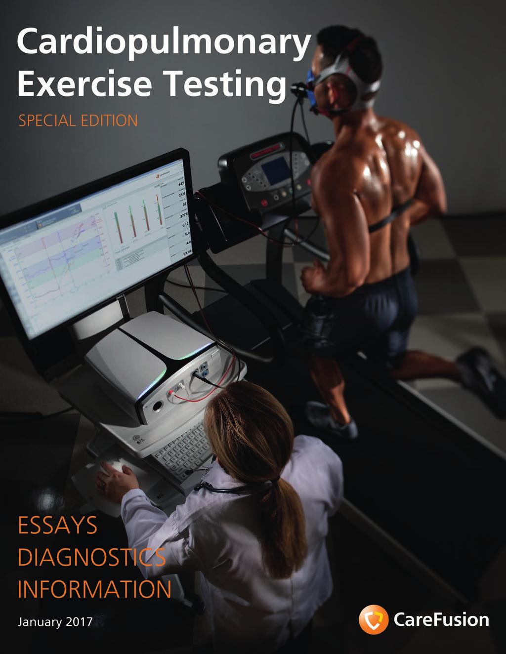
3 Editorial Relevance of Cardiopulmonary Exercise Testing Introduction Cardio Pulmonary Exercise Testing (CPET, sometimes abbreviated as CPX) is the determination of a person s performance during physical exercise by measuring, or calculating, the metabolic gas exchange alongside a number of other parameters. In order to perform a specific task, the body needs to provide the required energy. This energy is primarily produced by the breakdown of carbohydrates, fats and proteins in the presence of oxygen (aerobic metabolism). So in addition to fuel (through food intake), the body needs to provide the muscles with sufficient oxygen for this metabolic process. The rate of oxygen required increases with the intensity of the exercise. As with any other burning process, carbon dioxide (CO 2 ) is produced. This CO 2 is then transported from the muscle cells by the blood to the lungs, where it is removed from the body (via respiration). When exceeding a certain level of exercise, the body will not be able to provide sufficient oxygen to all of the exercising muscles. The additional required energy is then produced by means of the so-called anaerobic metabolism. Due to the limits of the anaerobic energy reserves, the body will only be able to exercise at this level for a short period of time until it is exhausted or these reserves are depleted. Anaerobic metabolism also results in the production of additional CO 2 which is discarded through the lungs driving increased rates of ventilation. Ultimately a number of finely tuned physiologic functions need to fit and interact together like a well-oiled machine, much like the impression the gear wheel model, created by Wasserman (simplified), illustrates so concisely below: Ambient air is inhaled via the lungs and some of the oxygen present in the air (oxygen uptake V O 2 ) diffuses through the lung membrane into the blood where it is absorbed by hemoglobin and delivered to muscles by the cardiovascular system (via the circulation blood). Once in the muscle, the actual breakdown of substrate takes place, providing the patient with energy and enabling the body to perform mechanical work (exercise). The CO 2 produced during that process is also absorbed into the blood, transported back to the capillary blood of the lungs, across the lungs membrane, and finally exhaled (carbon dioxide production V CO 2 ). Even from this simple description, it is easy to see that by measuring parameters such as ventilation, V O 2, V CO 2 and heart rate we can begin to determine the overall capacity of the system and start to pinpoint where any limitations may exist. The aim of a standard CPET protocol is for the individual to be exposed to a load using a bike ergometer or a treadmill and incrementally increase workload for about 8 to 12 minutes until they can go no further. These are often referred to as an incremental ramp protocol to a volitional maximum. During the test, the patient is connected via mask (or mouth piece) giving minute ventilation, breathing frequency, oxygen uptake, carbon dioxide production as well as other parameters and heart rate is measured from an ECG of the measuring system. This procedure makes it possible to determine the maximum exercise capacity as well as various thresholds such as the endurance capacity threshold (also see chapter Threshold Determination ). If an organ or organ-system is somehow impaired, the patient will fail to cope with the increasing load. In such a case, the characteristic patterns of the measured parameters can provide important information on which systems are affected by such impairment. Metabolic process V O 2 V CO 2 External Work Special Edition Cardiopulmonary Exercise Testing Page 3
4 Editorial In sports medicine / science, step protocols are often used in order to receive more precise information regarding the speed or power of the athlete. When measuring patients, on the other hand, a ramp protocol is usually preferred as this will allow the patient to approach the maximum load within an acceptable time range before the need to terminate the test due to exhaustion (and not due to the maximum exercise capacity). Thus, many aspects of cardiopulmonary exercise testing can be tailored to the individual s needs and capabilities. With CPET it is possible to receive significant information on single functions or limitations. This leads to the key application areas for cardiopulmonary exercise testing: Determining the individual exercise capacity Determining the severity of a performance limitation Determining the aerobic and anaerobic performance ranges Determining and analyzing the effect of therapeutic interventions and/or rehabilitation in patients with performance limitation Differential diagnosis regarding possible causes for a performance limitation such as - Pulmonary limitation - Malfunction of the gas exchange - Cardiac limitation - Peripheral limitation - Motivational limitation In contrast to simple stress tests, such as those performed with an ECG and a treadmill, CPET provides information on: test quality; allows for objective exercise capacity measurements; and points out causes for possible limitations. CPET also allows the possibility to assess the pre-operative risk for complications which may occur after a major surgery (such as lung or heart transplantation) more accurately. As a consequence, the postsurgical mortality rate can be reduced. The various parameters produced by a CPET can be broadly categorized into the following types: Measurement and Stress parameters Tidal volume (VT) Breathing frequency (BF) Inspired or expired oxygen concentrations (FIO 2, FEO 2 ) Inspired or expired CO 2 concentrations (FICO 2, FECO 2 ) Workload (Watt, respectively speed and elevation) Heart rate (HR, Stress ECG) Oxygen saturation (SpO 2 ) Calculated Parameters Respiratory minute ventilation (V E) Oxygen uptake (V O 2 ) Carbon dioxide production (V CO 2 ) Respiratory Exchange Ratio (RER) Oxygen pulse (O 2 -Pulse) Breathing equivalent (EqO 2, EqCO 2 ) Dead space ventilation (VD/VT) Breathing reserve (BR) Heart Rate Reserve (HRR) Further evaluation parameters such as Threshold determination (VT1, VT2, VT3) (for further information, please see chapter Threshold determination ) Maximum oxygen uptake (V O 2 max) Slope determination Aerobic capacity (dv O 2 /dwr) Ventilatory efficiency (V E(V CO 2 ) slope) Alveolar-arterial oxygen pressure difference (P(A-a)O 2 ) Page 4 Special Edition Cardiopulmonary Exercise Testing

5 Editorial Fields of Application The various parameters, measured and calculated, turn CPET into a comprehensive and highly informative method with applications in numerous fields of medicine: Respiratory Medicine Obstructive and restrictive ventilatory disorders Interstitial disorders Pulmonary hypertension Diffusion and distribution disorders Flow limitations Exercise related dyspnea of unknown origin Suspected limited exercise capacity due to circulatory or pulmonary vascular disorders Suspected exercise-induced asthma Trending for subtle respiratory disease changes Pre-operative risk assessment for lung transplant patients Cardiology Coronary heart disease Cardiomyopathy Heart disease, valvular heart failure Congenital cardiac defects Pre-operative risk assessment for heart transplant patients Cardiac insufficiency Occupational Medicine Exercise-related career proficiency tests Determining the degree of disability or work limitation/inability Fitness checkups (high altitude, air travel, tropical climate, diving) Intensive Care Pre-operative risk assessment Nutrition control (adjusting parenteral nutrition of intensive care patients) Rehabilitation Optimizing rehabilitative measures Assessing and documenting rehabilitative and therapeutic progress Nutrition Determination of Resting Energy Expenditure Energy Expenditure during Exercise Substrate utilization Nutritional counselling Dietary advice Sports Medicine / Science Measurement of physical exercise capacity Threshold determination Training management Quantification of training success Special Edition Cardiopulmonary Exercise Testing Page 5

6 Editorial CPET Evaluation Different evaluation and interpretation procedures are used depending on whether the subject is a healthy athlete or a patient with cardiac and/or pulmonary limitation. The following considerations cannot be considered comprehensive but are intended to describe only the main aspects of a CPET evaluation. For more detailed information, please refer to the list of additional literature at the end of this edition. When using CPET equipment it is desirable to be able to examine different parameters and graphs at different times, both during and after the measurement. The presentation of the data should be concise, comprehensive and systematic. The layout below uses the internationally recognized 9-Panel-plot according to Prof. Karlman Wasserman (Wasserman 2009). In 2012, the 9-Panelplot was updated (Wasserman (2012)). Some of the panels have been moved around, but the information content remains the same. The following 9-panel-graphic considerations refer to the original order. The software, of course, allows the user to choose between both alternatives, or create their very own 9-panel layout. The 9-Panel-plot offers a concise overview of the cardiovascular, ventilatory and gas exchange parameters: Ventilation 1, 4, Cardiovascular 2, 3, (4), Gas Exchange 4, 6, 9 The blue panels mainly illustrate ventilatory aspects, the red ones relate to the cardiovascular parameters and the green panels convey gas exchange information. Panel 1: V E and load against time Panel 6: EqO 2 and EqCO 2 against time Panel 2: HR and O 2 pulse against time Panel 7: VTex against V E Panel 3: V O2, V CO 2 and load against time Panel 8: RER and BR FEV% against time Panel 4: V E against V CO 2 Panel 5: HR and V CO 2 against V O 2 Page 6 Panel 9: PETO 2 and PETCO 2 as well as PaO 2 and PaCO 2 against time Special Edition Cardiopulmonary Exercise Testing
.")
 of a healthy individual increases linearly with the workload (in green) in an approximate ratio of 10 ml/w.")
7 Editorial Cardiology Aspects Panel 3 This panel illustrates the patient s general exercise performance and gives insight into oxygen delivery to, and utilization at, the exercising muscles. From this panel it is immediately apparent whether the subject has reached, or even exceeded, their expected exercise capacity (indicated by the hatched areas). If the expected exercise capacity is reached, it is safe to exclude a severe limitation. The oxygen uptake (in blue) of a healthy individual increases linearly with the workload (in green) in an approximate ratio of 10 ml/w. Providing the vertical axes are scaled to the same ratio (200 Watt = 2000 ml/min V O 2 or 200 Watt = 2.0 L/min V O 2 ) workload and V O 2 should increase in parallel to one another, though V O 2 will sometimes flatten upon reaching the peak work load. If flattening of the oxygen uptake occurs before the estimated exercise capacity (hatched area) is reached it is likely as a result of poor oxygen delivery to the muscles and evidence of cardiovascular limitation. It is not unusual for the V O 2 to flatten in athletes but this will occur at a level well above the expected V O 2 peak for a normal person. Furthermore, the panel provides information as to whether the peripheral muscle cells are utilizing sufficient oxygen. If this is not the case, the oxygen uptake will not increase linearly with increasing work load and will show a lower slope (less than 10 ml/w). Panel 2 This panel reveals information on the patient s heart rate (HR) and oxygen pulse (O 2 pulse). In healthy subjects, the heart rate is expected to rise with the increasing work load and will show a slight decline of the slope after some time, whereas patients with a cardiac impairment usually show a larger increase of the heart rate. With good cardiac function, the amount of oxygen transported per heart beat (O 2 pulse) is high and increases throughout the test. Patients with poor cardiac function, the oxygen transport can only be increased by additional oxygen extraction. The oxygen pulse will reach a plateau as soon as this maximal extraction is reached. Consequently, a further increase in work load will result in a disproportionate increase of the heart rate. Panel 2 of the 9-Panel-Graphics. Panel 5 Panel 3 of the 9-Panel-Graphics. The respective phases are marked by vertical lines, the dashed lines indicate the respective thresholds and the hatched areas indicate the predicted values to be reached. In healthy subjects, the heart rate against oxygen uptake trace (in pink) will increase linearly as illustrated in this panel. In general the main areas of aerobic conditioning (increased stroke volume; higher mitochondrial density in the exercising muscles; and increased capillarization of those muscle) result in increased oxygen delivery and utilization and as a result the heart has to beat less to deliver oxygen, this is seen as a lower HR versus V O 2 slope. In deconditioned subjects (e.g. with low stroke volume) the converse is true and if there is an acute cardiac impairment this will be reflected by a sudden, disproportionate increase in heart rate. Panel 5 also displays the V CO 2 against V O 2 slope allowing the user to determine the different thresholds via the V-Slope method (Beaver (1986)). Special Edition Cardiopulmonary Exercise Testing Page 7


. This will only occur provided there is sufficient breathing reserve to accommodate this hyperpnea.")
8 Editorial Panel 5 of the 9-Panel-Graphics Panel 4 Panel 4 provides cardiovascular information regarding, in particular, the pulmonary vascular circulation and for that reason will be described in detail both in the pulmonary as well as in the gas exchange section. Pulmonary Aspects With panels 1, 4 and 7 it is possible to assess the ventilatory performance. In order to determine the maximal ventilation, both tabular and individual predicted values are of significance. Panel 1 This plot presents minute ventilation (V E) and workload (Watts) against time. In healthy subjects, the ventilation initially increases in a linear fashion. As exercise continues the trace increases out of proportion as it passes the respective ventilatory thresholds, this is caused by the increase in anaerobically produced CO 2 and the ensuing metabolic acidosis (see Threshold Determination ). This will only occur provided there is sufficient breathing reserve to accommodate this hyperpnea. In subjects suffering from pulmonary disease it is useful to display the subject s maximum ventilation obtained by means of a forced spirometry measurement (usually calculated from 35 x FEV1) or a maximal voluntary ventilation maneuver (MVV) in order to detect ventilation limitation. Panel 1 of the 9-Panel-Graphics. The respective phases are marked by the vertical lines, the dashed lines indicate the respective thresholds and the hatched areas indicate the predicted values to be reached. A healthy subject requires an increase in ventilation (V E) of about 25 L per additional liter of CO 2. If dead space ventilation is increased and/or an impairment of the gas exchange is present, the ventilation must be increased in order to expel the same amount of CO 2. Increased dead space ventilation shifts this curve upwards without increasing the slope, while an impaired diffusion results in a steeper slope. In this panel, it is also possible to display the maximum ventilation (35*FEV1) next to the predicted values in order to easier determine whether there is any breathing reserve (BR). V E and V CO 2 are closely tied and as a result this relationship is highly linear for much of the test, but when it reaches the ventilatory thresholds VT2 and VT3 (provided VT3 has been reached) the slope increases due to the consequent hyperventilation. Panel 4 This panel demonstrates the relationship between minute ventilation (V E) and the carbon dioxide production (V CO 2 ). Page 8 Panel 4 of the 9-Panel-Graphics. Special Edition Cardiopulmonary Exercise Testing

9 Editorial Panel 7 This panel traces the changes in breathing pattern by plotting the expiratory tidal volume (V Tex ) against the minute ventilation (V E). Unusual values suggest the presence of an obstructive or restrictive disorder. Patients with flow limitations will try to breathe as deeply and as slowly as possible which will cause the trace to curve along the upper isopleth (the straight line from the origin, in this case representing a breathing frequency of 20 breaths per minute). If a restrictive lung disorder is present, the patient will quickly reach the maximum tidal volume due to the low vital capacity. Further increases in ventilation are through possible only by increasing the breathing frequency. As a result, the curve will reach an early plateau and then run horizontally to intersect the lower isopleth (representing 50 breaths per minute). In addition to the predicted value of V E, the MVV value (maximum voluntary ventilation) and / or the patient s predicted value calculated from FEV1*35 can be displayed in this panel to illustrate whether the patient has reached the maximum ventilation and whether a ventilatory impairment is present or not. Displaying the inspiratory capacity (IC) can also be useful as it corresponds to the maximum attainable tidal volume during the exercise. If the IC value has not been determined it can be approximated as 60% of the subject s vital capacity. By means of the EFVL measurement it is possible to immediately recognize a potential flow limitation of the subject which is indicated by the curve measured during exercise (blue) approaching or even slightly exceeding the maximum F/V curve (black) obtained from resting spirometry. F/V curve during exercise (blue) compared to the maximum F/V curve (black) If the EFVL curve is recorded several times during exercise, it is very easy to read if dynamic hyperinflation is present because the End-Expiratory Lung Volume (EELV) increases and/or if the subject exhausted his/her maximum tidal volume (End Inspiratory Lung Volume - EILV increases almost to TLC). Panel 7 of the 9-Panel-Graphics. EFVL Measurement Another pulmonary aspect is dynamic hyperinflation which can be clearly demonstrated by measuring the flow-volumecurve during exercise (Exercise Flow Volume Loop - EFVL). This feature was already implemented into our previous version of software at the beginning of However, it is unfortunately not (yet?) considered in the 9-Panel layout. Exercise Flow Volume Loops recorded during CPET displayed as a bar diagram Special Edition Cardiopulmonary Exercise Testing Page 9
10 Editorial Gas Exchange Panel 6 Both panel 4 as well as panel 6 provide important information on the gas exchange. Panel 6 displays the breathing equivalents for V O 2 and V CO 2 (EqO 2 and EqCO 2 ). Please note, although the equivalents are approximately the same, EqO 2 does not equal V E/V O 2 and EqCO 2 does not equal V E/V CO2. This is because the breathing equivalents need to be corrected for the apparative dead space, but despite this they are often incorrectly represented as V E/V O 2 or V E/V CO 2 in many publications. They give a measure of instantaneous ventilatory and gas exchange efficiency: How many L does the respective patient have to breath in order to uptake 1 L oxygen or to produce 1 L carbon dioxide? At the beginning of the measurement, the values are relatively high due to the high dead space to tidal volume ratio (VT low) and will decrease with the load as the tidal volume increases. EQO 2 will reach a minimum (indicating optimum efficiency) in the VT1 area, EQCO 2 between VT1 and VT2. Because of this, panel 6 can be used to help determining the ventilatory thresholds. A healthy person has a ventilatory demand of approximately L in order to absorb 1 L oxygen and needs to ventilate approximately L to release 1 L carbon dioxide. Elevated values indicate an inefficient gas exchange which can be caused by both an increased dead space ventilation and/or an impaired gas diffusion. released the higher the respiratory minute volume has to be). It corresponds (though not exactly) to the minimum EqCO 2 and is approximately 25 L/L for CO 2 in a healthy subject. Slopes of more than 40 L/L indicate additional cardiac impairment like pulmonary hypertension, thus this slope offers further valuable data for cardiologists. Panel 4 of the 9-Panel-Graphics. The respective thresholds are marked by the vertical lines. The inclining hatched area indicates the normal slope In addition to the slope, a shift of this curve is important as well: With an increased dead space ventilation the patient needs to breathe more frequently from the start. Since the dead space usually does not change with increasing ventilation (but rather decreases due to the higher breathing volume) an increased dead space ventilation is indicated by an upwards shift of the curve. Consequently, increased slopes indicate a diffusion impairment whereas a shift upwards is due to an increased dead space ventilation. Panel 9 Panel 6 of the 9-Panel-Graphics. The respective phases are marked by vertical lines, the dashed lines indicate the respective thresholds. Panel 4 The respiratory minute ventilation V E usually increases linearly until reaching VT2 as the respiratory drive is primarily determined by the production of CO 2 (the more CO 2 is Page 10 Further information on gas exchange as well as on threshold determination is provided in panel 9: This panel plots the end tidal partial pressure for O 2 (PETO 2 ) and CO 2 (PETCO 2 ), and if they are measured, the exercise blood gases (PaO 2 and PaCO 2 ). The end-tidal curves usually progress similarly (though reversed in case of PETCO 2 ) to the breathing equivalents displayed in panel 6: An initial decrease of PETO 2 is followed by an upwards defection at VT1 and VT2. PETCO 2 initially increases and flattens into a plateau at VT1 before decreasing at VT2 (and once again at VT3, provided that VT3 is reached). If blood gas values are taken, the end-tidal - arterial oxygen difference P(ET-a)O 2 can indicate a diffusion impairment. Special Edition Cardiopulmonary Exercise Testing
11 Editorial Positive P(a-ET)CO 2 values (which are normally negative during exercise) imply an increased dead space ventilation. Panel 8 of the 9-Panel-Graphics. Panel 9 of the 9-Panel-Graphics. Attention: 1. The PETO 2 progress is similar to PAO 2. However, the alveolar gas formula is required to accurately determine the P(A-a)O 2 gradient. 2. Some may approximate dead space ventilation and dead space ratio using PETCO 2 instead of using blood gas values. However, this method requires caution - it may work reasonably well in healthy subjects, but in patients with certain illness, it often can provide incorrect values (Lewis (1994)). Energy production, Metabolism Panel 8 Conclusions regarding metabolism can be drawn based on the RER (Respiratory Exchange Ratio, formerly known as Respiratory Quotient RQ) by means of the panel 8, which is the ratio of V CO 2 to V O 2. A mixed substrate metabolism of approximately 50% fat and 50% carbohydrates results in an RER value of around A value below suggests more fat oxidation, a value above suggests more carbohydrate oxidation. In the past, the anaerobic threshold was usually determined by means of RER=1. Today, however, this method is no longer used: VT1 is barely recognizable by means of the RER; VT2 can usually be found near RER=1. However, this value can be only used for a rough assessment of an upper limit of VT2 (please see Threshold determination section for more details). Note: In the past, RER (often also abbreviated as R) was usually termed RQ. The RQ, however, refers to the metabolism of the cell itself. Thus, the RER - which is measured at the mouth - replicates RQ only in Steady State due to the phase shift between V O 2 and V CO 2. References: Beaver W.L., Wasserman K., Whipp B.J.: A new method for detecting the anaerobic threshold by gas exchange. J Appl Physiology 60 (1986); Cooper C.B., Storer T.W.: Exercise testing and interpretation. Cambridge University Press (2001). ISBN: Jones N.L.: Clinical Exercise Testing. 4th Edition, W.B. Saunders Company (1997). ISBN: x Kroidl R.F., Schwarz S., Lehnigk B. Fritsch J.: Kursbuch Spiroergometrie - Technik und Befundung verständlich gemacht. 3. Auflage Thieme Verlag (2014). ISBN: Lewis D.A., Sietsema K.E., Casaburi R., Sue D.Y.: Inaccuracy of Noninvasive Estimates of VD/VT in Clinical Exercise Testing. Chest 106 (1994); Roca J., Whipp B.J.: Clinical Exercise Testing. ERS Monograph 6_2 (1997); ISSN x Rühle K.-H.: Praxisleitfaden der Spiroergometrie. 2. überarbeitete und erweiterte Auflage. Kohlhammerverlag (2008). ISBN: Ward S., Palange P.: Clinical Exercise Testing. ERS Monograph 40 (2007). ISBN: Wasserman K., Hansen J.E., Sue D.Y., Casaburi R., Whipp B..J.: Principles of Exercise Testing and Interpretation 3rd edition (1999) Lippincott Williams & Wilkins. ISBN: Wasserman K., Hansen J.E., Sue D.Y., Stringer W.W., Sietsema K.E., Sun X-G., Whipp B.J.: Principles of Exercise Testing and Interpretation 5th edition (2012). Lippincott Williams & Wilkins. ISBN-13: Weisman I.M., Zeballos R.J.: An Integrative Approach to the Interpretation of cardiopulmonary Exercise Testing. Clinical Exercise TestingProg Respir Res. Basel, Karger 32 (2002); Special Edition Cardiopulmonary Exercise Testing Page 11


12 Vyntus CPX - the Latest Product Generation The Vyntus CPX at a Glance Variable Configurations Variable Configurations are available such as: mobile cart configuration; table top configuration; and single or dual monitor setup, and as a result the system is easily customized to your individual needs. Combining it with a notebook will turn the system into a compact CPET station, reducing the footprint to a minimum. Vyntus CPX - Powered by SentrySuite The Vyntus CPX represents the new generation of Cardiopulmonary Exercise Testing and combines high measurement quality with ease-of-use and a workflow driven CPET evaluation. The Vyntus CPX is the result of over 50 years of experience in the development of CPET systems. The highly flexible system is suitable for various applications and can be easily used on a variety of subjects: from sick patients to top athletes and from children through to adults to old age. Thus, the Vyntus CPX can be used in a wide range of application fields. Furthermore, it ensures high-precision test results based on proven high-end sensor technology while its advanced functions offer useful support for interpretation of test results. The device is based on advanced technology and is the result of twelve generations of JAEGER, SensorMedics,... Viasys... CareFusion devices. It combines proven techniques with technical innovations and new medical results by offering additional measurement and evaluation procedures. Mobile Cart configuration with dual monitor setup Table top configuration with Notebook Vyntus CPX covers all essential CPET applications Breath-by-breath Cardiopulmonary Exercise Testing Slow and forced spirometry, MVV as well as Pre-/Post measurements and an animated incentive Flow/Volume loops during exercise (EFVL) with superimposed maximum flow volume loop New and original 9-panel-Wasserman-graph are both available along with Possible Limitation graph Ventilatory threshold determination (VT1, VT2 and VT3) Automatic slope calculation such as V O 2 (Watt), V E(V CO 2 ), V E(V O 2 ), HR(V O 2 /kg) Ability to edit all measurement ranges for baseline, warmup, peak, and recovery phases Indirect Calorimetry (REE, Fat...) using mask or mouthpiece Data input for RPE, blood pressure and comments Offline data input of blood gases with an automatic calculation of further parameters (P(A-a)O 2, VD/VT,....) Customizable workflow for CPET evaluation Comprehensive program for creating individual comments and interpretations including a helpful template manager Automatic control of bike, treadmill and blood pressure measurement Page 12 Special Edition Cardiopulmonary Exercise Testing


 The proven technology of the Digital Volume Transducer (DVT) meets the ATS/ERS guidelines for spirometry as well as passing all 24 wave forms.")
13 Vyntus CPX CPET Protocol Editor program for a flexible creation of ramp, step and weight dependent load protocols Report Designer program for a creation of customized reports including the possibility to export to Excel format at the touch of button Layout Editor to customize the graphical display during the measurement and after in evaluation mode. Combine Vyntus CPX with other devices or options: Integrated Nonin SpO 2 measurement with various sensor probes for the finger, forehead and ear Vyntus ECG: the fully integrated and wireless 12-Lead Bluetooth PC-ECG Polar Bluetooth Interface Choice of bike ergometers with/without integrated blood pressure measurements and treadmills Tango automated blood pressure monitor Indirect calorimetry using the dilution canopy method Measurement with high/low oxygen breathing Compatible with a large number of 3rd party ECGs Blood gas analyzer interface for serial import of blood gas data Digital Volume Transducer (DVT) The proven technology of the Digital Volume Transducer (DVT) meets the ATS/ERS guidelines for spirometry as well as passing all 24 wave forms. It is an accurate and reliable sensor for the complete flow range from low flows to maximum voluntary ventilation. Thanks to its compact and lightweight design (45 g only), the sensor has a very small dead space of only 30 ml. The DVT is insensitive both to water vapor and expired gas mix. As compared to a turbine, the flat vane system has no lag due to its small inertia. Patients and athletes will appreciate the fact that it adds minimal resistance to airflow and it is extremely comfortable to wear with both mask and mouthpiece. Different mask sizes and types (adult and pediatric, reusable or disposable) ensure best fit for each subject and ensure you can provide the highest level of clinical hygiene. Optional Workflow Applications Questionnaire Designer and patient questionnaire application for tablets Networking with further PFT systems and workstations for evaluation, interpretation and central data storage Web-based evaluation and interpretation of PDF reports via Sentry.NET Interface with hospital and medical practice systems Electronic Patient Records (EPR) interface through SentryConnect Interface DVT with mask DVT with mouthpiece Special Edition Cardiopulmonary Exercise Testing Page 13

 O 2 fuel cell can easily and quickly")
14 Vyntus CPX The heart of the system - the high-precision and proven O 2 /CO 2 Analyzer USB port to connect the PC and for in-field firmware upgrades Robust high value materials with long time resistance against disinfection fluids and easy to clean Port/Blower for unique, fully automatic volume calibration Additional built-in highly effective gas drying mechanism Status lights for continuous information about your system and automated self-check Port for future options Integrated SpO 2 measurement Proven Digital Volume Transducer (DVT) for exact determination of ventilation 2.4 m Twin Tube sample line for maximal freedom of movement Robust color coded medical connectors O 2 cell change - made easy The long-life (approximately 2 years) O 2 fuel cell can easily and quickly be exchanged at customer s site in only about a minute. All that is required is a coin to open the fuel cell door on the back of the Vyntus CPX. Take the old cell out and put the new one in. A fully automatic filter optimization system ensures measurement continuity after the cell is exchanged. and a volume sensor calibration will be automatically performed using the integrated blower. The special Twin Tube (TT) sample line and the fresh air flush system allow to perform a gas analyzer calibration without disconnecting the sample tube. Additionally, the easy and fully automatic click-and-play 2-point gas calibration of the O 2 / CO 2 analyzers determines the delay and response times for the exact synchronization with the volume signal in one procedure. High accuracy and stability Accurate and stable measurements, even during long exercise measurements, are guaranteed by: the special drying system with pre-drying via the Twin Tube; an additional arrangement inside the Vyntus CPX to remove the remaining humidity; and fast response gas analyzers (typical T 10 -T 90 = 75 msec). Calibration couldn t be easier The Vyntus CPX is equipped with a unique, fully automatic volume calibration unit - making a manual 3 Liter calibration syringe unnecessary. Just one click in the SentrySuite software Page 14 Flexibility The Twin Tube sample line with a length of 2.4 m offers maximum freedom of movement for the patient even with measurements performed on a treadmill. Special Edition Cardiopulmonary Exercise Testing


15 Vyntus CPX Software Vyntus CPX - the Software The Main Screen - a 360 working interface The SentrySuite CPET Software is designed for simplified CPET testing, which can be perfectly customized to the individual needs and capabilities of your current patient. The convenient and user-friendly software interface allows easy and effective control of the measurement procedure by providing a clear overview of test and equipment controls. Furthermore, it offers valuable support for an effective interpretation of the test results. All important programs such as patient data, calibration, measurements, and even reports can be selected directly from the same screen. Bring up personal data of the current patient, enter new patient data or search for specific patient data already available in the database Switch to other measurement programs Show, print and save reports Bring up various calibration programs Main screen after selecting the CPET measurement program. With just one click it is possible to directly switch to various programs such as patient data, calibration, spirometry, report, or to start a new measurement without leaving this window. Special Edition Cardiopulmonary Exercise Testing Page 15
 Automatically and individually calculated max.")
 and estimated maximum load")


16 Vyntus CPX Software The StartUp-Window All connected and activated devices are checked. (Green status icon: correct connection; red: incorrect connection) Automatically and individually calculated max. predicted values for the current patient. Interpretation of the final measurement results will be based on these values. Adjust masks and averaging settings Edit and create load profiles Estimated maximum minute ventilation (V Emax) and estimated maximum load calculated from values measured in a prior spirometry measurement ( Measured PFT Data ) to avoid the selection of an inappropriate load profile in case of a ventilatory limitation. If no measurement was performed previously, this field remains empty. Selection of default load profiles. Depending on the settings, the system automatically proposes the profile which comes closest to the patient s maximum load (predicted load value) - resulting from the comparison of the predicted load value with PFT maximum load - or the load profile set as standard. The types of devices and inputs supported by the SentrySuite CPET software are divided into the categories: Main Device, Ergometer, Heartrate, Blood Pressure, and O 2 -Saturation. These devices are set in the StartUp-Window prior to starting a measurement. In addition, the user can select from various masks and averaging methods. A wide range of preset load profiles containing many possible combinations of ergometer type (bike or treadmill) and load protocol (ramp or step load) as well as different settings for each are available from the same window. Furthermore, it is possible to create new protocols or to edit pre-existing profiles. Page 16 Special Edition Cardiopulmonary Exercise Testing


 in numbers Performance graph Display RPE Scale Spirogram This table provides a convenient")
17 Special Functions during Cardiopulmonary Exercise Testing Vyntus CPX Software It is easy to control all aspects of the test procedure during the measurement by selecting the appropriate button in the left-hand button bar. Furthermore, the user can quickly switch between various displays according to their individual preference, zoom in and out each graph, and/or manually advance to the next phase. If not already set in the pre-settings of the respective load profile, it is also possible to start an EFVL measurement, to activate the RPE scale or mark an blood-gas sampling event at any point of time during any phase with just one single click. Advance to the next phase 9-Panel-Graphics according to Wasserman Manual load change Start EFVL measurement Mark events Switch from the performance graph display to the load graph display. The hatched area indicates the patient s target performance range (determined from previously calculated individual predicted values). Currently measured values (predicted and actual values) in numbers Performance graph Display RPE Scale Spirogram This table provides a convenient overview of the measurement process. Upcoming events including their times are displayed enabling the user and the patient to get prepared. The displayed time is a countdown showing time (in minutes and seconds) remaining for the event to take place. The displayed events and phase duration depend on the settings in the selected load profile. Special Edition Cardiopulmonary Exercise Testing Page 17
), Breathing Reserve (BR FEV%) and Respiratory Exchange Ratio (RER) : The")
. If large reserves are available, the load can correspondingly be increased.")
18 Vyntus CPX Software Everything at a glance SentrySuite optimizes test efficiency by means of concise graphical overview. The performance graph clearly displays the degree of the maximum load with regard to Load, Heart Rate Reserve (HRR (B)), Breathing Reserve (BR FEV%) and Respiratory Exchange Ratio (RER) : The numerical display of parameters allowing the user to easily and quickly read both actual and predicted values is perfectly supplemented by the graphical display of the respective parameters. The filling bars indicate the current performance of the subject (grey areas). If large reserves are available, the load can correspondingly be increased. Graphical display of the actual and predicted value(s) In this graphic, the user is able to see the parameter values V O 2, V CO 2, load and HR from the current phase as well as from the previous phases at a glance. The vertical lines indicate the start of the respective phase. Additionally, the hatched area indicates the predicted target load range for a normal subject or patient. Actual and predicted load value (Watt) Actual and predicted heart rate Actual and predicted oxygen uptake It is also possible to record heart rate (with or without ECG) during the exercise test. With its fully integrated 12-lead Vyntus ECG for rest and stress ECG, CareFusion offers the optimum solution for this purpose: a complete ECG recording on a second monitor or as a single graph (using a single monitor). Alternatively, the heart rate can also be recorded via the integrated SpO 2 sensor, a Polar chest strap or other, combinable 3rd party ECG systems for a comprehensive CPET measurement. Page 18 Actual carbon dioxide production Actual Respiratory Exchange Ratio Actual systolic blood pressure Actual diastolic blood pressure Elapsed time (total) Numerical display of the actual and predicted values. It is possible to customize both parameter selection as well as parameter sequence. Special Edition Cardiopulmonary Exercise Testing


 EFVL")


19 Vyntus CPX Software - Evaluation Results and Interpretation The ability to switch between various graphical displays in the result screen provides the user both assistance and a selection of various approaches for interpretation. Many of the displays may also be customized to the users own preferences. 9-Panel-Graphics according to Wasserman (2012) EFVL measurement results Measurement results displayed numerically BR/HRR against time Possible Limitations V O 2, V CO 2, HR and Load against time The Guidance tab provides textual assistance Workload graph Automatic interpretation and classification of the measurement results User comments/interpretation Special Edition Cardiopulmonary Exercise Testing Page 19
.")
 and Heart Rate Reserve (HRR) against time If you move the dashed line (with diamond in the centre) to the left or to the right in one of the two windows, the values displayed in the tabular")
20 Vyntus CPX Software - Evaluation Graphical Display of the Results In the Result screen, the data is displayed numerically as well as graphically, and can be shown as either time or breath averaged. It is also possible to select additional displays in the right-hand graphic (BR/HRR). The left window displays: V O 2, V CO 2, Heart Rate (HR) and Load (Watt) against time The hatched areas mark the individual predicted areas of the subject The right window displays: Breathing Reserve (BR FEV%) and Heart Rate Reserve (HRR) against time If you move the dashed line (with diamond in the centre) to the left or to the right in one of the two windows, the values displayed in the tabular data will change according to the new position. If combined with Vyntus ECG, ECG tracing will also move to newly displayed position. The vertical lines in the charts display the different markers during the measurement; e.g.: W = Start of the warmup phase T = Start of the exercise phase R = Start of the recovery phase It is possible to add additional markers. The points VT1, VT2 and VT3 define the ventilatory thresholds. (For more information on the ventilatory thresholds please see the Threshold Determination chapter in this special edition.) Page 20 Special Edition Cardiopulmonary Exercise Testing
21 Vyntus CPX Software - Evaluation Every visible segment in a bar corresponds to a parameter. If the parameter reaches a limit value which is indicative of a limitation, the respective segment will be highlighted in red. If a limitation can be excluded due to the measured values, the respective segment in the respective limitation bar will be marked green. White segments indicate normal values whereas potential grey highlighted segments point out that an evaluation of the respective parameter is not possible, usually due to missing blood gas values (for example to calculate P(A-a)O 2 ). EFVL - Flow/Volume during exercise Besides the aforementioned display of predicted values, numerical and graphical measurement values, and 9-Panel- Graphics, the software offers further valuable graphics and tools which significantly simplify and support the evaluation of results. These additional features are: Possible Limitations The possible limitation chart indicates and excludes possible and specific diseases as the cause of cardiopulmonary limitations. However, it must be kept in mind that the possible limitations are only a suggestion based on the measurement data and need to be verified by the user (modified according to Weisman (2002)). The SentrySuite CPET Software checks the measurement results for the following possible limitations: Heart failure COPD Interstitial lung disease Pulmonary Vascular disease Obesity Deconditioning Subjects with limited lung function can only be subjected to physical exercise to a limited extent and therefore need to be observed carefully during a CPET measurement. An EFVL measurement (Exercise Flow Volume Loop) allows to supervise these patients and to decide whether to continue the exercise or terminate the test. It also indicates important aspects on when or whether a lung function disorder prevents further exercise. Measured Flow-Volume loops (superimposed) Flow-Volume Curves recorded during CPET displayed as a bar diagram Special Edition Cardiopulmonary Exercise Testing Page 21
. Just click Next to move from one step to another.")
22 Vyntus CPX Software - Evaluation Vyntus CPX Evaluation Workflow - easy to use from beginners to experts After the measurement, the evaluation workflow will automatically guide you step-by-step through post-test editing (also refer to Edit Mode ). Just click Next to move from one step to another. This procedure standardizes your evaluation/interpretation and reduces your time to produce a result. Depending on the use, it is possible to create different workflows for different users or user groups. The program will support you with threshold determination or slope calculation by means of intelligent evaluation. The final decision however, is up to the interpreting clinician. The entire workflow includes Entry of End of Test Criteria, manually or by means of predefined texts Editing the ranges of rest, warm-up, test and recovery phase Editing the ranges for slope determination purposes Editing the ventilatory threshold VT1 Editing the ventilatory threshold VT2 Editing the ventilatory threshold VT3 Editing the measured EFVL (Exercise Flow/Volume Loops) Editing RPE / Entering or editing markers, blood gases, RPE values... Editing Steady State measurements Editing the measured EFVL Example Workflow: Editing VT2 Editing the slope ranges Editing the phase ranges Page 22 Special Edition Cardiopulmonary Exercise Testing


23 Automatic Interpretation and Classification of the Measurement Results Vyntus CPX Software - Evaluation The Auto Interpretation tab provides an automatic textual interpretation of the measurement results: In addition to the textual interpretation, a classification of the test results is displayed. The classification is based on the predicted value of maximum oxygen uptake (Löllgen (2010)): With Auto Interpretation, it is possible to choose between several authors for a suggested interpretation. The respective measurement program saves the selected author as the standard author for the next examination. Among others, the authors CPET Eschenbacher, Mannina (1990) - Eschenbacher (1990) are available. Note that auto-interpretation does not substitute for medical advise, provides only support for qualified personnel and shall always be reviewed by a physician. Excellent = V O 2 max % Pred 120 Normal = 85 V O 2 max % Pred < 120 Mild = 70 V O 2 max % Pred < 85 Moderate = 50 V O 2 max % Pred < 70 Severe = V O 2 max % Pred < 50 User Comments/Interpretation The Interpretation/Comments tab allows the user to enter individual comments and/or interpretations manually. It is possible to load various templates and macros or to customize texts entered manually by choosing between various layout features. This allows the flexibility to quickly create comprehensive customized reports on the day of the test. This can be taken to the extent where patient clinic letters can be completed within the software without having to resort to additional dictation and letter writing. Choose standard text modules under Templates or compose an individual text. According to the template, the corresponding measurement values will be imported directly from the measurement and incorporated into the text. Thus, an entire summary can be created with one single click, which - if necessary - can be edited or extended. Both graphics as well as measurement and evaluation parameters can be transferred to predefined or user-generated reports. Easy export of the data into Excel for further processing is also possible. Special Edition Cardiopulmonary Exercise Testing Page 23

24 Vyntus CPX Software - Edit Mode Edit Mode It is not only possible to edit the automatic workflow at the end of a measurement, but single sections may also be edited by means of the Edit Mode, which shall only be described as an example in the following. Threshold determination As pointed out in the later section Threshold Determination (see chapter Basics and Diagnostics ), there are different procedures for the determination of the respective thresholds. These shall be discussed by means of VT1: At the end of a measurement the program tries to mathematically determine the different thresholds (break points) within the specified white area and marks them: Orange: Break point in the V-Slope Graphic (V CO 2 (V O 2 )) Light blue: Break point in EqO 2 (Time) - in this example superimposed by the red point. Red: Break point in V CO 2 (Time) As those break points are usually not identical, the average of all determined break points is displayed (vertical blue line). Furthermore, the program tries to confirm the break points by means of the regression line. In case the users do not agree, they are free to modify the white areas to initiate a recalculation or alternatively manually shift the blue line to the position they believe the threshold to be. The corresponding data will also be displayed numerically in the table at the top. For a better evaluation, each graph can be expanded to full screen just by one mouse click. Page 24 Special Edition Cardiopulmonary Exercise Testing
-Slope). This calculation is automatically performed already at the end of a measurement. Via the edit mode, it can be checked, and if applicable, edited.")
25 Vyntus CPX Software - Edit Mode Slope Calculation As already outlined in the introduction, various evaluations also require the calculation of the dynamic behavior of parameters (e.g. V E(V CO 2 )-Slope). This calculation is automatically performed already at the end of a measurement. Via the edit mode, it can be checked, and if applicable, edited. The 4 most important slopes calculated by means of the white areas are the following: Top left: Aerobic capacity (V O 2 (Watt)) Top right: Respiratory efficiency for CO 2 (V E(V CO 2 )) Lower left: Respiratory efficiency for O 2 (V E(V O 2 )) Lower right: Cardiovascular efficiency (HR(V O 2 /kg)) In this mode, the user can modify the pre-set white areas and thus initiate a recalculation. The corresponding data will also be displayed numerically in the table. References: Eschenbacher W.L., Mannina A.: An algorithm for the interpretation of cardiopulmonary exercise tests. Chest 97 (1990); Löllgen H., Erdmann E., Gitt A.K.: Ergometrie, 3. Edition. SPRINGER (2010). Weisman I.M., Zeballos R.J.: An Integrative Approach to the Interpretation of cardiopulmonary Exercise TestingClinical Exercise Testing Prog Respir Res. Basel, Karger 32 (2002); Special Edition Cardiopulmonary Exercise Testing Page 25


26 Vyntus CPX - Options Vyntus ECG The Vyntus ECG is wirelessly connected to a PC or notebook via Bluetooth and allows for a recording of a 12-lead resting as well as stress ECG. Excellence in diagnostic and prognostic value in a powerful combination Exposing the heart to increased workloads is often the only way to detect cardiac abnormalities. Consequently, the combination of heart and lung parameters is essential for comprehensive cardiopulmonary exercise testing. The Vyntus ECG allows for a 12-lead stress ECG recording while automatically evaluating and analyzing the signals. Detected abnormalities such as extrasystoles or pacemaker control are displayed on screen during the measurement. The user can modify Speed Gain Lead selection Define print areas Print an on-line report during the measurement Additionally, the Vyntus ECG provides a Full Disclosure feature for saving the unfiltered, continuous ECG signals. Via SentrySuite, the 12-lead Vyntus ECG integrates fully and seamlessly with the Vyntus CPX system. This enables laboratories to leverage their medical devices as well as Healthcare IT investments and provides an easy and clear interpretation of the measurement results. Further benefits: One user interface One program to train One central database One combined report One network interface One HIS-connection Vyntus ECG - the art of diagnostic integration The Vyntus ECG is intended for measuring the surface ECG of the patient. It communicates wirelessly and directly via Bluetooth and integrates conveniently with the Vyntus CPX system. Patients will appreciate the wireless technology, the small and light design of the amplifier and the short electrode cables which improves comfort and providing maximum freedom of movement. If an untoward event occurs during testing, the wireless connection of the ECG permits easier movement of the patient to a table or chair, while maintaining constant ECG collection and display. Additionally, the all-inone view ensures a user-friendly interface. The acquired ECG can be displayed on the screen or conveniently be printed on paper. Page 26 Special Edition Cardiopulmonary Exercise Testing


) is compiled.")
27 Vyntus CPX - Options ECG Recording As soon as all electrodes are connected, the minimal fast potential differences originating from the heart can be detected on the body surface and subsequently be recorded by the Vyntus ECG. At the beginning of the measurement, the electrode contacts are checked automatically. Stress ECG The stress ECG application offers an attractive graphical user interface and leaves nothing to be desired: Stress ECG during the measurement The green electrodes displayed in the screen indicate good contact. If a contact is poor, the respective electrode flashes orange. The ECG recording starts automatically as soon as the electrodes and the skin are in good contact. Resting ECG If required, several resting ECG trials can be recorded and compared to each other (similar to spirometry). In addition, a proposed interpretation according to HES (Hanoverian ECG Interpretation system, Willems (1991)) is compiled. As well as the continuous recording of the single leads, the complexes, including the appropriate ST values, are displayed on the left. The lower left area of the screen shows a full disclosure recording with potentially present abnormality markers. Both recordings can be paused and scrolled back during the measurement offering a close look at previous signals. The complex shown at the lower right screen section is displayed with the reference signal including the appropriate numerical abnormalities and can be customized by the user for speed, gain and lead selection. All ECG raw data is recorded and saved during the entire exercise measurement. Resting ECG after recording with an interpretation according to HES as well as a classification The HES program was part of the Common Standards for Quantitative Electrocardiography project, CSE. The results were independently analyzed in Willems J.L et al.: The diagnostic performance of computer programs for the interpretation of electrocardiograms. N Engl J Med. 25_325 (1991); Special Edition Cardiopulmonary Exercise Testing Page 27


28 Vyntus CPX - Options Vyntus CPX - High / Low FIO 2 Option (Not available in US) A powerful extension This option allows the user to make measurements while the subject breathes increased or decreased concentrations of inspired oxygen. For this, a Y-valve is connected to the volume sensor (which is also connected to the gas sample tube) permitting the subject to inhale the prescribed oxygen concentration from a reservoir and allowing a CPET measurement to be performed simultaneously. Measurement principle; the breathing bag containing the oxygen concentration to be inspired can either be refilled via a gas cylinder or by means of an appropriate blender. Arrangement of the individual parts; the measurement can either be performed with a mask or with a mouthpiece. A head-gear for support is available as well. High FIO 2 Subjects suffering from a ventilation-perfusion disorder (e.g. transplant patients, idiopathic pulmonary fibrosis, severe COPD) are often not able to handle everyday life without supplemental oxygen. In order to at least perform some simple tasks, such as moving around home or taking a walk, they are often equipped with a portable nasal oxygen supply. To examine those subjects exercise capacity, the patient must be supplied with additional oxygen during the measurement. However this is not possible using nasally supplied oxygen, because as ventilation increases with exercise, it would have the effect of diluting the oxygen and consequently lowering FIO 2. In this situation wash in or wash out effects would cause the user to measure the superimposition of oxygen uptake and wash out effects (or wash in effects, respectively) rather than the actual oxygen uptake. In order to avoid this problem, the subject is provided with a constant FIO 2 concentration (typically 30 % - 40 %) via the breathing bag during the measurement. The measurement procedure is otherwise similar to a normal BxB measurement. Additionally, this option considers the additional dead space caused by the Y-valve. With these measurements, special attention must generally be paid to the prior washing in of the lung and the blood (this is evident when V O 2 is too high and RER is consequently too low) until balance is reached. If a severe ventilationperfusion disorder is present, this can take up to 10 minutes. Only then the actual measurement and exercise should be started. Behind the scenes, however, the software applies the Eschenbacher transformation (Eschenbacher (2016)) for the calculations as the Haldane transformation (Haldane, (1912)) does not provide plausible and reliable data, especially at high FIO 2 values (also refer to chapter Haldane and Eschenbacher transformation ). Page 28 Special Edition Cardiopulmonary Exercise Testing


29 Vyntus CPX - Options Low FIO 2 The same procedure can be used to reduce the inhaled oxygen concentration. The oxygen uptake strongly depends on the oxygen partial pressure PAO 2 in the lungs and therefore on the environmental partial pressure PIO 2. Instead of performing a measurement at an altitude of 2500m, an identical PIO 2 can be established in a laboratory by reducing the FIO 2 (at sea level) to approximately 15.5 %. Doing this offers the ability to simulate altitude and examine a patient s response to altitude within the safe environment of a laboratory, for example: will they desaturate on an airplane during a flight; or will an athlete respond well to the effects of high altitude training. Low FIO 2 used to simulate high altitudes The current air pressure Pbar and the oxygen concentration FIO 2 result in PIO 2 [kpa] = Pbar [kpa] * FIO 2 [%] /100 Consequently, the partial pressure of environmental oxygen (and, with it, the oxygen partial pressure in the lungs) strongly depends on the environmental air pressure and thus on the altitude. From the above equation a low PIO 2 can be achieved by reducing either the air pressure or the inspired oxygen concentration. The relationship between altitude and FIO 2 can easily be estimated and is shown in following table: Altitude Pressure FiO 2 [m] [hpa] [%] % % % % % % % % % % % % % % % % % % Correlation between altitude, air pressure and oxygen concentration A modern passenger plane has a pressure complying with about 2500 m (or ca % O 2 ). The measurement procedure is almost identical to a BxB measurement with an increased oxygen supply; the only difference being that the breathing bag is filled with a reduced oxygen concentration. Again, special attention must be paid to the prior washing in of the lung and blood (V O 2 too low, RER too high) until equilibrium is reached. Only then the exercise should start and accurate measurement can be taken. Furthermore, it should be noted that with both a high and low FIO 2 the adjusted oxygen concentration needs to remain constant during the entire measurement in order to avoid wash in and wash out effects during the measurement. References: [1] Haldane J.S.: Methods of air analysis. Charles Griffin & Co., Ltd., JB Lippincott Co., Philadelphia (1912). [2] Eschenbacher H.: Haldane and Eschenbacher transformation. White Paper RD5693A (0716/PDF). CareFusion (2016). Special Edition Cardiopulmonary Exercise Testing Page 29


, the plug-in rings (3) used to attach the")
, used to draw the sample gas through the system, can be disassembled and cleaned (7) but the")


30 Vyntus CPX - Options Vyntus CPX - Canopy Option (Not available in US) Canopy - Indirect Calorimetry Indirect calorimetry measurements can be made with a face mask or a mouth piece using breath by breath technology, however the dilution canopy option improves subjects comfort: This dilution canopy method is a proven and patient-friendly method to determine the resting energy expenditure. During the development of this option, increased focus was placed on our cleaning and hygiene concept, which is becoming a progressively more important topic within the hospital and laboratory environment. By means of an innovative, patented dilution system including a single use canopy (1), the plug-in rings (3) used to attach the transparent foil to the holder (2) are the only parts which need to be cleaned and disinfected between the measurements. Parts of the powerful Canopy module (6), used to draw the sample gas through the system, can be disassembled and cleaned (7) but the design allows for the additional application of a bacterial filter (4) to avoid contamination of all downstream parts (5, 6 and 7) including the DVT The dilution system flow can be varied over a wide range reaching from approx. 25 L/min to approx. 80 L/min. It can also be adapted to a specified value or automatically be controlled by the software. Thus, the dilution flow can automatically be adapted to the patient by means of the measured CO 2 concentration. The blower-system is constructed so, that the exhaled air from the patient does not come into contact with the blower itself. Page 30 Special Edition Cardiopulmonary Exercise Testing


31 Vyntus CPX - Options Measurement and Evaluation The measurement procedure for Canopy mode is similar to a BxB measurement. However, the CPET-startup window also shows the predicted values for resting energy expenditure as well as fan control. In the section beneath, the SentrySuite software uses patient data to propose the initial flow to be used for the measurement. Additionally, the user can select whether to perform the measurement using a specified value for V E or if the software should control the flow automatically (these settings can also be modified during the measurement). As with BxB, you can select various profiles to automatically run additional features such as: zeroing of the gas analyzers during longer measurements; taking blood pressure; or blood gas entry during the test. During the measurement, flow, FECO 2 and SpO 2 are constantly monitored and controlled. If a deviation from the predicted values is detected, a window will point this out and offers the user the option to intervene. As soon as the measurement is finished, the data is clearly displayed both numerically and graphically. The user has the ability to discard or summarize single areas for evaluation purposes. If required, the measured urinal nitrogen value can be entered for further calculation. Among the usual parameters such as V O 2, V CO 2, RER and V O 2 /kg, the following parameters are also of interest: EE (Energy Expenditure) nprer (non protein RER) Division of energy production and substrate consumption in - Carbohydrates - Fats - Proteins (with the entry of urinary nitrogen) regarding g/day, percentage of the substance, percentage of energy As outlined in chapter Indirect Calorimetry, there are various calculation formulas the user can generate via the software settings. Special Edition Cardiopulmonary Exercise Testing Page 31
32 Basics and Diagnostics Threshold Determination An important aspect of cardiopulmonary exercise testing is the determination of the different thresholds in order to identify, for example the anaerobic threshold, the respiratory compensation point or the Steady State. The respective thresholds are usually displayed as break points in the CPET graphical displays. Unfortunately, ambiguous terms and abbreviations exist in literature which can lead to confusion, misunderstanding (Binder (2008)) or even an incorrect interpretation. As an example, Wasserman (2012) refers to the first break point as the anaerobic threshold (AT), whereas it is called the aerobic threshold (AE or AeT) in the field of sports medicine/ science (for example Kindermann (2004)). The same applies to the second break point: Wasserman describes it as the Respiratory Compensation Point (RCP), sports medicine, however, usually refers to it as the anaerobic threshold (AT, IAT or AnT). The classification according to Weber (1997) was also referred to as the anaerobic threshold but was defined to be the point at which RER = 1. This point is located near the Maximal Lactate Steady State (MLSS) (or RCP, according to Wasserman) as well rather than near the anaerobic threshold according to Wasserman (AT). In sports medicine, lactate is often used to identify the different aerobic and anaerobic areas, since it is easy to determine. This offers the ability to quickly evaluate training success (even in the field) and to compare laboratory measurements with field tests by combining CPET and lactate determination. Figure 1 schematically shows the classification of the lactate and ventilatory thresholds. Unfortunately, confusion about the terminology is present here as well: Wasserman defines the beginning of the lactate increase as the so-called lactate threshold LT (and as anaerobic threshold), whereas e.g. Mader (1976) refers to the anaerobic threshold with a lactate value of greater than 4 mmol. This has been improved upon with methods such as the determination of MLSS or further anaerobic lactate thresholds (e.g. Heck (1985), Dickhut (1991), Stegmann (1981), Pokan (2004) and others). Accordingly, LT is located near the first break point (VT1 or AT, according to Wasserman), MLSS near the second break point (VT2 or RCP, according to Wasserman). Unfortunately, neither the second nor the third (if reached) break point can be recognized directly in the lactate curve. Lactate LT MLSS Load/Time Fig. 1: Connection between lactate and ventilatory thresholds Training ranges are usually specified by means of VT2 (AT according to sports medicine, RCP according to Wasserman). However because of the confusion mentioned above some therapists/trainers accidently use the anaerobic threshold according to Wasserman for the creation of a training schedule which are consequently at too low intensity and effectively useless. In 2012, a major CPET working group (Westhoff 2013) decided to refer to the thresholds as VT1 and VT2 according to their emergent order rather than continuing to name them aerobic, anaerobic or respiratory compensation point in order to avoid this confusion in the future. These terms seem to have gained wider international acceptance which prompted us to implement these definitions into all our current software versions. Additionally, it is possible to recognize a third break point (VT3), primarily in high-performance athletes. So far, this point is hardly described in literature and its meaning is still not entirely clear (see some notes further below). Nevertheless we implemented this point as VT3 in our software. Sport medicine/science occasionally refers to this point as Respiratory Compensation Point, Panic breathing or as Hot ventilation. Ventilation VT1 VT2 VT3 Load/Time Page 32 Special Edition Cardiopulmonary Exercise Testing
33 Following table shows a summary of the various terms and how they are implemented in our programs: SeS, JLAB JLAB <V 5.72 Binder Wasserman Sports medicine Lactate VT1 AE AE AT AeS, AeT, 1. Lactate threshold (Beginning of the lactate to increase), LT VT2 AT AT RCP AnT, IAS, MLSS, IAT, Lactate threshold, MLSS (ca. 4 mmol) or various other methods such as Dickhut, Stegmann etc. VT3 RCP???? Sometimes referred to as Hot Ventilation (HV), sometimes labelled RCP Fig. 2: Summary of the various threshold terms Basics and Diagnostics -- The respective abbreviations stand for: VT1, VT2, VT3 = First, second and third ventilatory threshold AE, AeS, AeT = Aerobic threshold AT, AnT = Anaerobic threshold IAS, IAT = Individual anaerobic threshold LT = Lactate threshold (beginning of the lactate to increase) MLSS = Maximum Lactate Steady State RCP = Respiratory Compensation Point HV = Hot Ventilation ( extreme hyperventilation) As already mentioned and as it can be seen in the table above, different and partially misleading terms are used for the lactate thresholds as well. It is therefore recommended to use the term (analogically for the ventilatory thresholds) LT1 for the beginning and LT2 for the second threshold. Furthermore, it should be noted which of the more than 60 different concepts have been used for the lactate threshold determination. With Cardiopulmonary Exercise Testing it is possible to recognize the respective thresholds by means of more or less distinctive break points. Depending on the selected parameters they can vary from: obvious; to ambiguous; to not apparent, in the various graphical displays. For this reason, it is recommended to observe several plots simultaneously. For clarity, it should be noted that the thresholds we usually refer to are in fact transition areas which do not appear as an individual data point but appear as a transition area due to the body s different control mechanisms. Therefore, it is possible that these points can seem to occur in slightly different positions from one graph to another. The averaging of the individual parameter is significant as well (for example: over 8 breaths; 30 seconds or no averaging at all). According to our experience, a moving average over 8 to 10 breaths has proven its worth. Details of the appearance of the respective break points shall be given in the following. Special Edition Cardiopulmonary Exercise Testing Page 33
34 Basics and Diagnostics Ventilatory Threshold VT1: Typically, we burn a mixture of approx. 50 % fat and 50 % carbohydrates during rest and low intensity exercise. (As proteins only contribute a little to energy production, they will be disregarded for the purpose of this discussion). Metabolic oxidation of fats produces approx. 700 ml CO 2 from 1000 ml oxygen. Thus, the RER (=V CO 2 /V O 2 ) is ~0.70. The breakdown of carbohydrates results in 1000 ml of CO 2 being produced of 1000 ml O 2 resulting in an RER of ~1.00. The average RER with a mixed metabolization is consequently approx. ( )/2 = The V-Slope graphic V CO 2 (V O 2 ) Fig. 3 therefore shows a linear increase with a slope of less than 1 in the lower area (approx. 0.85). If the same amount of CO 2 is produced as oxygen was utilized the RER would be 1.00, as represented by the dashed line. V CO2 [ml/min] VT3 Slope>>>1 Thus, the break point VT1 is also evident in several other graphics (Fig. 4), such as V CO 2 (V O 2 ) (V-Slope graphic) EqO 2 (Time, Load) V E (Time, Load, V O 2 ) V CO 2 (Time, Load, V O 2 ) PETO 2 (Time, Load) For verification, it is often helpful to consult further parameters. EqCO 2 (Time, Load) decreases and subsequently turns into a plateau PETCO 2 (Time, Load) increases and subsequently turns into a plateau As previously mentioned, anaerobic metabolism starts to contribute a larger proportion of energy causing increased production of CO 2 and lactate. However, the CO 2 and lactate amount is still so small that the body is able to metabolize it (if the load does not increase any further). V CO2 EQ O2 VT1 VT2 VT3 VT1 VT2 VT VT2 RER =1 Slope = Slope >> VT1 V O2 Load/Time Slope > V E VT1 VT2 VT3 P ET,O2 VT1 VT2 VT Slope < V O2 [ml/min] Load/Time/V O2 Load/Time Fig. 3: V-Slope Graphic illustrating the emergence of the break points An increased load results in an increased energy requirement. The body recognizes the demand for a more efficient performance so it tries to metabolize more carbohydrates and less fat. As a consequence, more CO 2 per oxygen uptake is produced. At the same time, the anaerobic glycolysis is initiated which causes CO 2 (and lactate) to be released as well. As a result, the curve is slowly approaching the RER=1 line and the slope is > 1 from that moment on. The resulting increase in CO 2 production drives a proportionate increase in ventilation due to the causal relationship between these two parameters. Fig. 4: Graphics for the determination of VT1 Ventilatory Threshold VT2: When the subject is no longer able to provide the muscles with enough oxygen for the generation of energy and the (still relatively low) additional anaerobic metabolism does not provide enough energy any more, the body will intensify the anaerobic metabolism. Thus, further lactate and, with it, further CO 2 is produced. This transition becomes visible in form of another break point (VT2, Fig. 3 and Fig. 5), since the body will disproportionally increase ventilation due to the increasing Page 34 Special Edition Cardiopulmonary Exercise Testing
 further break point upwards PETO 2 (Time, Load) further break point upwards V E V E VT1 VT1 VT2 VT2 VT3 V CO2 VT3 EQ CO2 VT1 P ET,CO2 VT1 EQ O2 VT2 VT3 Load/Time P VT2 VT3 ET,O2 V")
35 Basics and Diagnostics metabolic acidosis: V E (V CO 2 ) EqCO 2 (Time, Load) V E (Time, Load, V O 2 ) V CO 2 (Time, Load, V O 2 ) PETCO 2 (Time, Load) Further parameters can be used for verification: EqO 2 (Time, Load) further break point upwards PETO 2 (Time, Load) further break point upwards V E V E VT1 VT1 VT2 VT2 VT3 V CO2 VT3 EQ CO2 VT1 P ET,CO2 VT1 EQ O2 VT2 VT3 Load/Time P VT2 VT3 ET,O2 V E VT1 VT2 VT3 EQ CO2 VT1 VT2 VT3 Load/Time/V O2 Load/Time V CO2 Load/Time Fig. 6: Graphics for the determination of VT3 V E VT1 VT2 VT3 P ET,CO2 VT1 VT2 VT3 Load/Time/V O2 Load/Time Fig. 5: Graphics for the determination of VT2 Ventilatory Threshold VT3: If the subject is able to continue to exercise far beyond VT2, a third break point (VT3, Fig. 3) can be observed. This threshold, however, can only rarely ever be reached: Due to the continually increasing acidosis, the subject is no longer able to control his breathing and starts to hyperventilate (sometimes also referred to panic breathing or hot ventilation ). Now, the breathing only aims to compensate the metabolic acidosis and to eliminate the accumulated CO 2 as quickly as possible. This can often be noticed by the breathing sound and can be recognized in different graphics (Fig. 6): V E (V CO 2 ) EqO 2, EqCO 2 (Time, Load) PETO 2, PETCO 2 (Time, Load) V E (Time, Load, V O 2 ) or V CO 2 (Time, Load, V O 2 ) However, the usage of this third break point regarding further evaluation, interpretation or exercise prescription is still investigated and will be addressed in the future. Fig. 7: Measurement example for all three thresholds Comparison between Ventilatory and Lactate Thresholds The thresholds VT1 and VT2 mentioned above can be assigned to the respective lactate thresholds LT and MLSS provided that the measurements were performed under identical conditions, e.g. identical load profile, identical load device. With such comparisons, however, it needs to be observed that measurements including lactate determination are usually performed with a step protocol CPET measurements are usually performed with a ramp protocol, causing V E, V O 2, V CO 2 and further parameters to follow the load Special Edition Cardiopulmonary Exercise Testing Page 35
36 Basics and Diagnostics the lactate determination is often interrupted in order to take blood samples. During this interruption period, a short rest occurs (V E, V O 2, HR and other parameters decrease) and the lactate will partially be metabolized. when resuming the exercise, the subject must provide a large amount of anaerobic energy at first in order to reach the steady state (or quasi-steady-state) again. Threshold Application As already outlined, the respective threshold can be used to determine various ranges such as aerobic exercise (below VT1) or the different aerobic-anaerobic transitions (VT1 and VT2). A reduced VT1 with unhealthy subjects can be used for further differential diagnosis. In particular, the area between VT1 and VT2 or VT2 itself can serve exercise prescription purposes (e.g. Kindermann (2004)). As the length of this chapter is limited, further information on this topic is available in the respective literature (e.g. Jones (1999), Cooper (2001), Pokan (2004), Rühle (2008) Wasserman (2012), Kroidl (2013)). Furthermore, conclusions regarding the potential endurance capacity can be made from the different VT s: This capacity is important in the context of occupational medicine, retiring or end of working life-span. Comparing these values with tables on task specific exercises can inform decisions for retirement or occupational redeployment. However it should be noted, that - similar to the thresholds - misunderstandings regarding the term endurance capacity exist: Some publications (especially in sports medicine) refer to MLSS (consequently, VT2 in CPET) as the endurance limit whereas other publications choose VT1 as the limitation (e.g. Hollmann (1959) point of best efficiency). Although it is obvious that exercising at the MLSS without completely exploiting the anaerobic reserves is possible for a specific amount of time (approx minutes), a subject cannot be expected to be able to continue this throughout an entire day. Unfortunately, various policies (for example retirement) do not mention the thresholds at all but focus on V O 2 max, or only discuss the anaerobic threshold. The question of which threshold (VT1 or VT2) is actually meant often goes unanswered. In my opinion, the steady state limit (for an 8-hours-day) is most closely approximated by VT1, whereas it is located near VT2 with regards to short term exercise (less than 1 hour). References: Binder R.K., Wonisch M., Corra U., Cohen-Solal A., Vanhees L., Saner H., Schmid J.-P.: Methodological approach to the first and second lactate threshold in incremental cardiopulmonary exercise testing. Eur J of Cardiovascular Prevention and Rehabilitation 6_15 (2008); Cooper C.B., Storer T.W.: Exercise testing and interpretation. Cambridge University Press (2001). ISBN: Dickhuth H.-H., Huonker M., Munzel T. et al.: Individual anaerobic threshold for evaluation of competitive athletes and patients with left ventricular dysfunctions. In: Bachl N, Graham TE, Löllgen H (Hrsg.): Advances in ergometry. Berlin, Heidelberg, New York, Springer (1991); Heck H., Mader A., Hess G.,Mücke S., Müller R., Hollmann W.: Justification of the 4-mmol/l lactate threshold. Int J Sports Med 6 (1985); Hollmann W.: The relationship between ph, lactic acid, potassium in the arterial and venous blood, the ventilation, PoW and puls frequency during increasing spirometric work in endurance trained and untrained persons. Chicago, 3rd Pan-American Congress for Sports Medicine ( ). Jones N.L.: Clinical Exercise Testing. 4th Edition, W.B. Saunders Company (1997). ISBN: x. Kindermann W.: Anaerobe Schwelle. Salzburg, Austria (1986) Bd.-Hrsg. ISBN: Dt Zeitschrift für Sportmedizin 55 (2004); Kroidl R.F., Schwarz S., Lehnigk B. Fritsch J.: Kursbuch Spiroergometrie - Technik und Befundung verständlich gemacht. 3. Auflage Thieme Verlag (2014). ISBN: Mader A, Liesen H, Heck H., Philippi H., Rost R., Schuerch P., Hollmann W.: Zur Beurteilung der sportartspezifischen Ausdauerleistungsfähigkeit im Labor. Dtsch Z Sportmed 27 (1976); Pokan R., Förster H., Hofmann, P., Hörtnagl H., Ledl- Kurkowski, Wonisch M.: Kompendium der Sportmedizin. SpringerWienNewYork (2004). ISBN: Rühle K.-H.: Praxisleitfaden der Spiroergometrie. 2. überarbeitete und erweiterte Auflage. Kohlhammerverlag (2008). ISBN: Stegmann H., Kindermann W., Schnabel A.: Lactate kinetics and individual anaerobic threshold. Int J Sports Med 2 (1981) Wasserman K., Hansen J.E., Sue D.Y., Stringer W.W., Sietsema K.E., Sun X-G., Whipp B.J.: Principles of Exercise Testing and Interpretation. 5th edition (2012). Lippincott Williams & Wilkins. ISBN-13: Weber K.T.: What can we learn from exercise testing beyond the detection of myocardial ischemia. Clin Cardiol 20 (1997); Westhoff M., Rühle K.-H., Greiwing A., Schomaker R., Eschenbacher H., Siepmann M., Lehnigk B.: Ventilatorische und metabolische (Laktat-)Schwellen. Dtsch Med Wochenschrift 138 (2013); Page 36 Special Edition Cardiopulmonary Exercise Testing
37 Basics and Diagnostics Indirect Calorimetry Basics Parameters measured or calculated with cardiopulmonary exercise testing (V O 2, V CO 2 as well as RER) and the additional determination of urinary nitrogen (if applicable), can be used to obtain information on the energy expenditure and the relative contribution of the individual substrates (carbohydrates, fat and proteins). Calculations are based on the respective chemical equations for the oxidation of the various substrates. However, it must be noted that there are different substances (e.g. glucose, disaccharides or polysaccharides for carbohydrates). Consequently, oxygen consumed and carbon dioxide produced varies according to the basic substance. As a result, the energy production as well as the RER can be slightly different depending on the substrates. Different examples of substances and chemical equations are listed below: 1. Carbohydrates: Glycogen: C 6 H 12 O O 2 Á 6 H 2 O + 6 CO 2 + ca kj/g Disaccharides: C 12 H 22 O O 2 Á 11 H 2 O + 11 CO 2 + ca kj/g Polysaccharides: C 6n H 10n+2 O 5n+1 + ca kj/g In this case, a specific amount of energy is released per mole with the amount of O 2 required and CO 2 produced being identical (RER = 1.00): RER = 6/6 = 11/11 =. = Fats: Palmitine: C 16 H 32 O O 2 Á 16 H 2 O + 16 CO 2 + ca kj/g Average : C 55 H 104 O O 2 Á 52 H 2 O + 55 CO 2 + ca kj/g This results in an average RER of about 0.70 (16/23 = 0.696; 55/78 = 0.705). 3. Proteins: Collagen: 2 C 10 H 19 N 3 O O 2 Á 13 H 2 O + 12 CO CH 4 N 2 O + ca kj/g Average : C 31 H 56 N 8 O 10 + ca kj/g This results in an RER of about 0.80 (=12/15). The protein metabolism even though it contributes only little to energy production and is consequently often ignored can be derived from the urinal nitrogen value: Approximately 16% of all converted proteins is excreted via the urinal nitrogen which allows for the calculation of the total protein oxidation rate (dp): dp = 6.25 *UN Furthermore, the chemical equations listed above can be used to calculate the amount of oxygen required as well as carbon dioxide produced per mole (or g), e.g. for Average fats: C 55 H 104 O O 2 Á 52 H 2 O + 55 CO 2 + ca kj/g 1 mole C 55 H 104 O 6 weighs about 860 g. 78 mole O 2 weigh about 78 * 32 g and occupy about 78 * 22.4 L. 55 mole CO 2 weigh about 55 * 44 g and occupy about 55 * 22.4 L. Thus, in order to metabolize 860 g fat, about 1747 L O 2 and 1232 L CO 2 are required. To break down 1 g fat, respectively, about 2.03 L O 2 and 1.43 L CO 2 are required. As already mentioned, carbohydrates, fats as well as proteins consist of different molecules. Consequently, the metabolization is the respective average of the single molecules. Depending on the mixture, slightly different values for the caloric value as well as the respective gas proportion (V O 2 and V CO 2 ) therefore exist. Gas volume required for 1 g of the substrate Caloric Value Substrate O 2 [L] CO 2 [L] RER [kj/g] [kcal/g] CHO FAT Protein UN Average indirect calorimetry values according to Takala (1989) According to those average values of Takala (1989), the total amount of V O 2 or V CO 2, respectively, is calculated as follows: V O 2 = * CHO * FAT * Prot = * CHO * FAT * UN (1) V CO 2 = * CHO * FAT * Prot = * CHO * FAT * UN (2) Special Edition Cardiopulmonary Exercise Testing Page 37
![Basics and Diagnostics Solving both equations for CHO, FAT and Proteins provides the following: CHO [g/day] = 4.12 * V CO 2-2.91 * V O 2-2.54 * UN (3) FAT [g/day] = 1.69 * V O 2-1.69 * V CO 2-1.](/docs-images/91/107355518/images/38-0.jpg "94 * UN (4) Prot [g/day] = 6.25 * UN (5) The total energy results of EE[kcal/d] = 4.18 * CHO + 9.46 * FAT + 27 * UN (6) Using the individual contingents (3, 4, 5), (6) becomes: EE[kcal/d] = 4.18 * (4.")
38 Basics and Diagnostics Solving both equations for CHO, FAT and Proteins provides the following: CHO [g/day] = 4.12 * V CO * V O * UN (3) FAT [g/day] = 1.69 * V O * V CO * UN (4) Prot [g/day] = 6.25 * UN (5) The total energy results of EE[kcal/d] = 4.18 * CHO * FAT + 27 * UN (6) Using the individual contingents (3, 4, 5), (6) becomes: EE[kcal/d] = 4.18 * (4.12 * V CO * V O * UN) * (1.69 * V O * V CO * UN) + 27 * UN EE[kcal/d] = 3.82 * V O * V CO * UN (7) Correspondingly, the single energies for CHO, FAT and Proteins can be calculated. As already mentioned, literature partially uses different caloric values resulting in slight differences in calculations. Other formulas frequently used are those from Frayn (1983) or e.g. those from de V. Weir (1949): Note 2: Those calculations for CHO, FAT and proteins are actually intended for steady state measurements only. Consequently, they cannot be used for RER < 0.70 and RER > 1! Fields of Application Indirect calorimetry is used in various fields of application. Therefore, it can only be briefly discussed in the context of this special edition. For more detailed information please refer to the appropriate literature (e.g. Ferrannini (1988), De Lorenzo (2001), AARC (2004), Schols (2014)). The proper relation between food intake and energy expenditure is essential for a proper, balanced nutrition. By means of indirect calorimetry, the energy expenditure can be determined to adjust the nutrition according to the individual goal (weight gain, weight loss, weight maintenance, or activating the fat burning process, for example). Food intake Energy expenditure WEIGHT MAINTENANCE EE [kcal/d] = 3.94 * V O * V CO * UN Note 1: With the formulas derived above, V O 2 and V CO 2 are used in L/day. For the conversion into the units used in cardiopulmonary exercise testing (ml/min), the factor results in 24 * 60 / 1000 = 1,44. As an example, the de Weir formula becomes: EE [kcal/d] = 3.94 * V O * V CO * UN (V O 2, V CO 2 in L/day; UN in g/day) EE [kcal/d] = 5.67 * V O * V CO * UN (V O 2, V CO 2 in ml/min; UN in g/day) Page 38 Special Edition Cardiopulmonary Exercise Testing
: As all required units are available in BxB-Mode as well as with the mixing chamber, a standard BxB measurement already provides the respective parameters.")
 3.")
39 Basics and Diagnostics 1. BxB (or mixing chamber): As all required units are available in BxB-Mode as well as with the mixing chamber, a standard BxB measurement already provides the respective parameters. Thus, the energy production can already be graphically displayed during an exercise test and, as an example, the optimum load for the fat burning process can be determined (Note: the subject needs at least to be in a Quasi Steady State. A ramp profile is the best approach for the patient to achieve this state.) 3. Patients with additional oxygen supply: Indirect calorimetry is of great benefit especially for ventilated patients: As a patient s healing process is delayed in case of both supernutrition as well as malnutrition, a detailed knowledge of the resting energy expenditure is necessary, especially as the ventilator carries out a part of the work of breathing, which is unknown. By means of indirect calorimetry, the ventilated patient s resting energy expenditure can be determined as well. Furthermore, energy production can be divided according to fats, carbohydrates and proteins in order to optimally adjust enteral nutrition. Due to the large variety of ventilators and ventilation modes (for example bias flow with increased oxygen concentration), the adaptation to a ventilator may differ from case to case. Especially with an increased oxygen concentration, the Haldane transformation turns out to be questionable (Eschenbacher (2016)). Indirect Calorimetry during exercise 2. Canopy (also see Option Canopy ): The canopy dilution method is a patient friendly test procedure and the gold standard to determine the resting energy expenditure: The subject lies for ca min while relaxing (or even sleeping) under a canopy. The gas exhaled is analyzed for V O 2, V CO 2, RER via a blower system and is evaluated regarding resting energy expenditure, carbohydrates, fats and proteins according to the formulas mentioned above. Indirect calorimetry using the canopy dilution method References: AARC: Clinical Practice Guideline - Metabolic Measurement Using Indirect Calorimetry During Mechanical Ventilation - Revision and Update. Respiratory Care 9_49 (2004).pdf De Lorenzo A., Tagliabue A., Andreoli A.,T estolin G., Comelli M., Deurenberg P.: Measured and predicted resting metabolic rate in Italian males and females, aged 18 ± 59 y. Eur Journal of Clin Nutr 55 (2001); de V. Weir J.B.: New methods for calculating metabolic rate with special reference to protein metabolism. J Physiol. 109 (1949); 1 9. Eschenbacher H.: Haldane and Eschenbacher transformation. White Paper RD5693A (0716/PDF). Carefusion (2016) Ferrannini E.: The Theoretical Bases of Indirect Calorimetry: A Review. Metabolism 3_37 (1988); Frayn K.N.: Calculation of substrate oxidation rates in vivo from gaseous exchange. J Appl Physiol 2_55 (1983); Schols A.M., Ferreira I.M., Franssen F.M., Gosker H.R., Janssens W., Muscaritoli M., Pison C., Rutten-van Mölken M., Slinde F., Steiner M.C., Tkacova R., Singh S.J.: Predicting basal metabolic, new standards and review of previous work. Eur Respir J 44 (2014); Takala J., Merilläinn P.: Handbook of Gas Exchange and Indirect Calorimetry. Libris Oy, Datex Finland (1989); Doc No Special Edition Cardiopulmonary Exercise Testing Page 39
40 Basics and Diagnostics Haldane and Eschenbacher Transformation* Background and History Over 100 years ago, Haldane (1912) proposed the Haldane transformation (HT) as the calculation basis for cardiopulmonary exercise tests which has been used ever since. However, notes can repeatedly be found in literature at least questioning the validity of this calculation. Prieur (2002), for example, reports that increased oxygen uptakes with hyperoxia measurements can only partially be explained. Whereas Stanek (1979) describes that the measurement of oxygen uptake with elevated oxygen concentrations calculated by means of the HT is incomprehensively increased whereas calculations via blood gas analysis and the direct Fick method, however, do not confirm this. In 1986 I attended a congress, Methodische Fragen zur Indirekten Kalorimetrie, in Austria, where the methods of indirect calorimetry measurements were discussed (Kleinberger (1986)). Of particular interest: Why during cardiopulmonary exercise testing measurements seem accurate with normal breathing but are implausible with elevated FIO 2 concentrations? The group reached following conclusions: When FIO 2 < 40%, measurements seem accurate. With FIO 2 between 40% and 60%, a careful calibration is required to achieve results that are plausible, although not always. When FIO 2 is between 60% and 80%, most values are not plausible. For FIO 2 above 80%, all values are implausible. At FIO 2 of 100%, no calculation of VO 2 is possible. Similar conclusions can be found in the Handbook of Gas Exchange and Indirect Calorimetry published by the Finnish company Datex (Takala (1989)). During the same period, in the late 1980s, one manufacturer of cardiopulmonary exercise testing even withdrew the system from the market due to similar concerns regarding results that were either implausible and/or not reproducible. In 1987 JAEGER, the predecessor company to CareFusion, received similar complaints from customers in Italy and South Africa that the values delivered from our EOS-Sprint were implausible at elevated FIO 2 concentrations. I carefully repeated the tests and got the same results as those reported from Italy and South Africa. I discovered that these inaccurate results seemed to be a general problem with the Haldane transformation. I set about to solve this problem by creating a new set of formulas. These new formulas delivered plausible results over the whole range, even at FIO 2 = 100%. Note: The inspired and expired volumes (V I, V E) are expressed in BTPS, while V O 2 and V CO 2 are expressed in STPB. For simplification of the formulas, the conversion factors are ignored in the following discussions. The change in the water vapor content between inspiration and expiration can be ignored as the analyzed gases are conditioned (dried) before analysis. Furthermore, FICO 2 (normally ca %) is ignored as well. The Haldane Transformation (HT) (see e.g. Consolazi (1963)) Oxygen uptake (V O 2 ), carbon dioxide output (V CO 2 ) as well as nitrogen exchange (V N 2 ) are calculated as the difference between inspired and expired volumes. The following basic calculations are used: V O 2 = FIO 2 x V I - FEO 2 x V E (1) V CO 2 = FECO 2 x V E - FICO 2 x V I (2) V N 2 = FIN 2 x V I - FEN 2 x V E (3) with: FI = mean inspired gas fractions of O 2, CO 2 and N 2 FE = mean expired gas fractions of O 2, CO 2 and N 2 V I = inspired volume V E = expired volume *The term Eschenbacher Transformation was coined by our Italian representative after verifying that the formula I developed was giving plausible values over the whole range of FIO 2, even at 100%. Page 40 Special Edition Cardiopulmonary Exercise Testing
41 Basics and Diagnostics During ergospirometry, traditionally only the expired volume is measured, while the inspired volume is calculated via the Haldane transformation. Haldane made the assumption that there is no nitrogen exchange: V N 2 = 0 (4) With this assumption, equation (3) leads to: V I = V E x (FEN 2 /FIN 2 ) (5) The low concentration gases in the air (e.g., helium or argon) act like nitrogen and can be neglected or added to the nitrogen content. This results in the following two equations: FIN 2 + FIO 2 + FICO 2 = 1 (6) FEN 2 + FEO 2 + FECO 2 = 1 (7) or: FIN 2 = 1 - FIO 2 - FICO 2 (6a) FEN 2 = 1 - FEO 2 - FECO 2 (7a) (6a) and (7a) in (5) leads to the following result: V I = V E x (1 - FEO 2 - FECO 2 ) / (1 - FIO 2 - FICO 2 ) (8) (8) in (1) therefore gives: V O 2 = V E x kh x FIO 2 - V E x FEO 2 (9) with the Haldane correction factor: kh = (1 - FEO 2 - FECO 2 ) / (1 - FIO 2 - FICO 2 ) (10) Discussion of the Haldane transformation Under normal conditions we can expect for a constant workload, that below the ventilatory threshold 2 (VT2) the same oxygen uptake is needed as well as the same carbon dioxide is produced, independent of the inspired FIO 2. So the difference of the gas fractions should be constant: DFO 2 = FIO 2 - FEO 2 respectively DFCO 2 = FECO 2 (with FICO 2 = 0) Example: At a load of 40 W, the following measuring results are expected: V E = 20 L/min DFCO 2 = 4 % DFO 2 = 4.8 % Using the Haldane transformation with these values and a varying FIO 2, we will get the following results: V O 2 and RER as a function of FIO 2 with the Haldane transformation for a typical 40 W exercise. Note the scaling. Left side: V O 2 increases from ca ml/min at 20% FIO 2 to around 1200 ml/min at 60% FIO 2, while RER decreases from 0.80 down to <0.70. Right side: V O 2 increases to > 5000 ml/min at 97% FIO 2 and goes to infinity, while RER goes down to zero. The following conclusions can be derived: With FIO 2 approaching 100% the calculated V O 2 goes to infinity. This is due to the Haldane transformation, which is obviously not valid. That also seems to be the reason why in Principles of Exercise Testing and Interpretation (Wasserman (2012)) all cases with oxygen breathing do not show any data for V O 2, RER and other depending parameters. For FIO 2 going to 0% the oxygen uptake gives the same value as if V I = V E. This, however, is for example with V O 2 = 960 ml/min V CO 2 = 800 ml/min in contradiction to: V I - V E = V O 2 - V CO 2 = 160 ml/min or V I V E Due to the Haldane transformation, the V I (and therefore also the inspiratory tidal volume VTin) should increase dramatically with a high FIO 2, for example above to VTin > 2 x VTex at 99.2 % FIO 2. However, such differences could not be measured and would cause an enormous drift in the spirogram, which could not be observed as well. Special Edition Cardiopulmonary Exercise Testing Page 41
42 Basics and Diagnostics As the formula is neither valid for FIO 2 nearing 0%, nor for FIO 2 going to 100%, and measurements often show, that the results are already questionable at FIO 2 of about 50% (1000 ml/min at 20%, 1125 ml/min and RER = 0.7 at 50%) the question is: For which FIO 2 can the Haldane transformation be applied at all? Last but not least, many publications indicate that there is also a nitrogen exchange during the respiration (both a retention as well as a production, is possible depending for example on the content of the last meal and measurement time after the last meal - Wilmore (1973)). This of course is in contradiction to the assumption (4), that V N 2 = 0. Also the Bland-Altman comparison shows a good agreement. In order to facilitate the comparison, the following graphic does not show the difference between both methods but the respective difference to the mean value: New considerations for Eschenbacher transformation (ET) Both the implausible values at elevated oxygen breathing as well as the fact that the Haldane transformation cannot be applied at 100% oxygen breathing made it necessary to develop a new calculation which is not based on the assumption that V N 2 = 0. still takes into account that for RER unequal to 1, V I is different to V E. calculates plausible values also at elevated FIO 2. even allows to calculate V O 2 at FIO 2 = 100%. Measurements at normal room air At normal ambient conditions (FIO 2 = 20.93%), both calculations deliver the same values within the measurement accuracy. V O 2 deviation from the mean value for HT (blue) and ET (red) at FIO 2 = 20.93%. RER = 1 is reached at ca. V O 2 = 2000 ml/min. While the HT V O 2 is a bit higher for RER < 1 and lower for RER > 1, the ET is a bit lower for RER < 1 and higher for RER > 1. Both calculations, however, are within the measurement accuracy (solid lines). The V O 2 with the HT is a bit higher at RER < 1 and a bit lower at RER > 1, while the ET shows the opposite tendency. Therefore, RER will also show small differences between HT and ET, but both deviations are within the measurement accuracy. Measurements at elevated FIO 2 More obvious are the differences at higher FIO 2 values for V O 2 and RER: V O 2 at normal conditions (FIO 2 = 20.93%), evaluated with the HT (blue) and ET (red). No significant differences between both calculations can be observed. V O 2 measurement at ca. FIO 2 = 60%; while the HT (blue) seems ot overestimate V O 2 (e.g ml/min at 90 W) whereas the ET (red) delivers more plausible values (1505 ml/min at 90 W). Page 42 Special Edition Cardiopulmonary Exercise Testing
43 Basics and Diagnostics According to Wasserman (2012), for V O 2 the following is usually expected: V O 2 = 5.8 x BW * W which leads in a measurement to a value of ca ml/min at 90 W (BW = 79 kg). While the ET calculates a V O 2 value close to this expected value (1505 ml/min), the HT seems to overestimate the V O 2 at 90 W by ca. 250 ml/. The difference between the ET and the HT is even more obvious when comparing the resulting RER: RER measurement at ca. FIO 2 = 60%; due to the overestimation of V O 2 via the HT (blue), the RER remains implausible below 0.70 almost over the whole measurement, while the ET (red) delivers plausible RER values (between ca and 0.85). Conclusion The HT seems to be limited to FIO 2 values close to room air. Higher FIO 2 values will create significant deviations and HT cannot be used at 100% oxygen breathing. In contrast, the ET delivers plausible values over the whole FIO 2 range, even when breathing 100% oxygen. This therefore raises the question, if the Haldane transformation should actually be used due to this limitation, or if it should be skipped from now on? During my investigations I also had the following impression (though this needs to be investigated in detail, even if it can already be explained by the HT assumption): In the case of nitrogen production, the HT calculation is already implausible at FIO 2 < 50%, while in case of nitrogen retention the HT seems to deliver more plausible values even at higher FIO 2. A change between nitrogen retention and nitrogen production strongly depends on the last meal itself as well as on the time between the meal and the measurement. Therefore, at least at higher FIO 2 the HT will lead to large fluctuations and non-reproducible results, even with the same patient. While the ET delivers plausible values for RER (between ca und 0.85), the HT calculates RER values which are unrealistic: The RER with the HT remains below 0.70 (entirely fat burning) even after the wash-in period, which is physiologically impossible. Measurements at 100% FIO 2 breathing While the HT does not permit calculating V O 2 at 100% FIO 2 at all, the ET delivers plausible values even at 100% oxygen breathing. At the moment, several studies are conducted regarding this issue in order to confirm those aspects and to compare the HT and ET with an elevated FIO 2 regarding their plausibility. References: Consolazi CF, Johnson RE, Pecora LJ.: Physiological Measurements of Metabolic Functions in Man. New York: McGraw-Hill (1963). Haldane JS.: Methods of air analysis. Charles Griffin & Co Ltd., JB Lippincott Co., Philadelphia (1912). Kleinberger G, Eckart J.: Methodische Fragen zur indirekten Kalorimetrie. Salzburg, Austria (1986), Bd.-Hrsg. ISBN: Prieur F1, Benoit H, Busso T, Castells J, Geyssant A, Denis C.: Effects of moderate hyperoxia on oxygen consumption during submaximal and maximal exercise. Eur J Appl Physiol. 3_88 (2002); Stanek KA, Nagle FJ, Bisgard GE, Byrnes WC.: Effect of hyperoxia on oxygen consumption in exercising ponies. J Appl Physiol Respir Environ Exerc Physiol. 6_46 (1979); Takala J., Merilläinn P.: Handbook of Gas Exchange and Indirect Calorimetry. Datex Finland (1989); Doc No Wasserman K, Hansen JE, Sue DY, et al.: Principles of Exercise Testing and Interpretation. 5th ed. Lippincott Williams & Wilkins (2012). ISBN-13: Wilmore JH, Costill DL.: Adequacy of the Haldane transformation in the computation of exercise VO 2 in man. J Appl Physiol. (1973);1-35. Special Edition Cardiopulmonary Exercise Testing Page 43


![[1965] JAEGER](/docs-images/91/107355518/images/44-5.jpg "Laufergotest")
![[1984] JAEGER](/docs-images/91/107355518/images/44-6.jpg "EOS-Sprint [1989]")


![[1990] JAEGER](/docs-images/91/107355518/images/44-9.jpg "-Pneumotest")
![[1976]](/docs-images/91/107355518/images/44-10.jpg "SensorMedics SMC")


44 Our CPET History - Examples from JAEGER Ergometer [1956] SensorMedics MMC Horizon [1981] JAEGER Portable [1988] JAEGER Ergo Glockenspirometer [1965] JAEGER Laufergotest [1984] JAEGER EOS-Sprint [1989] Mijnhardt Oxycon Portabe [1973] JAEGER Ergo-Oxyscreen [1986] Mijnhardt Oxycon Sigma [1990] JAEGER Ergo-Pneumotest [1976] SensorMedics SMC 2900 [1988] JAEGER Oxycon Alpha [1994] Page 44 Special Edition Cardiopulmonary Exercise Testing


![Mobile [2002] JAEGER](/docs-images/91/107355518/images/45-2.jpg "VIASprint [2004]")
![JAEGER LE 200 [1999]](/docs-images/91/107355518/images/45-3.jpg "Encore [2004]")

![Pro [2000] Spectra](/docs-images/91/107355518/images/45-5.jpg "[2001] CareFusion")
![Vyntus CPX [2014]](/docs-images/91/107355518/images/45-6.jpg "The date of the")


45 Examples from SensorMedics Vmax [1995] JAEGER Oxycon Mobile [2002] JAEGER VIASprint [2004] JAEGER LE 200 [1999] SensorMedics Vmax Encore [2004] CareFusion MS-CPX [2008] JAEGER Oxycon Pro [2000] SensorMedics Vmax Spectra [2001] CareFusion Vyntus CPX [2014] The date of the picture does not necessarily indicate the device s first release. Special Edition Cardiopulmonary Exercise Testing Page 45


46 Device Presentation Vyntus WALK Vyntus WALK is the mobile solution to easily, safely and comfortably perform 6-Minute Walk Test for assessing the functional exercise capacity of your patients. It can be seamlessly integrated with the diagnostic software platform of SentrySuite for central data management and reporting. Smart solutions for an ideal test procedure With Vyntus WALK the 6-minute walk test becomes truly mobile. To perform the test, only a pulse oximeter and a tablet PC are required. Real-time heart rate and SpO 2 data are transferred and recorded via Bluetooth. The manual entry of: blood pressure; oxygen supply; type of oxygen supply; as well as the rate of perceived exertion, allows for supplemental measurements and a plausibility check of your data. The tablet application has an intuitive workflow which smoothly moves you through the test procedure enabling you to focus on your patient. To ensure an optimal test process, handy pop-ups help you to guide and motivate the patient. The user-friendly interface lets you keep track of the measured values and the patient s stability. By means of the event button, events can be easily added during the test phase. As soon as the test is completed, they can be saved and transferred to the SentrySuite database together with the test data. 6-Minute Walk Test: Small test with a great impact A 6-minute walk test measures the total distance a patient is able to walk in a period of 6 minutes. This type of exercise testing has consistently been proving value in various medical fields ranging from respiratory, cardiology, physical therapy and rehabilitation medicine to rheumatology, geriatrics and even neurology. The test is especially useful for preoperative and postoperative evaluation, for measuring the response to a therapeutic intervention and for monitoring the medical condition of a patient.* Additionally, studies show that the 6-minute walk test highly correlates with the maximum oxygen uptake (V O 2 max) with COPD and cardiac insufficiency and therefore contributes significantly to evaluation. Due to the integration of the latest mobile technology, the 6-minute walk test is quick and easy to perform almost anywhere, making it the preferred test method of both patients and healthcare professionals. High reproducibility, as well as validated and reliable test results and proven diagnostic and prognostic value are only some of the aspects which caused the American Thoracic Society (ATS) to recommend this type of exercise testing.* * Page 46 Special Edition Cardiopulmonary Exercise Testing

47 The Last Page CPET Seminars and Workshops (Germany) Promotion Material CareFusion regularly offers different kind of workshops and seminars: CPET Basic principles and measuring methods of resting and stress ECGs. Cardiopulmonary Exercise Testing, device preparation and practical measurements. Evaluation and simple interpretation. Practical Training Reliable settings, preparing and operating the measuring system. Calibration, screen display, creating reports, load profiles, text editor, offline entries, predicted values as well as basic evaluation. Cleaning the system and hygiene. Interpretation Principles of CPET. Assessment, evaluation and interpretation of measurements. Ventilatory thresholds, pulmonary and/or cardiac limitations, blood gases and cardiac output as well as metabolism. Case studies. Please ask for available appointments: CareFusion Germany 234 GmbH Training Center Monika Schleyer Leibnizstrasse Hoechberg +49 (0) tel +49 (0) fax training.eu@carefusion.com CPET Training and Education (USA) CareFusion offers training and education at our Yorba Linda, CA training facility or on-site at your facility. Deliver comprehensive operational knowledge of the Vyntus CPX medical device and software. Emphasis placed on device understanding, measurement applications, American Thoracic Society (ATS) and American College of Sports Medicine (ACSM) clinical guidelines, customization, and quality control. Participants will be able to operate the Vyntus CPX device and system properly, safely, and effectively. Continuing Respiratory Care Education (CRCE) Credits Many of our courses have been approved for CRCE credits by the American Association for Respiratory Care (AARC). CareFusion Savi Ranch Parkway Yorba Linda, CA Tel training.diagnostics@carefusion.com Poster: Cardiopulmonary Exercise Testing; Typical normal CPET curves and curves in different diseases Format: 60 x 80 cm Available in English and German language. Please call your local rep for assistance. Published by: CareFusion Germany 234 GmbH Leibnizstrasse Hoechberg +49 (0) tel +49 (0) fax Responsible: Dr. Hermann Eschenbacher Hermann.Eschenbacher@carefusion.com Text and graphics have been compiled very carefully. CareFusion does not assume liability for mistakes and any damages resulting thereof. Subject to technical alterations. Nothing in this publication is intended to be or substitute medical advise which may be provided only by qualified medical personnel. Special Edition Cardiopulmonary Exercise Testing Page 47
CARDIOVIT AT-104 ergospirometry
 E D E R I T Z M A N U F A C T U R E D I N A N D S W I G N S E D L A N D CARDIOVIT AT-104 ergospirometry Cardiopulmonary exercise tests (CPET) with touch screen by SCHILLER it s the experience that counts.
Lung Volumes and Capacities
 Lung Volumes and Capacities Normally the volume of air entering the lungs during a single inspiration is approximately equal to the volume leaving on the subsequent expiration and is called the tidal volume.
The physiological functions of respiration and circulation. Mechanics. exercise 7. Respiratory Volumes. Objectives
 exercise 7 Respiratory System Mechanics Objectives 1. To explain how the respiratory and circulatory systems work together to enable gas exchange among the lungs, blood, and body tissues 2. To define respiration,
bespoke In general health and rehabilitation Breath-by-breath multi-functional respiratory gas analyser In human performance
 Introduction Expired Gas Analysis or indirect calorimetry, can be used to measure ventilation and the fractions of oxygen and carbon dioxide in expired air. From these measurements, the body's oxygen consumption
RESPIRATORY REGULATION DURING EXERCISE
 RESPIRATORY REGULATION DURING EXERCISE Respiration Respiration delivery of oxygen to and removal of carbon dioxide from the tissue External respiration ventilation and exchange of gases in the lung Internal
PICU Resident Self-Study Tutorial The Basic Physics of Oxygen Transport. I was told that there would be no math!
 Physiology of Oxygen Transport PICU Resident Self-Study Tutorial I was told that there would be no math! INTRODUCTION Christopher Carroll, MD Although cells rely on oxygen for aerobic metabolism and viability,
iworx Sample Lab Experiment HE-5: Resting Metabolic Rate (RMR)
") Experiment HE-5: Resting Metabolic Rate (RMR) Before Starting 1. Read the procedures for the experiment completely before beginning the experiment. Have a good understanding of how to perform the experiment
Experiment HE-5: Resting Metabolic Rate (RMR) Before Starting 1. Read the procedures for the experiment completely before beginning the experiment. Have a good understanding of how to perform the experiment
Experiment HE-9: Resting, Active, and Exercising Metabolic Rates
 Experiment HE-9: Resting, Active, and Exercising Metabolic Rates Before Starting 1. Read the procedures for the experiment completely before beginning the experiment. Have a good understanding of how to
MasterScreen PFT. Powered by SentrySuite
 MasterScreen PFT Powered by SentrySuite MasterScreen PFT powered by SentrySuite, is an effective combination of robust hardware technology and innovative software platform. A complete, stabile and durable
Experiment HE-9: Resting, Active, and Exercising Metabolic Rates
 Experiment HE-9: Resting, Active, and Exercising Metabolic Rates Before Starting 1. Read the procedures for the experiment completely before beginning the experiment. Have a good understanding of how to
iworx Sample Lab Experiment HE-4: Respiratory Exchange Ratio (RER)
") Experiment HE-4: Respiratory Exchange Ratio (RER) Before Starting 1. Read the procedures for the experiment completely before beginning the experiment. Have a good understanding of how to perform the experiment
Experiment HE-4: Respiratory Exchange Ratio (RER) Before Starting 1. Read the procedures for the experiment completely before beginning the experiment. Have a good understanding of how to perform the experiment
Understanding the Issues Affecting the Accuracy of Measuring Oxygen Consumption Utilizing Metabolic Carts
 Understanding the Issues Affecting the Accuracy of Measuring Oxygen Consumption Utilizing Metabolic Carts Thank you for joining us. The webinar will begin shortly. Tweet #LifeScienceWebinar #ISCxAEITech
Lab 3. The Respiratory System (designed by Heather E. M. Liwanag with T.M. Williams)
") Name Lab Partners Lab 3. The Respiratory System (designed by Heather E. M. Liwanag with T.M. Williams) Part 1. Lung Volumes and Capacities Objectives 1. Obtain graphical representation of lung capacities
Name Lab Partners Lab 3. The Respiratory System (designed by Heather E. M. Liwanag with T.M. Williams) Part 1. Lung Volumes and Capacities Objectives 1. Obtain graphical representation of lung capacities
BREATH-BY-BREATH METHOD
 BREATH-BY-BREATH METHOD COR-MAN-0000-005-IN / EN Issue A, Rev. 2 2013-07 INNOISION ApS Skovvænge DK-5620 Glamsbjerg Denmark Tel.: +45 65 95 91 00 Fax: +45 65 95 78 00 info@innovision.dk www.innovision.dk
Capnography in the Veterinary Technician Toolbox. Katie Pinner BS, LVT Bush Advanced Veterinary Imaging Richmond, VA
 Capnography in the Veterinary Technician Toolbox Katie Pinner BS, LVT Bush Advanced Veterinary Imaging Richmond, VA What are Respiration and Ventilation? Respiration includes all those chemical and physical
CHAPTER 3: The cardio-respiratory system
 : The cardio-respiratory system Exam style questions - text book pages 44-45 1) Describe the structures involved in gaseous exchange in the lungs and explain how gaseous exchange occurs within this tissue.
Respiration. Exercise 1A: Breathing in Resting Volunteers Aim: To measure breathing parameters in a resting individual.
 Respiration Background The amount of air that moves in or out of the lungs during any one breathing cycle is called the tidal volume. Above and beyond normal inspiration, it is possible to breathe in additional
12. Laboratory testing
 12. Laboratory testing The performance lab of a Sports Medical Center offers various tests. In this paper we elaborate the testing of the aerobic system of a runner on a treadmill. To test the aerobic
Introduction. Respiration. Chapter 10. Objectives. Objectives. The Respiratory System
 Introduction Respiration Chapter 10 The Respiratory System Provides a means of gas exchange between the environment and the body Plays a role in the regulation of acidbase balance during exercise Objectives
PROBLEM SET 9. SOLUTIONS April 23, 2004
 Harvard-MIT Division of Health Sciences and Technology HST.542J: Quantitative Physiology: Organ Transport Systems Instructors: Roger Mark and Jose Venegas MASSACHUSETTS INSTITUTE OF TECHNOLOGY Departments
PULMONARY FUNCTION TESTING CORE
 PULMONARY FUNCTION TESTING CORE NHLBI Pulmonary Vascular Disease Phenomics Program Funded by the National Heart, Lung, and Blood Institute of the National Institutes of Health with support from the Pulmonary
AQAI SIS MODULES AND MODELS PAGE 1. AQAI Simulator Interface Software AQAI SIS
 AQAI SIS MODULES AND MODELS PAGE 1 AQAI Simulator Interface Software AQAI SIS AQAI SIS MODULES AND MODELS PAGE 2 AQAI SIS CONTENTS: AQAI SIS General information AQAI SIS Models and Modules Combining SIS
O 6 = 6 CO H 2 O H 2 = 16 CO 2. consumed during the oxidation of fatty acids, a ratio of 0.7.
 Experiment HE-4: Respiratory Exchange Ratio (RER) Background The two sources of energy available for human metabolism are carbohydrates (CHO) and fats. These molecules are broken down, or catabolized,
Solutions for transcutaneous monitoring
 Solutions for transcutaneous monitoring About Radiometer Radiometer is a leading provider of technologically advanced acute care solutions that simplify and automate all phases of acute care testing. By
TESTCHEST RESPIRATORY FLIGHT SIMULATOR SIMULATION CENTER MAINZ
 TESTCHEST RESPIRATORY FLIGHT SIMULATOR SIMULATION CENTER MAINZ RESPIRATORY FLIGHT SIMULATOR TestChest the innovation of lung simulation provides a breakthrough in respiratory training. 2 Organis is the
PVDOMICS: Cardiopulmonary Exercise Testing (CPET) Training
 Training") PVDOMICS: Cardiopulmonary Exercise Testing (CPET) Training Cardiovascular Physiology Core Cleveland Clinic, Cleveland OH March 16, 2018 NHLBI Pulmonary Vascular Disease Phenomics Program Funded by the
PVDOMICS: Cardiopulmonary Exercise Testing (CPET) Training Cardiovascular Physiology Core Cleveland Clinic, Cleveland OH March 16, 2018 NHLBI Pulmonary Vascular Disease Phenomics Program Funded by the
Diffusing Capacity: 2017 ATS/ERS Standards for single-breath carbon uptake in the lung. Susan Blonshine RRT, RPFT, FAARC, AE-C
 Diffusing Capacity: 2017 ATS/ERS Standards for single-breath carbon uptake in the lung Susan Blonshine RRT, RPFT, FAARC, AE-C Joint ATS/ERS Taskforce Recent literature reviewed Surveyed current technical
VENTIlogic LS VENTIlogic plus. 100 % Mobility and Reliability in IV and NIV
 VENTIlogic LS VENTIlogic plus 100 % Mobility and Reliability in IV and NIV VENTIlogic LS VENTIlogic plus Your requirements for reliability and mobility are our benchmark. VENTIlogic LS and VENTIlogic plus
GE Healthcare. CARESCAPE R860 Nutritional Assessment Tools Appliguide
 GE Healthcare CARESCAPE R860 Nutritional Assessment Tools Appliguide 1 Contents Introduction... 3 Scope of this appliguide... 3 Structure of this appliguide... 3 Indirect Calorimetry... 4 Limitations of
Figure 1. A schematic diagram of the human respiratory system.
 Introduction to Respiration In this experiment, you will investigate various aspects of normal breathing, hyperventilation, rebreathing the effect of changing airway resistance and ways in which to measure
Panther 5 Acute Care Ventilator
 1 HIGHLIGHTS High performance and advanced features target the ICU environment Utilizes an internal blower with a specially designed flow control valve removing the need for using compressed air without
Biology 347 General Physiology Lab Basic Pulmonary Functions: Respirometry and Factors that Effect Respiration
 Biology 347 General Physiology Lab Basic Pulmonary Functions: Respirometry and Factors that Effect Respiration Objectives Students will measure breathing parameters in a resting subject. Students will
MEDUMAT Standard a MEDUMAT Standard. The Ventilation Standard for Professionals
 a The Ventilation Standard for Professionals a The Ventilation Standard for Professionals a offers you everything you need for reliable emergency ventilation. Users around the world are impressed by its
The centerpiece of a comprehensive anaesthesia workstation
 The centerpiece of a comprehensive anaesthesia workstation IntelliSave AX700 anaesthesia machine A slim design with rich functionality The IntelliSave AX700 is a comprehensive anaesthesia machine from
Intelligent Ventilation solution from ICU to MRI
 Intelligent Ventilation solution from ICU to MRI www.hamilton-medical.com/mr1 Supporting you in saving lives! HAMILTON-MR1 ventilators feature a compact, powerful design that increase the availability
Chapter 4: Ventilation Test Bank MULTIPLE CHOICE
 Instant download and all chapters Test Bank Respiratory Care Anatomy and Physiology Foundations for Clinical Practice 3rd Edition Will Beachey https://testbanklab.com/download/test-bank-respiratory-care-anatomy-physiologyfoundations-clinical-practice-3rd-edition-will-beachey/
Experiment AMe-1: Small Animal Respiratory Exchange Ratio (RER)
") Experiment AMe-1: Small Animal Respiratory Exchange Ratio (RER) Background There are two main sources of energy available for animal metabolism: carbohydrates (CHO) and fats. These molecules are broken
Experiment AMe-1: Small Animal Respiratory Exchange Ratio (RER) Background There are two main sources of energy available for animal metabolism: carbohydrates (CHO) and fats. These molecules are broken
VENTIlogic LS VENTIlogic plus. 100 % Mobility and Reliability in IV and NIV
 VENTIlogic LS VENTIlogic plus 100 % Mobility and Reliability in IV and NIV VENTIlogic VENTIlog LS Your Requirements for Reliability and Mobility are Our Benchmark. VENTIlogic LS and VENTIlogic plus are
Respiration (revised 2006) Pulmonary Mechanics
 Pulmonary Mechanics") Respiration (revised 2006) Pulmonary Mechanics PUL 1. Diagram how pleural pressure, alveolar pressure, airflow, and lung volume change during a normal quiet breathing cycle. Identify on the figure the
Respiration (revised 2006) Pulmonary Mechanics PUL 1. Diagram how pleural pressure, alveolar pressure, airflow, and lung volume change during a normal quiet breathing cycle. Identify on the figure the
Experiment AMe-1: Small Animal Respiratory Exchange Ratio (RER)
") Experiment AMe-1: Small Animal Respiratory Exchange Ratio (RER) Background There are two main sources of energy available for animal metabolism: carbohydrates (CHO) and fats. These molecules are broken
Experiment AMe-1: Small Animal Respiratory Exchange Ratio (RER) Background There are two main sources of energy available for animal metabolism: carbohydrates (CHO) and fats. These molecules are broken
HAMILTON-C2 HAMILTON-C2. The universal ventilation solution
 HAMILTON-C2 HAMILTON-C2 The universal ventilation solution The universal ventilation solution HAMILTON-C2 - The compact ventilation solution The HAMILTON-C2 mechanical ventilator is a universal ventilation
Public Assessment Report Scientific discussion. Lung test gas CO (He) AGA, 0.28%, inhalation gas, compressed (carbon monoxide, helium) SE/H/1154/01/MR
 AGA, 0.28%, inhalation gas, compressed (carbon monoxide, helium) SE/H/1154/01/MR") Public Assessment Report Scientific discussion Lung test gas CO (He) AGA, 0.28%, inhalation gas, compressed (carbon monoxide, helium) SE/H/1154/01/MR This module reflects the scientific discussion for
Public Assessment Report Scientific discussion Lung test gas CO (He) AGA, 0.28%, inhalation gas, compressed (carbon monoxide, helium) SE/H/1154/01/MR This module reflects the scientific discussion for
Let s talk about Capnography
 Let s talk about Capnography This is one of a series of articles by Keith Simpson BVSc MRCVS MIET (Electronics) discussing the practical aspects of some common monitoring techniques. Capnometry is the
Supporting you in saving lives!
 Minimum size maximum performance www.hamilton-medical.com/c1 Supporting you in saving lives! HAMILTON-C1 ventilators bring powerful features in a compact design that increases the availability of appropriate
Part 1: Inspiratory and expiratory pressures
 BIOEN 327 Autumn 2013 Experimental procedures Throughout these experiments, record in your notebook the purpose of the experiments, the methods you used, and the results. Where possible, make predictions
Experiment B-3 Respiration
 1 Experiment B-3 Respiration Objectives To study the diffusion process of oxygen and carbon dioxide between the alveoli and pulmonary capillaries. To determine the percentage of oxygen in exhaled air while
New A7 Anesthesia Workstation
 New A7 Anesthesia Workstation Visible Precision Pls refer to user manual for manufacturer information BeneVision Central Monitoring System-210X285X12PX20150329 Since 2006 Mindray has successfully installed
Airox Supportair Ventilator
 Airox Supportair Ventilator A versatile hospital ventilator with a titration platform that enables a smooth transition to the patient s home care ventilator Titration Platform The Supportair ventilator
Mechanical Ventilation
 Mechanical Ventilation Understanding Modes Rob Chatburn, RRT-NPS, FAARC Research Manager Respiratory Therapy Cleveland Clinic Associate Professor Case Western Reserve University 1 Overview Characteristics
Homecare Pneumology NEONATOLOGY ANAESTHESIA INTENSIVE CARE VENTILATION Step beyond the future
 Homecare Pneumology NEONATOLOGY ANAESTHESIA INTENSIVE CARE VENTILATION Step beyond the future Sleep Diagnostics Service Patient Support The future starts now! For over 30 years, Heinen + Löwenstein has
INTELLiVENT -ASV. The world s first Ventilation Autopilot
 INTELLiVENT -ASV The world s first Ventilation Autopilot Intelligent Ventilation since 1983 We live for ventilation technology We live for ventilation technology that helps caregivers improve the lives
Experiment B-18 Heart Rate and Coughing
 1 Experiment B-18 Heart Rate and Coughing Objectives To study the changes in heart rate at rest and after coughing. Modules and Sensors PC + NeuLog application USB-200 module NUL-208 Heart rate & pulse
Basics of Mechanical Ventilation. Dr Shrikanth Srinivasan MD,DNB,FNB,EDIC Consultant, Critical Care Medicine Medanta, The Medicity
 Basics of Mechanical Ventilation Dr Shrikanth Srinivasan MD,DNB,FNB,EDIC Consultant, Critical Care Medicine Medanta, The Medicity Overview of topics 1. Goals 2. Settings 3. Modes 4. Advantages and disadvantages
Physiology of the Respiratory System
 Biology 212: Anatomy and Physiology II Physiology of the Respiratory System References: Saladin, KS: Anatomy and Physiology, The Unity of Form and Function 8 th (2018). Required reading before beginning
How Ventilators Work. Chapter 3
 How Ventilators Work Chapter 3 To care for a ventilator patient, you need to know: The various functions of the ventilator used How the ventilator interacts with the patient How changes in lung condition
Pulmonary Function I (modified by C. S. Tritt, April 10, 2006) Volumes and Capacities
 Volumes and Capacities") I. Introduction Pulmonary Function I (modified by C. S. Tritt, April 10, 2006) Volumes and Capacities The volume of air a person inhales (inspires) and exhales (expires) can be measured with a spirometer
I. Introduction Pulmonary Function I (modified by C. S. Tritt, April 10, 2006) Volumes and Capacities The volume of air a person inhales (inspires) and exhales (expires) can be measured with a spirometer
Selecting the Ventilator and the Mode. Chapter 6
 Selecting the Ventilator and the Mode Chapter 6 Criteria for Ventilator Selection Why does the patient need ventilatory support? Does the ventilation problem require a special mode? What therapeutic goals
New Generation System M, leading the World in the Non-Invasive Measurement of Critical Real-Time Parameters.
 New Generation System M, leading the World in the Non-Invasive Measurement of Critical Real-Time Parameters. System M Spectrum Medicals total commitment to continuous product improvement is demonstrated
Emergency Transport and Ventilation
 Emergency Transport and Ventilation When you get a call and the patient is not breathing, are you and your equipment ready? Can you make the difference? Pneupac portable gas powered ventilators (PGPVs)
Gas exchange measurement in module M- COVX
 Gas exchange measurement in module M- COVX This document explains how the gas exchange measurement in the gas module M-COVX works. 1. Basic equations To obtain the oxygen consumption VO2 of a patient,
Chapter 53: Clinical Center Single-Breath Carbon Monoxide Diffusing Capacity (DLCO) MOP 53.1 Introduction
 MOP 53.1 Introduction") Chapter 53: Clinical Center Single-Breath Carbon Monoxide Diffusing Capacity (DLCO) MOP 53.1 Introduction The DLCO will determine the degree of pulmonary impairment in participants enrolled in the PVDOMICS
Chapter 53: Clinical Center Single-Breath Carbon Monoxide Diffusing Capacity (DLCO) MOP 53.1 Introduction The DLCO will determine the degree of pulmonary impairment in participants enrolled in the PVDOMICS
ALVEOLAR - BLOOD GAS EXCHANGE 1
 ALVEOLAR - BLOOD GAS EXCHANGE 1 Summary: These notes examine the general means by which ventilation is regulated in terrestrial mammals. It then moves on to a discussion of what happens when someone over
Experiment HS-2: Breathing and Gravity
 Experiment HS-2: Breathing and Gravity Background Over a long time, the amount of oxygen (O 2 ) taken up and carbon dioxide (CO 2 ) given off at the tissues is matched with the amount of O 2 taken up and
Medical Instruments in the Developing World
 2.2 Ventilators 2.2.1 Clinical Use and Principles of Operation Many patients in an intensive care and the operating room require the mechanical ventilation of their lungs. All thoracic surgery patients,
GA-300 Gas Analyzer. Technical Note. Overview. Front Panel. iworx Systems, Inc. GA-300
 Technical Note GA-300 Overview The GA-300 CO2 and O2 Gas Analyzer is easy to use, robust, and adaptable to human, animal, and plant applications. The GA-300 has two analog outputs to allow recording and
Completed downloadable Test Bank for Pilbeams Mechanical Ventilation Physiological and Clinical Applications 5th Edition by Cairo
 Completed downloadable Test Bank for Pilbeams Mechanical Ventilation Physiological and Clinical Applications 5th Edition by Cairo Link full download: http://testbankcollection.com/download/pilbeams-mechanicalventilation-physiological-and-clinical-applications-5th-edition-test-bank-cairo
CHAPTER 3: The respiratory system
 CHAPTER 3: The respiratory system Practice questions - text book pages 56-58 1) When the inspiratory muscles contract, which one of the following statements is true? a. the size of the thoracic cavity
The HumiPyc - Model 1 - Gas Pycnometer; Density, Moisture, Permeation Analyzer; RH sensor Calibrator
 The HumiPyc - Model 1 - Gas Pycnometer; Density, Moisture, Permeation Analyzer; RH sensor Calibrator Designed, built, and supported by InstruQuest Inc. Temperature controlled, multi-technique volumetric
AFG FITNESS APP OWNER S MANUAL AFG MANUEL DU PROPRIÉTAIRE DU TAPIS ROULANT AFG MANUAL DEL PROPIETARIO DE LA CAMINADORA
 AFG FITNESS APP OWNER S MANUAL AFG MANUEL DU PROPRIÉTAIRE DU TAPIS ROULANT AFG MANUAL DEL PROPIETARIO DE LA CAMINADORA Read the GUIDE and OWNER S MANUAL before using this CONNECTED FITNESS MANUAL. Lisez
Lung Volumes and Capacities
 BIOL242 Lung Volumes and Capacities Measurement of lung volumes provides a tool for understanding normal function of the lungs as well as disease states. The breathing cycle is initiated by expansion of
The HumiPyc ( Model 2) - Gas Pycnometer; Density, Moisture, Permeation Analyzer; Filter Integrity Tester; RH sensor Calibrator
 - Gas Pycnometer; Density, Moisture, Permeation Analyzer; Filter Integrity Tester; RH sensor Calibrator") The HumiPyc ( Model 2) - Gas Pycnometer; Density, Moisture, Permeation Analyzer; Filter Integrity Tester; RH sensor Calibrator Designed, built, and supported by InstruQuest Inc. Universal pycnometer, no
The HumiPyc ( Model 2) - Gas Pycnometer; Density, Moisture, Permeation Analyzer; Filter Integrity Tester; RH sensor Calibrator Designed, built, and supported by InstruQuest Inc. Universal pycnometer, no
Flight Medical presents the F60
 Flight Medical presents the F60 Reliable Ventilation Across the Spectrum of Care Adult & Pediatric Pressure/Volume Control Basic/Advanced Modes Invasive/NIV High Pressure/Low Flow O2 Up to 12 hours batteries
Organis TestChest. Flight Simulator for Intensive Care Clinicians
 Organis TestChest Flight Simulator for Intensive Care Clinicians Organis TestChest Critical Care challenges Training on ventilation modes with simulation is crucial for patient safety The equipment and
Cornell Institute for. Biology Teachers. Respirometry Part I: Lung Volumes and Capacities. Lab issue/rev. date: 12/12/96. Title:
 Cornell Institute for Biology Teachers Copyright Cornell Institute for Biology Teachers, 1999. This work may be copied by the original recipient from CIBT to provide copies for users working under the
Unit II Problem 4 Physiology: Diffusion of Gases and Pulmonary Circulation
 Unit II Problem 4 Physiology: Diffusion of Gases and Pulmonary Circulation - Physical principles of gases: Pressure of a gas is caused by the movement of its molecules against a surface (more concentration
Device overview. 6 Breathing circuit connection. Note: When the remote control foot pedal is connected, the manual switch is disabled.
 CoughAssist E70 Quick start guide 1 2 3 4 5 Device overview Starting therapy Modifying therapy settings Monitoring view and icons 6 7 Suggested guidelines for therapy Device menu Mask fitting 1 Device
Mechanical Ventilation
 PROCEDURE - Page 1 of 5 Purpose Scope Physician's Order Indications Procedure Mechanical Artificial Ventilation refers to any methods to deliver volumes of gas into a patient's lungs over an extended period
Intelligent SUNTEX DC-5310(RS) Dissolved Oxygen Transmitter
 Dissolved Oxygen Transmitter") Intelligent SUNTEX DC-5310(RS) Dissolved Oxygen Transmitter Overview C % ppm 4~20mA Analog Output ppb Power Supply 100~240 VAC mg/l Dimensions 96 x 96 x 132mm RS-485 Digital Output (for DC-5310-RS only)
Intelligent SUNTEX DC-5310(RS) Dissolved Oxygen Transmitter Overview C % ppm 4~20mA Analog Output ppb Power Supply 100~240 VAC mg/l Dimensions 96 x 96 x 132mm RS-485 Digital Output (for DC-5310-RS only)
Operating instructions for. For visualization of your measurements
 Operating instructions for For visualization of your measurements FermVis operating instructions Important message In the interest of preventing operator errors and incorrect measurement results, you must
Why limit your studies to the length of the cable? Pulmonary Gas Exchange (VO 2, VCO 2 ) Breath by Breath Technology
 Breath by Breath Technology") Cardio Pulmonary Exercise Testing Why limit your studies to the length of the cable? Pulmonary Gas Exchange (VO 2, VCO 2 ) Breath by Breath Technology Telemetry Data Transmission up to 800m Indirect Calorimetry
Cardio Pulmonary Exercise Testing Why limit your studies to the length of the cable? Pulmonary Gas Exchange (VO 2, VCO 2 ) Breath by Breath Technology Telemetry Data Transmission up to 800m Indirect Calorimetry
April KHALED MOUSA BACHA. Physiology #2. Dr. Nayef AL-Gharaibeh. Pulmonary volumes & capacities
 25 th April Physiology #2 Pulmonary volumes & capacities Dr. Nayef AL-Gharaibeh KHALED MOUSA BACHA We will start this lecture by explaining an important concept from the previous one: Intrapleural pressure
Hospital and Transport for Controlled Breathing
 Hospital and Transport for led Breathing When transporting a critically ill patient you need a ventilator that can go anywhere in any situation. Smiths Medical Pneupac small portable gas powered ventilators
Respiratory system & exercise. Dr. Rehab F Gwada
 Respiratory system & exercise Dr. Rehab F Gwada Objectives of lecture Outline the major anatomical components & important functions of the respiratory system. Describe the mechanics of ventilation. List
HumiSys HF High Flow RH Generator
 HumiSys HF High Flow RH Generator Designed, built, and supported by InstruQuest Inc. Versatile Relative Humidity Generation and Multi-Sensor System The HumiSys HF is a high flow version of the previously
SmartMan Code User Manual Section 5.0 Results
 SmartMan Code User Manual Section 5.0 Results For SmartMan Code, Megacode and Megacode Low Volume Table of Contents SmartMan Code User Manual Section 5.0 Results... 1 SMARTMAN CODE MEGACODE MEGACODE LOW
Collin County Community College. Lung Physiology
 Collin County Community College BIOL. 2402 Anatomy & Physiology WEEK 9 Respiratory System 1 Lung Physiology Factors affecting Ventillation 1. Airway resistance Flow = Δ P / R Most resistance is encountered
Disclosures. The Pediatric Challenge. Topics for Discussion. Traditional Anesthesia Machine. Tidal Volume = mls/kg 2/13/14
 2/13/14 Disclosures Optimal Ventilation of the Pediatric Patient in the OR Consulting Draeger Medical Jeffrey M. Feldman, MD, MSE Division Chief, General Anesthesia Dept. of Anesthesiology and Critical
The Physiologic Basis of DLCO testing. Brian Graham Division of Respirology, Critical Care and Sleep Medicine University of Saskatchewan
 The Physiologic Basis of DLCO testing Brian Graham Division of Respirology, Critical Care and Sleep Medicine University of Saskatchewan Objectives Review gas transport from inhaled gas to the rest of the
Astral in AirView: Improving patient care through connectivity. ResMed.com
 Astral in AirView: Improving patient care through connectivity ResMed.com Using Astral in AirView via the ResMed Connectivity Module (RCM) Astral is ResMed s portable, invasive and non-invasive life support
Series Environmental Chambers
 3119-600 Series Environmental Chambers Challenges in Non-Ambient Testing Testing at non-ambient temperatures adds another layer of challenges to your testing laboratory. Ensuring you get accurate and stable
Guidelines on Monitoring in Anaesthesia
 Page 1 of 8 Guidelines on Monitoring in Anaesthesia Version Effective Date 1 OCT 1992 2 FEB 2002 3 APR 2012 4 JUL 2013 5 MAY 2017 Document No. HKCA P1 v5 Prepared by College Guidelines Committee Endorsed
Thermal Profiling the Reflow Process
 Thermal Profiling the Reflow Process The Nomadics TCProfile system is a cost-effective instrument to measure the temperature characteristics of any process where the thermal profile is important to the
PERFORMANCE EVALUATION #34 NAME: 7200 Ventilator Set Up DATE: INSTRUCTOR:
 PERFORMANCE EVALUATION #34 NAME: 7200 Ventilator Set Up DATE: 1. **Identify and name the filters on the 7200ae. 2. **Explain how each filter is sterilized. 3. **Trace the gas flow through the ventilator
O2100C Oxygen Measurement Module Technical Use Notes do not use other wall adapters with the O2100Cmodule. 10% / V 5% / V 2% / V 1% / V 10% / V
 O2100C Oxygen Measurement Module The O2100C module measures the partial pressure of O2 and thus the module output is proportional to the pressure in the sample cell. Gas sampled must be free of liquids
Pulmonary Circulation Linda Costanzo Ph.D.
 Pulmonary Circulation Linda Costanzo Ph.D. OBJECTIVES: After studying this lecture, the student should understand: 1. The differences between pressures in the pulmonary and systemic circulations. 2. How
Independent Health Facilities
 Independent Health Facilities Assessment Protocol for Pulmonary Function Studies - Technologist Assessor INSTRUCTIONS: Please complete ( ) the attached protocol during the assessment. Ensure that all the
Circulation and Respiration: Vital Signs Student Version
 Circulation and Respiration: Vital Signs Student Version In this lab, you will learn about the circulatory and respiratory systems. You will test the capacity of your lungs, measure your blood pressure
ekom Compressor for Medical Air Sonata with Ekom
 Compressor for Medical Air The compressor A40 is an ideal accessory for the intensive ventilators elisa and sonata. The A40 can be used either as the main or as a back-up gas supply. In the event of a
Supporting your patients every breath
 Supporting your patients every breath ResMed.com/Astral Stay informed of patient changes throughout the therapy journey Improve patient care through connectivity. Astral s connectivity to AirView gives