Myodocopid Ostracoda from Exuma Sound, Bahamas, and from Marine Caves and Blue Holes in the Bahamas, Bermuda, and Mexico
|
|
|
- Edwina Summers
- 5 years ago
- Views:
Transcription


1 Myodocopid Ostracoda from Exuma Sound, Bahamas, and from Marine Caves and Blue Holes in the Bahamas, Bermuda, and Mexico LOUIS S. KORNICKER and THOMAS M. 1LIFFE W 9\ I SMITHSONIAN CONTRIBUTIONS TO ZOOLOGY NUMBER 606

2 SERIES PUBLICATIONS OF THE SMITHSONIAN INSTITUTION Emphasis upon publication as a means of "diffusing knowledge" was expressed by the first Secretary of the Smithsonian. In his formal plan for the institution, Joseph Henry outlined a program that included the following statement: "It is proposed to publish a series of reports, giving an account of the new discoveries in science, and of the changes made from year to year in all branches of knowledge." This theme of basic research has been adhered to through the years by thousands of titles issued in series publications under the Smithsonian imprint, commencing with Smithsonian Contributions to Knowledge in 1848 and continuing with the following active series: Smithsonian Contributions to Anthropology Smithsonian Contributions to Botany Smithsonian Contributions to the Earth Sciences Smithsonian Contributions to the Marine Sciences Smithsonian Contributions to Paleobiology Smithsonian Contributions to Zoology Smithsonian Folklife Studies Smithsonian Studies in Air and Space Smithsonian Studies in History and Technology In these series, the Institution publishes small papers and full-scale monographs that report the research and collections of its various museums and bureaux or of professional colleagues in the world of science and scholarship. The publications are distributed by mailing lists to libraries, universities, and similar institutions throughout the world. Papers or monographs submitted for series publication are received by the Smithsonian Institution Press, subject to its own review for format and style, only through departments of the various Smithsonian museums or bureaux, where the manuscripts are given substantive review. Press requirements for manuscript and art preparation are outlined on the inside back cover. I. Michael Heyman Secretary Smithsonian Institution

3 S M I T H S O N I A N C O N T R I B U T I O N S T O Z O O L O G Y N U M B E R Myodocopid Ostracoda from Exuma Sound, Bahamas, and from Marine Caves and Blue Holes in the Bahamas, Bermuda, and Mexico Louis S. Kornicker and Thomas M. lliffe Smithsonian Institution Press Washington, D.C. 2000

4 ABSTRACT Komicker, Louis S., and Thomas M. Iliffe. Myodocopid Ostracoda from Exuma Sound, Bahamas, and from Marine Caves and Blue Holes in the Bahamas, Bermuda, and Mexico. Smithsonian Contributions to Zoology, number 606, 98 pages, 56 figures, 6 maps, 5 tables, Sixteen species in four families of myodocopine ostracodes were collected from a submarine escarpment on the SW edge of Exuma Sound at water depths of 62 m to 142 m. Seven new species from this collection are described and illustrated: Vargula exuma, Eurypylus eagari, E. hapax, Eusarsiella ryanae, Rutiderma schroederi, Diasterope procax, and Synasterope browni. Danielopolina kakuki, a new species of troglobitic (cave-limited) halocyprid ostracod in the family Thaumatocyprididae from Oven Rock Cave, Great Guana Cay, Great Bahama Bank, Bahamas, is described and illustrated. New records are presented for three troglobitic halocyprid ostracodes: Spelaeoecia bermudensis Angel and Iliffe, 1987 (from Church and Bitumen caves, Bermuda), Spelaeoecia mayan Kornicker and Iliffe, 1998 (from Cenote 27 Steps, Quintana Roo, Yucatan Peninsula, Mexico), and Danielopolina mexicana Kornicker and Iliffe, 1989 (from Cenote 27 Steps and Cenote Ponderosa in Quintana Roo, Yucatan Peninsula, Mexico). The collecting localities are described in detail, and a general discussion is included on marine caves and their biota. OFFICIAL PUBLICATION DATE is handstamped in a limited number of initial copies and is recorded in the Institution's annual report, Annals of the Smithsonian Institution. SERIES COVER DESIGN: The coral Montastrea cavernosa (Linnaeus). Library of Congress Cataloging-in-Publication Data Kornicker, Louis S. Myodocopid Ostracoda from the Exuma Sound, Bahamas, and from marine caves and blue holes in the Bahamas, Bermuda, and Mexico / Louis S. Kornicker and Thomas M. Iliffe. p. cm. - (Smithsonian contributions to zoology ; no. 606) Includes bibliographic referecnces (p. 95). 1. Myodocopida-Bahamas-Exuma Sound. 2. Myodocopida-Bahamas. 3. Myodocopida-Bermuda Islands. 4. Myodocopida-Mexico. I. Iliffe, Thomas M. II. Title. HI. Series. QL1.S54 no. 606 [QL444.O85] 59Os-dc21 [595.3'3] The paper used in this publication meets the minimum requirements of the American National Standard for Permanence of Paper for Printed Library Materials Z

5 Contents Page Introduction 1 Terminology 2 Abbreviations 4 Sampling Methods 4 Sampling Results 4 Cave Zonation 7 Disposition of Specimens 7 Acknowledgments 7 Description of Collection Localities 8 Physical and Chemical Parameters of the Escarpment and Caves 15 Biogeographic Comparisons 15 Temporal Effect 16 Superorder MYODOCOPA Sars, Order MYODOCOPIDA Sars, Suborder MYODOCOPINA Sars, Superfamily CYPRIDINOIDEA Baird, Family CYPRIDINIDAE Baird, Subfamily CYPRIDININAE Baird, Vargula Skogsberg, Vargula exuma, new species 16 Vargula species indeterminate 26 Skogsbergia Koraicker, Skogsbergia lerneri (Kornicker, 1958) 30 Superfamily SARSIELLOIDEA Brady and Norman, Family SARSIELLIDAE Brady and Norman, Subfamily SARSIELLINAE Brady and Norman, Eusarsiella Cohen and Kornicker, Eusarsiella ryanae, new species 30 Eusarsiella costata (Kornicker, 1958) 45 Eusarsiella species indeterminate 45 Eurypylus Brady, Eurypylus hapax, new species 45 Eurypylus eagari, new species 49 Family RUTIDERMATIDAE Brady and Norman, Subfamily RUTIDERMATIDINAE Brady and Norman, Rutiderma Brady and Norman, Rutiderma schroederi, new species 53 Superfamily CYLINDROLEBERIDOIDEA Muller, Family CYLINDROLEBERIDIDAE Muller, Subfamily CYLINDROLEBERIDINAE Muller, Diasterope Kornicker, Diasterope procax, new species 61 Synasterope Kornicker, Synasterope browni, new species 72 Parasterope Kornicker, Parasterope muelleri (Skogsberg, 1920) 77 Genus and Species Indeterminate 81 in
6 iv SMITHSONIAN CONTRIBUTIONS TO ZOOLOGY Subfamily ASTEROPTERONINAE Kornicker, Actinoseta Kornicker, Actinoseta chelisparsa Kornicker, Asteropella Kornicker, Asteropella species indeterminate 85 Order HALOCYPRIDA Dana, Suborder HALOCYPRIDINA Dana, Superfamily HALOCYPRIDOIDEA Dana, Family HALOCYPRIDIDAE Dana, Subfamily DEEVEYINAE Kornicker and Iliffe, Spelaeoecia Angel and Iliffe, Spelaeoecia bermudensis Angel and Iliffe, Spelaeoecia capax Kornicker in Kornicker et al., Spelaeoecia styx Kornicker in Kornicker et al., Spelaeoecia mayan Kornicker and Iliffe, Superfamily THAUMATOCYPRIDOIDEA Muller, Family THAUMATOCYPRIDIDAE Muller, Danielopolina Kornicker and Sohn, Danielopolina mexicana Kornicker and Iliffe, Danielopolina exuma Kornicker and Iliffe, Danielopolina kakuki, new species 86 Appendix: Station Data with Specimens Collected 92 Literature Cited 95

, the Yucatan Peninsula of Mexico (Kornicker and Iliffe, 1989a; Iliffe, 1993), the Canary Islands (Iliffe et al.")
7 Myodocopid Ostracoda from Exuma Sound, Bahamas, and from Marine Caves and Blue Holes in the Bahamas, Bermuda, and Mexico Louis S. Kornicker and Thomas M. Iliffe Introduction Within the last 15 to 20 years, the development of specialized cave diving techniques has opened the doors to a new and previously unsuspected biological realm in anchialine caves. Faunal surveys of anchialine caves in Bermuda (Sket and Iliffe, 1980; Iliffe et al., 1983; Iliffe, 1994; Kornicker and Iliffe, 1989b), the Yucatan Peninsula of Mexico (Kornicker and Iliffe, 1989a; Iliffe, 1993), the Canary Islands (Iliffe et al., 1984; Kornicker and Iliffe, 1995); the Galapagos Islands (Kornicker and Iliffe, 1989; Iliffe, 1991), the Balearic Islands (Jaume, 1995; Jaume and Boxshall, 1995, 1996a,b), the Cape Range Peninsula of Western Australia (Humphreys, 1993; Humphreys and Adams, 1991), and Croatia (Sket, 1986), among other locations, have resulted in the discovery of a diverse array of new marine taxa. These discoveries have included a new class of crustaceans (Yager, 1981), two new orders of Peracarida and Copepoda (Bowman et al., 1985; Fosshagen and Iliffe, 1985), and six new families or subfamilies of Isopoda, Ostracoda, Caridea, Harpacticoida, Cyclopoida, and Calanoida (Sket, 1979; Kornicker and Iliffe, 1985; Hart and Manning, 1986; Huys, 1988; Rocha and Iliffe, 1991; Louis S. Kornicker, Department of Invertebrate Zoology, National Museum of Natural History, Smithsonian Institution, Washington D.C Thomas M. Iliffe, Department of Marine Biology, Texas A&M University at Galveston, P.O. Box 1675, Galveston, Texas 77553; iliffe@tamug.tamu.edu. Review Chairman: Robert Hershler, Department of Invertebrate Zoology, National Museum of Natural History, Smithsonian Institution. Reviewers: John R. Holsinger, Old Dominion University, Norfolk, Virgina, and Boris Sket, University of Ljubljana, Yugoslavia. Suarez-Morales and Iliffe, 1996), in addition to more than 50 new genera (Sket, 1997). It is of interest to note that crustaceans dominate the lists of fauna exclusively occurring in anchialine habitats, and some crustacean taxa are much better represented than others. For example, among the Ostracoda, halocyprids are far more important as anchialine troglobites than either myodocopids or polycopids that include mostly troglophilic species. Halocyprids include regressive and convergent characters (depigmentation, loss of eyes), expected adaptations (increase in tactile and chemical sensitivity, increase in metabolic economy, development of pedomorphic forms), certain trends in life cycles (such as K strategies, which include longer duration of phases of the life cycle, lack of reproductive cycles, tendency toward direct development), and development of certain reproductive strategies (low fecundity, increase in egg size, a decrease in number of eggs) (Camacho et al., 1992:181). These features, however, are not exclusively found in cave animals, as they also occur in species inhabiting other lightless, environmentally stable environments, such as the deep sea. Indeed, there are considerable faunal similarities between anchialine cave and deep-sea habitats. A rich variety of troglobitic taxa, most notably polychaetes, remipedes, amphipods, ostracodes, isopods, mysids, thermobaenaceans, copepods, shrimp, and fishes inhabit extensive anchialine cave systems in the Bahama Islands (Juberthie and Iliffe, 1994). Among the ostracodes, 11 species of troglobitic halocyprids come from three genera: Danielopolina, Spelaeoecia, and Deeveya (Kornicker and Iliffe, 1985, 1989a, 1998; Kornicker et al., 1990; Kornicker and Palmer, 1987; Kornicker and Barr, 1997).

 within islands.")
8 SMITHSONIAN CONTRIBUTIONS TO ZOOLOGY The waters of anchialine caves in the Bahamas and elsewhere have marine salinities and experience tidal fluctuations, indicating a connection with the sea. These caves, however, have exceptionally transparent waters, little suspended particulate matter, and no typical open-water marine species, which suggests that connections to the sea are hydrologically remote. At first approximation, hydrologically detached anchialine caves on geographically remote islands would appear to be very isolated habitats. From the island biogeography viewpoint, they would seem to be islands (isolated caves) within islands. Despite the apparent hydrological and geographical isolation imposed by the anchialine cave habitat, these cave-limited ostracodes have a surprisingly wide and anomalous distribution. Species of Danielopolina inhabit caves in the Canary Islands, Bahamas, Cuba, Jamaica, Yucatan Peninsula (Mexico), Galapagos Islands, and Cape Range Peninsula (Western Australia), in addition to 3459 m deep waters in the South Atlantic. Species of Spelaeoecia are found in caves in the Bahamas, Bermuda, Cuba, and the Yucatan Peninsula, and species of Deeveya occur in caves in the Bahamas and the adjacent Turk and Caicos Islands. The anomolous distribution, primitive nature, and troglomorphic adaptations (i.e., morphological adaptations to the lightless cave habitat, such as eye and pigment reduction of anchialine species exclusively limited to this habitat) of these taxon suggest a long history in an aquatic cave environment. During the last period of Pleistocene glaciation, however, sea level was depressed at least 100 m. As most known anchialine caves occur in relatively shallow waters, they would have been dry just 18,000 years ago. This conclusion is supported by the presence of large stalactites and stalagmites, which can only form in air by dripping waters. Thus, an alternate, deeper habitat must have provided a refuge for anchialine fauna for considerable periods of time. In the Canary Islands, anchialine taxa, including specimens of Danielopolina, have been collected from brackish, inland ground water accessible through wells (Wilkens et al., 1986:225). This suggests that cave species may disperse, at least over short horizontal distances, through water-filled cracks and crevices within the bedrock, i.e., the crevicular environment. Similarly, vertical dispersal through the crevicular medium also may be possible. This could account for the colonization of previously dry caves as they became flooded by rising sea levels at the end of the Pleistocene. In addition, it has been proposed that the anomalous distribution of anchialine taxa may be attributed to dispersal down submerged seamount or island slopes along the mid-ocean ridges through deep-sea crevicular habitats (Hart et al., 1985:291; Iliffe, 1990:95). The close taxonomic affinities of some anchialine species to deep sea taxa, such as is the case with Danielopolina, provide additional evidence for this theory. An alternate hypothesis suggests a shallow water origin for anchialine fauna from widely dispersed, open water ancestors (Stock, 1986; Danielopol, 1990); however, no deep water cave or crevicular (crevice-dwelling) species have ever been observed or collected. One of the purposes of the present study was to document and compare faunistic characteristics of crevicular habitats (e.g., micro cave-like crevices or fissures) on the submerged, cliff-like escarpments with those of anchialine caves. To this end, submersible and deep scuba diving collections were made from ledges along the near vertical submarine escarpment bordering the 1800 m deep Exuma Sound (Figure 1). Although numerous smaller (0.5-2 m diameter) openings and vertical fissures were observed at 90 m to 100 m depths on the escarpment, it was not possible to get close enough in the submersible to sample these habitats. Thus, trap, grab, and suction samples were made from ledges within a few meters from the outer end of these openings. No halocyprids previously identified from anchialine caves were collected from the escarpment. The collection contained 16 species of Myodocopina, which include three left in "open nomenclature" and two species that are described by Cohen et al. (in press) (Table 1; Appendix). Seven new species from the escarpment are described and illustrated herein. New localities for halocyprids collected from anchialine caves in the Bahamas, Bermuda, and Mexico (Yucatan Peninsula) are also reported upon herein, and a new species of Danielopolina is described and illustrated from Oven Rock Cave, Great Guana Cay, Exuma Cays, Bahamas. TERMINOLOGY. Under the classification scheme of Barr and Holsinger (1985:314), cave-dwelling (cavernicolous) TABLE 1. Species collected from Exuma Sound in baited tube trap, plankton net, and grab and suction samplers. (X = one sample.) CYPRIDINIDAE Xavrm loauil CYPRIDININAE Jimmorinia gamma Jimmorinia gunnari Skogsbergia lerneri Vargula exuma Vargula sp. indet. SARSIELLIDAE SARSIELLINAE Eurypylus eagari Eurypylus hapax Eusarsiella costata Eusarsiella ryanae Eusarsiella sp. indet. RUTIDERMATIDAE Rutiderma schroederi CYLINDROLEBERIDIDAE CYLINDROLEBERIDINAE Diasterope procax Parasterope muelleri Synastervpe browni ASTEROPTERONINAE Actinoseta chelisparsa Asteropella sp. indet. Tube trap X XXXX XX XX X Plankton Grab Suction net sampler tube X X X X XX XX X X X X X X X X
 of the submarine escarpment off Lee Stocking Island (adapted from Kendall et al., 1990). organisms can be ecologically summarized as follows.")

9 NUMBER 606 Lee Stocking Island reef hard grounds - - SL sand sand - 50 m m FIGURE 1. Profile view (with exaggerated vertical scale) of the submarine escarpment off Lee Stocking Island (adapted from Kendall et al., 1990). organisms can be ecologically summarized as follows. Troglobites are obligate cavernicoles, whereas troglophiles are facultative cavernicoles able to live inside or outside caves. Trogloxenes are regular cave inhabitants that return periodically to the surface for food. Accidentals are epigean species that involuntarily enter caves by falls, by flood waters, etc. Troglobites are typically found only in isolated and environmentally stable deep cave interiors, whereas troglophiles, trogloxenes, and accidentals generally occur in entrance-to-middle zones. In practice, the decision as to which of the above four categories a particular ostracode species should be referred to is an interpretation subject to change, especially when indicated by pertinent additional data. No ostracodes have been reported to be trogloxenes. Unless it is known that an ostracode can or cannot complete its life cycle within a cave, it is not possible to designate with certainty a species as either troglophilic or accidental. If the area outside the cave has not been sampled, so that the possible presence of a cave species is unknown, the designation as a troglobite must depend on other factors, such as location of the species within the cave. Examples of ostracodes designated as either troglobites, troglophiles, or accidentals may be found in Kornicker and Iliffe (1989b:6). Some myodocopid and podocopid ostracodes may border between troglophile and troglobite, but halocyprids like Deeveya, Spelaeoecia, and most Danielopolina spp. are almost certainly troglobites because they have been found exclusively in caves and, even then, only from the deep cave interior, where normal nontroglobitic species are absent. In addition, they are locally abundant in such habitats, and various stages of the life cycle are present. Accidental cave species are infrequently observed; they are present in low numbers and are usually in poor physical condition. An example of an accidental species would be a frog that falls into a dry cave entrance; it usually stays around the illuminated parts of the entrance area, but it eventually starves to death. In underwater caves directly connected to the sea, emaciated open water surface fish that apparently became lost in the cave have been observed by the junior author; they soon starve and their bodies become food for cave animals. Troglophiles can also live and carry out their life cycle in suitable habitats outside caves. Examples are sponges and hydroids present in cave entrances as well as under rocks and in cracks outside of caves. The "dissolution caves" referred to in Juberthie and Iliffe, 1994:452) would include typical anchialine caves like Oven Rock (described in Kornicker and Iliffe (1998) and Hatchet Bay (described in Kornicker and Iliffe, 1989a:4, 19, fig. 4). Such caves have extensive horizontal development, abundant speleothems (stalactites and stalagmites), and marine waters with long residence times in the caves. The term "Blue Hole"

10 SMITHSONIAN CONTRIBUTIONS TO ZOOLOGY refers to the deep blue water in these sinkholes. "Inland blue holes" are circular, often deep, water-filled shafts that bell out beneath the surface into a wide underwater cavern. Few have solutional cave passages associated with them. "Oceanic blue holes" are openings to extensive, strongly tidal, submerged cave systems. Most of the caves associated with oceanic blue holes are fault caves with parallel walls and collapsed rock on the floor. Norman's Pond Cave, Norman's Pond Cay, is a fault cave, but it lacks strong tidal currents. Mystery Cave, Stocking Island, and Master Harbour Blue Hole, Great Exuma Island, are strongly tidal oceanic blue holes. A new section of Mystery Cave has recently been discovered that extends under Stocking Island to an area lacking in water currents and contains remipedes and possibly other anchialine troglobites (Brian Kakuk, in litt., 1997). ABBREVIATIONS. In the figures, Arabic numbers indicate limbs 1-7, as well as the individual joints of each limb (the location of the numeral indicating whether a limb or joint is indicated); the number 5 is also used to designate the sensory bristle of the 5th joint of the 1st antenna. Roman numerals indicate the endites. Arrows indicate the anterior. All measurements are in millimeters unless otherwise noted. The following abbreviations are used in the illustrations and legends. am ant ap av bas Bo br CO ex end epip esop ex fu gen gird gl im iv le 1ft 11 IP lv me mnd mv mx ov precx pro pv rt rv ul up central adductor muscle attachments antenna anterior process anterior view basale Bellonci organ brush-like organ copulatory organ coxale endopodite epipodite esophagus exopodite furca genitalia girdle gland inner margin of infold inside view lateral eye left lower lip lamellar prolongation of selvage lateral view medial eye mandible medial view maxilla outside view precoxale protopodite posterior view right right valve upper lip unpaired bristle of furca SAMPLING METHODS. Ostracodes collected along the escarpment off Lee Stocking Island at m water depth were obtained with plankton nets, baited traps, and grab and suction samplers placed on ledges (Table 1; Appendix). The plankton net was 30 cm in diameter and had a 93 (im mesh; a scuba diver fanned up the top layer of sediment and then strained this material through the plankton net. The grab sampler used from the Nekton Gamma submersible consisted of a clam-shell type device with a mouth opening of about 30 cm. Surface samples on sandy substrates were obtained from depths of approximately 5 to 10 cm and were dumped into a wire collecting basket lined with a plastic bag; about 3 to 5 scoops of sediment were collected at each site. The traps consisted of a clear plexiglass cylinder 18 cm long by 11 cm in diameter. A 15 cm diameter funnel placed at one end allowed entry of small animals into the trap. A 93 im mesh plankton screen at the opposite end permitted water flow through the trap but retained the animals. A 1.5 m long PVC handle was attached to the trap with duct tape. Traps were baited with lobster legs or sandwich meat and placed on ledges with the robot arm of the manned submersible Nekton Gamma; one or two days later (in one sample, two years later), the traps were recovered by the submersible and taken to the 25 m water depth where a scuba diver placed the trap in a plastic bag before bringing it to the surface. The trap rested horizontally on the bottom so that one side of the funnel mouth was in contact with the substrate, providing a ramp that benthic animals potentially could use to enter the trap. Swimming animals could enter the trap directly through the funnel. A sample from the submarine escarpment off Great Exuma Cay was collected with a suction sampler operated from the submersible Clelia. The suction sampler consisted of a tube to suck up sediments that was extended by a mechanical arm from the submersible. A water jet was used to create the suction. The suspended sediments were deposited in an external chamber for transport. The sample was obtained from sandy ledges in m depth. Cave samples were collected with a 93 im mesh plankton net. One sample from Oven Rock Cave was collected with a suction bottle. Salinity, temperature, ph, and dissolved oxygen profiles within the water column at the submarine escarpment off George Town, Great Exuma Island, Bahamas, in Norman's Pond Cave, Norman's Pond Cay, Bahamas, in Church Cave, Hamilton Parish, Bermuda, and in Cenote Maya Blue, Tulum, Quintana Roo, Mexico, were obtained with a Hydrolab Recorder multiprobe logger. This device was carried by divers with the sensors held down in front of the diver's body so as to sample undisturbed water (Figures 2,3). SAMPLING RESULTS. A plankton net drawn across the substrate captures specimens burrowed into the upper few centimeters of sediment as well as specimens that are either crawling on the substrate or swimming just above it. A grab sampler collects a similar array of specimens, and, in addition, might collect more deeply burrowed specimens, if the sampler should
 FIGURE 2.")
11 NUMBER 606 Escarpment Dissolved Oxygen (mg/1) Maya Blue Cenote Salinity (g/l) Temperature (deg. C) -10; Dissolved Oxygen (mg/l) FIGURE 2. Salinity, temperature, ph, and dissolved oxygen profiles within the water column of the submarine escarpment off George Town, Great Exuma Island, Bahamas, and Cenote Maya Blue, Tulum, Quintana Roo, Mexico.
 Norman's Pond Cave 2 4 6 Dissolved Oxygen (mg/l) FIGURE 3.")
12 SMITHSONIAN CONTRIBUTIONS TO ZOOLOGY Church Cave 4-8 L Salinity (g/l) L L_ Temperature (deg. C) i PH Dissolved Oxygen (mg/1) Norman's Pond Cave Dissolved Oxygen (mg/l) FIGURE 3. Salinity, temperature, ph, and dissolved oxygen profiles within the water column of Church Cave, Hamilton Parish, Bermuda, and Norman's Pond Cave, Norman's Pond Cay, Exuma Cays, Bahamas.

13 NUMBER 606 penetrate more deeply into the sediment than the plankton net. A grab sample, however, generally obtains fewer specimens than a plankton net because it collects from a smaller area. A baited trap having the edge of the funnel mouth on the sediment collects only specimens that are able to swim or climb into the mouth of the trap and that are attracted to the bait. A baited trap with its mouth some distance above the sediment collects only specimens capable of swimming. The trap collects specimens that must exert themselves in order to be captured, whereas specimens are passively collected in a net or grab sampler. A suction tube generally collects specimens on or in the sediment. Cohen (1983:242) placed traps baited with dead fish on coral reefs in the vicinity of Belize. Traps consisted of one-liter plastic jars. The jars had a funnel with a diameter of 10 cm in the open end. A tube having a diameter of 7 mm extended from the funnel almost to the bottom of the jar. One jar was placed on its side on the bottom, whereas two jars were attached in an upright position to a rod so that they were 45 cm and 70 cm above the substrate (Cohen, 1987:16, fig. 5B; in litt., 1996). The only ostracodes collected in the traps were specimens of Skogsbergia lerneri (Kornicker, 1958). Skogsbergia lerneri and many other species were collected in net samples drawn through the sediment (Cohen, 1989b:329), but many more specimens of 5. lerneri were collected in traps than in sediment; it was concluded that most collecting methods (other than traps) have underestimated this species' abundance (Cohen, 1983:242). In the present collection, the baited traps attracted five species, whereas 12 species were collected in the plankton net and grab sampler and three species were collected in a suction tube (Table 1). No members of either the Sarsiellidae or Rutidermatidae were collected in the traps even though they are capable swimmers. The members of both families are carnivores and, apparently, are not attracted to dead bait. Perhaps prey is located by its movement rather than odor. Skogsbergia lerneri was collected in two traps but not in either the plankton net or the grab sampler, which agrees with the finding of Cohen (1983:242) that the species may be more abundant in an area than is indicated by substrate samples. Cohen (1989b:330) estimated that, apparently, at night, S. lerneri may swim as far as 400 m to feed. In addition to S. lerneri, Synasterope browni, new species, was collected only in the trap samples. If those species are capable of traveling some distance to feed, it is possible that they do not live precisely where they were collected. Jimmorinia gamma Cohen et al., in press, was collected both in a baited trap and in a suction tube; the latter capture indicates that the species lives at the collecting site. CAVE ZONATION. Three distinct stages are involved in cave colonization and adaptation: (1) the capacity to enter and survive in caves; (2) the capacity to reproduce, spread, and compete in caves; and (3) the capacity to evolve lineages of increasing troglomorphic and competitive species within caves (Christiansen, 1992:464). A considerable winnowing process goes on for organisms passing from one stage to the next. These stages in cave colonization may occur in distinct ecological zones that typically are found in anchialine caves (Iliffe, 1986:5). The coastal or open-sea entrance zone has an environment intermediate between cave and open water. Owing to the proximity to the open sea and to strong tidal currents passing through this part of the cave, residence times are on the order of hours. This region contains a rich biota with especially large numbers of sponges, hydroids, bryozoans, and other encrusting organisms. Most of these species also can be found in shaded sites outside the cave. A middle zone shows a marked decrease in species abundance, but it begins to include animals not normally found outside caves. As the strength of tidal currents decreases, residence time increases from several days to a week. The far interior of anchialine caves has a lower diversity, but organisms inhabiting this part of the cave are almost exclusively troglobiants. No noticeable tidal currents are observed, and residence times may range from months to years. Local alteration of this zonation can result from cave collapse, producing surface openings and anchialine pools. DISPOSITION OF SPECIMENS. With the exception of most specimens of Danielopolina mexicana, specimens have been deposited in the National Museum of Natural History, Smithsonian Institution (S.I.), and have been assigned USNM catalog numbers. All except one specimen of D. mexicana have been sent to William F. Humphreys, Western Australia Museum, Perth, Australia, who plans to analyze their DNA. ACKNOWLEDGMENTS. This Bahamian research was supported by grants from the National Oceanographic and Atmospheric Administration (NOAA) Caribbean Marine Research Center at Lee Stocking Island, Bahamas, and from the National Science Foundation (NSF # ). We thank Brian Kakuk (Caribbean Marine Research Center) and John Pohlman and Brett Dodson (Texas A&M University) for assisting with these collections. Bermuda cave studies were carried out as part of an environmental impact survey relating to a proposed resort development on land directly above Church and Bitumen Caves. The study was funded by grants from Save Open Spaces, a Bermudan conservation organization, and the Bermuda Cave Diving Association. We thank Brett Dodson and John Pohlman for helping with the cave-diving collections and Mike Madden and Steve Gerrard for providing logistical assistance in Yucatan. We also thank Jack Schroeder (Jack Schroeder Associates) and Dianet Giraldo (S.I. volunteer) for inking shell and appendage drawings, Molly Ryan (S.I.) for rendering shaded drawings of carapaces, and Betty Smith (S.I. volunteer) for assisting in preparing figures. We also thank Craig Warren, Smithsonian Institution Press, for editing and preparing the manuscript.


14 SMITHSONIAN CONTRIBUTIONS TO ZOOLOGY Description of Collection Localities BAHAMAS. Collections from the Bahamas were from a submarine escarpment in Exuma Sound, Great Bahama Bank; an anchialine cave on Great Guana Cay and another on Norman's Pond Cay, Exuma Cays, Great Bahama Bank; and from caves and blue holes in the Bahamas and Mexico. Submarine Escarpment in Exuma Sound: The Great Bahama Bank is part of a series of large segmented fossil carbonate platforms that include parts of Florida, Cuba, Hispaniola, and Mexico's Yucatan Peninsula (Kendall et al., 1990: 10-13) (Map 1). Shallow waters covering the Great Bahama Bank have a surface area of more than 100,000 km 2. The islands, cays, and hard grounds consist of Pleistocene limestone covered by a thin veneer of Holocene carbonate reefs and sediments. Underlying these younger limestones is a continuous section of Tertiary and Cretaceous limestones and dolomites exceeding 11,000 m in thickness. Water depths on the I Little Florida^ Bahama broad, flat-floored interior of the platform are typically less than 10 m. The platform is dissected by several steep-sided submarine canyons, including Exuma Sound, which reach oceanic depths. The upper rim of the southwestern edge of Exuma Sound off Lee Stocking Island, Exuma Cays, is a steep escarpment (Figure 1). At the top of the escarpment, shallow submarine cliffs at 20 m or less have overhanging surfaces cut by still stands of sea level. Pronounced sea-level notches and ledges are especially evident on the nearly sheer rock face at 80 to 120 m depths. A talus slope begins at the base of the escarpment in about 250 m depths. This slope grades downward to a smooth, often gullied, rock surface with a thin sediment veneer. The slope angle of the escarpment is variable. Rock overhangs exist such that before ascending in some areas, it was necessary to move the submersible away from the escarpment in order to avoid rock outcrops. N A y Nassau j \ Cat ^ ^v^bank o j Island 150 km Vk Exumax v_ Great Guana Lee Stocking Island Long Island MAP 1. Great Bahama Bank showing the location of Lee Stocking Island, Great Guana Cay, and Exuma Sound. (Map based on Caribbean Marine Research Center, Research Opportunities and Proposal Guidelines for 1995, NOAA National Research Program (fig. 2).)

15 NUMBER 606 MAP 2. Part of Great Bahama Bank showing the location off Lee Stocking Island of sampling transects AB, AA, BA, and BB along the submarine escarpment. "Shelf Edge" on the map indicates the location of the upper edge of the submarine escarpment The phrase "possible cave entrances," used in station descriptions in the Appendix, refers to numerous holes and undercut ledges on the escarpment. These are particularly common at m depths, which was the approximate low point of Pleistocene sea level (according to data from drilled coral reefs off Barbados, sea level was at 131 m below present during the last glacial maximum (Fairbanks, 1989:639)). As the last Pleistocene low sea level stand was only about 18,000 years BP, the contribution of subsidence of the Bahamian Platform (about 1 cm per 1000 years (Sealey, 1994:46)) would not have been significant. These holes and ledges may represent wave-cut notches or even erosional sea caves. Many of these angle upwards so that it was not possible to determine from the submarine whether they continued or simply ended in a blind pocket. No water currents were noted, and no unusually profuse sponge growth (a typical sign of water movement in underwater caves) was observed in any of these. Perhaps these should more correctly be referred to as simply holes and undercut ledges on the escarpment until proven otherwise. Along with ostracodes, the samples in total yielded small fish, copepods, cumaceans, tanaidaceans, amphipods, polychaetes, isopods, and nebaliaceans. At greater depths, less macrofauna was visible from the submersible, perhaps the result of the presence of more sediment and less exposed rock in deeper water where the slope is more gradual. The samples were taken along transects AA, AB, BA, and BB, that began at the top of the escarpment (also referred to as the "Wall") in approximately 30 m depth and ended on a downward trending sand and rubble slope at 300 m (Map 2). The transect locations were used by the submersible pilots to navigate along the Wall and for a point of reference. Depth contours are not shown in Map 2, but they would be roughly parallel to the approximately 30 m deep shelf edge (top of the escarpment), which is shown in Map 2. Additional samples were collected on the escarpment off George Town, Great Bahama Bank (Map 3). Oven Rock Cave, Great Guana Cay: The entrance to this cave is situated in a hillside about 1 km from the tip of the island (Map 4). The cave was described and four species of tro-


16 10 SMITHSONIAN CONTRIBUTIONS TO ZOOLOGY Lee Stocking ~\ Island 10 km Georgetown Little Exuma Island MAP 3. Part of Great Bahama Bank showing the location of suction samples collected on the submarine escarpment off George Town, Great Exuma Island. (Map based on Defense Mapping Agency Hydrographic Chart ) globitic ostracodes were reported in the cave by Kornicker and Iliffe (1998): Spelaeoecia capax Kornicker, 1990; S. styx Kornicker, 1990; Deeveya exleyi Kornicker and Iliffe, 1998; and Danielopolina species A, Kornicker and Iliffe, Danielopolina kakuki, new species, is described herein. Also, additional specimens of S. capax are described herein from the cave. Norman's Pond Cave, Norman's Pond Cay: The entrance is a 2 m wide by 8 m long sinkhole located near the northern end of Norman's Pond Cay (Maps 2,4). The cave extends horizontally 210 m and reaches a depth of 86 m (Kornicker and Iliffe, 1998). Waters in the cave are fully marine. Two species of Ostracoda, Spelaeoecia styx and Danielopolina exuma, were reported in the cave by Kornicker and Iliffe (1998), and additional specimens of those species are reported herein. A specimen of S. capax also was collected in the cave. Mystery Cave, Stocking Island: Mystery Cave is located on the southwest side of Stocking Island in an almost totally enclosed bay that faces Elizabeth Harbour (Map 4). The cave entrance is situated at 6 m depth in a vertical rock wall forming one edge of the island. The cave trends gradually downward, extending under the island, as a 5 m diameter fissure passage, somewhat similar in configuration to Norman's Pond Cave. Very strong reversing tidal currents sweep through this section of the cave, and all exposed rock surfaces are covered by sponges and other encrusting species. At depths exceeding 50 m, the passage becomes much narrower and partially filled with breakdown boulders. Recently, an upper-level passage has been discovered that leads to more hydrologically isolated sections of the cave where anchialine fauna, including remipedes, have been observed (Kakuk in litt., 1997). The isolated sections were not sampled herein. Ostracodes were collected by sweeping a plankton net across the walls and coarse sand bottom of the cave at 50 m water depth. None of the ostracodes are troglomorphs. Tanaidaceans, cumaceans, mysids, amphipods, and nebaliaceans none apparently troglomorphic also were collected. Salinity in the cave where the ostracodes were collected is fully marine. Master Harbour Cave, Great Exuma Island: Master Harbour Cave is located in Master Harbour, about 4 km southeast of George Town on the northeast coast of Great Exuma Island (Map 4). The cave consists of an elongate fissure at 6-8 m depths in a submerged sinkhole. The fissure becomes roofed over at one end and continues as a narrow 1-3 m wide by up to 10 m high rift. Strong tidal currents move through the cave so that it can only be entered by divers at slack tide when the water flow changes direction. Like Mystery Cave, all exposed rock surfaces are covered with encrusting organisms. The cave has been explored horizontally for over 300 m, reaching water depths of 30 m. Ostracodes were collected with a plankton net used to scrape samples from the ceiling in m water depths. None of the ostracodes are troglomorphs. Copepods, tanaidaceans, mysids, brachiopods, polychaetes, amphipods, shrimp, bryozoans and cumaceans none apparently troglomorphic also were collected. Salinity in the cave is fully marine.
, Norman's Pond Cave (Norman's Pond Cay), Sugar Cay Blue Hole (Sugar Cay), Mystery Cave (Stocking Island), and Master")
17 NUMBER voven Rock Cave N t 10 km.norman's Pond Cave Mystery Cave Cay Master Harbour' Blue Hole MAP 4. Part of Great Bahama Bank showing the location of Oven Rock Cave (Great Guana Cay), Norman's Pond Cave (Norman's Pond Cay), Sugar Cay Blue Hole (Sugar Cay), Mystery Cave (Stocking Island), and Master Harbour Blue Hole (Great Exuma Island). (Map based on Defense Mapping Agency Hydrographic Chart ) Sugar Cay Blue Hole, Sugar Cay: Sugar Cay Blue Hole is located about 50 m offshore from the small island of Sugar Cay, near Barraterre, Great Exuma Island (Map 4). The entrance is a submerged sinkhole about 10 m in diameter and 6 m deep from which extends a 60 cm high by 6 m wide passage floored with deep silt. The water flow in the entrance passage is very low due to its larger cross sectional profile, but it becomes very strong in the subsequent, smaller diameter passageways. Two small, phreatic passages located past the entrance section lie over, and are connected to, a very large and deep fracture type cave system. Entry into the main fracture is accomplished by squeezing through a tiny vertical crack at the end of the largest phreatic tube passage, 180 m from the entrance. Inside the main fracture cave, the tidal flow becomes almost undetectable, and breakdown rocks on the floor are covered with large, deep, silt mounds. This passage is more than 6 m wide and may reach depths in excess of 60 m. Due to the strong tidal flow in the upper, phreatic level section, visually observed animals are generally limited to sessile filter feeders, and the bottom of the passage is usually made up of large shell fragments. Specimens were collected in a very small phreatic tube, 70 m from the entrance, where a small alcove in the rock allowed accumulation of fine sediment away from the strong tidal flow. On 5 August 1995, specimens were collected (by Brian Kakuk) with a 93 urn mesh plankton net from the surface of a silt mound in 31 m depth. None of the ostracodes are troglomorphs. Conch Sound Blue Hole, Andros Island: Conch Sound Blue Hole is located near the northeastern tip of Andros Island on the Great Bahamas Bank (Map 1). The cave was first dived by Dr. George Benjamin in the 1960s and was further explored by Rob Palmer in the 1980s (Palmer, 1986:271). At the far limits of present explorations, Brian Kakuk (pers. comm., 18 Apr 1996) discovered a large chamber with a passage that continues for more than 1300 m horizontal penetration. Maximum water depth in the cave is 30 m. The cave is fully marine and strongly tidal, with measured current velocities of up to 0.5 m/sec (Warner and Moore, 1984:31). It has a single entrance in shallow, 1-2 m deep, algal and seagrass flats close to shore. The large-entrance sinkhole drops to 10 m depth, whereupon the main passage (2 m high by 4-6 m wide) leads southwest, dropping rapidly to m depth and then leveling out. A sessile invertebrate community consisting of hydroids, sponges, ahermatypic corals, anemones, and ascidians covers almost 100% of the rock surface in the main passage (Warner and Moore, 1984:32). Farther into the cave, the sessile community
.")
18 12 SMITHSONIAN CONTRIBUTIONS TO ZOOLOGY becomes much less dense. Ostracod specimens were collected from a side passage approximately 700 m from the entrance at a depth of 22 m. The side passage has very little tidal flow and has been partially filled in with fine sediments (silt). Due to low flow conditions, this passage contains only a restricted sessile fauna consisting of a few serpulid worms. Wall rock is very fretted and brittle, with occasional remnants of eroded speleothem. A light dusting of red microbial mat typically covers all horizontal surfaces. This blue hole is equivalent to "Conch Sound #1" referred to in Warner and Moore (1984:30). Also collected in this sample were cumaceans, copepods, polychaetes, tanaidaceans, and isopods. Specimens were collected (by Brian Kakuk) on 6 June 1996 from the surface of a silt mound on the wall of the cave passage in 22 m depth using a 93 Urn mesh plankton net. None of the ostracodes are troglomorhs. Angelfish Blue Hole, Stocking Island: Ostracodes collected in the vicinity of this cave were mentioned by Kornicker and Iliffe (1998), but as the cave was not described in that paper, it is described herein (Map 4). Angelfish Blue Hole is located on the southwest side of Stocking Island in the northwest end of the same bay containing Mystery Cave. The cave entrance is a large submerged sinkhole at 10 m depth in the center section of the bay. The 2 m diameter cave passage leading off from the bottom of the entrance pit trends to the west at depths of 27 to 30 m for at least several hundred meters. Salinity in the cave is fully marine. Very strong tidal currents sweep through the cave. All rock surfaces are covered by encrusting sponges, hydroids, etc. Ostracodes were collected with a suction bottle from a ledge outside the cave entrance at 8 m depth and thus represent an open water, rather than a cave sample. Cumaceans, mysids, tanaidaceans, copepods, and a shrimp also were collected. Crab Cay Crevasse, Crab Cay: Ostracodes collected in this cave were mentioned by Kornicker and Iliffe (1998), but as the cave was not described in that paper, it is described herein (Map 4). Crab Cay Crevasse is a submarine blue hole located in the central section of the bay between Great Exuma Island and Crab Cay. The entrance is a crescent-shaped collapse depression leading into a 5-10 m wide, boulder-floored passage at 40 m depth. Salinity in the cave is fully marine. Strong tidal currents are present, and abundant encrusting sponges, hydroids, etc. cover all rock surfaces. Ostracods in sample were collected with a plankton net from a sand and shell hash on the bottom, about 100 m inside the cave at 35 m depth. Copepods, tanaids, and cumaceans also were collected in this sample. Sample was collected with a suction bottle from algae covered rocks outside the cave entrance in 3 m depth and yielded copepods, mysids, cumaceans, and tanaidaceans, but no ostracods. MEXICO. Collections were made from two anchialine cenotes in the Yucatan Peninsula, which is a flat limestone plain with no surface streams or rivers. All drainage is subterranean through extensive networks of submerged cave systems. Kornicker and Iliffe (1989a: 15; 1998) described Danielopolina mexicana and Spelaeoecia mayan from the Cenote Maya Blue, which is one of the two main entrances of the Systema Naranjal Cave System located about 5 km inland from the Caribbean coast near Tulum. They also reported D. mexicana from the Cenote Temple of Doom (also known as Cenote Esqueleto) near Tulum (Map 5). The two cenotes reported upon herein, Cenote Ponderosa and Cenote 27 Steps, are located along the eastern coast in the state of Quintana Roo, about 20 km northeast of both Tulum and Cenote Maya Blue (Map 5). Cenote Ponderosa: This cenote is part of an 11.5 km long cave (Systema Ponderosa) located 4 km south of Puerto Aventuras and 1 km inland (west) of the main coastal highway. The main cenote is a 100 m long by 40 m wide collapse depression with a large lake covering most of the bottom. A spacious cavern, m wide by 6 m high, extends 90 m back to the Cenote Corral. From the Cenote Corral, an underwater passage extends for more than 600 m to an air-filled cave chamber called the Chapel. The halocline in the cave is at 11.5 m. Specimens of D. mexicana were collected in the submerged passages between Cenote Corral and the Chapel. Cenote 27 Steps: This cenote was named for the 27 cement steps that lead down the sinkhole slope to the edge of the pool. The steps were constructed within the last 10 years as part of an unsuccessful attempt to develop this cave for use by tourist divers. This cave is located about 1.5 km west of the main highway at Akumal along a dirt road that intersects a large powerline. The cave is about 200 m north and 30 m west of the powerline. The cenote consists of a 30 m long by 2 m wide and 8 m deep sinkhole with a pool extending along the undercut edges around three sides of the cenote. Three underwater passages extend from the pool. The left and center passages connect below the halocline in a series of large rooms that have walls that have been highly etched by solution. The far right passage off the main pool extends into a 12 m diameter air-filled dome room containing many tree roots. Total surveyed length of the cave is 760 m. The halocline in the cave is at 13 m depth. Specimens of D. mexicana and S. mayan were collected in the cave. BERMUDA. Collections were made from Church and Bitumen Caves, two anchialine caves located adjacent to one another on the grounds of Castle Harbour Hotel in Hamilton Parish (Map 6). Angel and Iliffe (1987:545) and Kornicker and Iliffe (1989b:46) reported Spelaeoecia bermudensis Angel and Iliffe, 1987, from nine other anchialine caves in Bermuda. The same species is reported herein from Church and Bitumen Caves. Of interest is that each of the two caves contained at least one adult male of S. bermudenis. Previously, an adult male was known from only Wonderland Cave, Bermuda (Kornicker, 1989:313). Church Cave: This cave contains the largest underground lake in Bermuda (Map 6). This breakdown-floored lake measures approximately 40 m by 35 m and covers an area of 1400 m 2 The lake contains tidal seawater reaching a maximum depth of 23 m. Considering that the cave entrance is 44 m above sea level, the total depth of this cave is thus 67 m, mak-
.")
19 NUMBER YUCATAN Valledolid Cenote Ponderosa /Cenote I 27 Steps X QUINTAN A ROO MAP 5. Northeastern coast of Yucatan Peninsula, Mexico, showing the locations of Ponderosa, 27 Steps, Temple of Doom, and Maya Blue cenotes. (Map derived from Reddell (1977, fig. 11).) ing it the deepest cave in Bermuda (Courbon et al., 1989:45). Tides in the lake are 40% that of the open ocean and occur 106 minutes later (Iliffe, unpublished data). The reduced amplitude and relatively long delay for the tides indicate a rather indirect connection with the sea and a corresponding long residence time for the cave waters. An underwater passage at depths from 7.5 to 14 m was surveyed extending away from the pool in a westwardly direction for 92 m. The main entrance to the cave consists of a 30 m diameter collapse sinkhole, with a breakdown slope leading down to the lake. On the far side of the lake, a steep breakdown slope ascends to a second entrance on the far side of the hill. The second entrance had at one time been used as a dump, and it contains a large amount of broken bottles, rusting metal, and other debris. Bitumen Cave: This cave lies just north of Church Cave (Map 6). The entrance is located in an area of thick undergrowth bordered by the main hotel road on the west and a golf cart track on the south. The entrance consists of a collapse sinkhole floored with broken glass and other debris (Map 6). At the base of the sink is a small, very unstable opening to a


20 14 SMITHSONIAN CONTRIBUTIONS TO ZOOLOGY Sinkhole Entrance N A 50 m Bitumen Cave Poison Ivy Entrance Church Cave cave pool underwater cave breakdown MAP 6. Plan view of Bitumen and Church caves, two adjacent anchialine caves located on the grounds of Castle Harbor Hotel in Hamilton Parish, Bermuda. (Map derived from a 1974 map by A.N. Palmer, M.V. Palmer, J.M. Queen, and J.E. Mylroie that was later published in Mylroie et al. (1995:261, fig. 6).) passage covered by glass and metal debris that descends into a large chamber with a sloping floor. This chamber contains hundreds of rusted and crumbling metal drums still containing residues of what appears to be bitumen. Although these barrels fill the bottom two-thirds of the chamber below the entrance, they have become wedged in such a way that only one was found in the pool at the lower level of the cave. 1 At the south end of the main chamber, a vertical 7.5 m pit reaches a tidal salt water pool. This pool connects to an underwater room well decorated with speleothems. A deep rift follows the overall trend of the cave down to a depth of 25.5 m where a low unexplored passage heads in the direction of Church Cave. This depth is the deepest yet reached in any of Bermuda's underwater caves and is less than 5 m away from the limestone-basalt interface that averages 30 m below sea level. Based on the similar overall orientation of the breakdown 'Any further disturbances or collapse in the cave could dislodge the barrels and cause them to slide into the pool. The barrels presumably date from the early days, about 1930, of the Castle Harbour Hotel. Several areas of recent subsidence are evident in the parking lot next to the hotel cycle shop. As this site is situated directly above the 1.5 m wide by 30 m long main chamber, further collapse could result in the entire ceiling of the cave and anything sitting on top of it dropping into the void.


21 NUMBER slopes in Church and Bitumen Caves and the observation that these caves end in breakdown, the two are most likely connected, at least hydrologically. On a rising tide, silt stirred up from the pool in Bitumen Cave was observed drifting in the direction of Church Cave. Physical and Chemical Parameters of the Escarpment and Caves Along with ecological zonation patterns, well-developed vertical zonation of chemical and physical parameters occur within the cave water column. These were less evident on the submarine escarpment. Submarine Escarpment: According to Hydrolab profiles taken in September, 1996, with the submersible along the escarpment of Exuma Sound, physical and chemical parameters within the water column (Figure 2) varied as follows: Minimum Depth 0m Salinity 35.5 g/1 Temperature 25.2 C PH 8.10 Dissolved Oxygen (percent saturation) 92 Redox potential (mv) 372 Maximum 105 m 36.3 g/ C Caves: Well-developed vertical zonation of chemical and physical parameters occur within the cave water column (Figures 2, 3). Surface salinities of the three caves range from 2.9 g/1 in Cenote Maya Blue (Figure 2) to 23.3 g/1 in Church Cave (Figure 3) and 35.3 g/1 in Norman's Pond Cave (Figure 3). Deeper waters in the three caves approach open ocean salinities. In Cenote Maya Blue, salinity increases from 3.3 to 33.8 g/1 across an abrupt halocline situated between 17.4 and 19.9 m depth. Salinity gradually increases with depth in both Church and Norman's Pond caves, which lack significant haloclines. In Cenote Maya Blue and Church Cave, water temperature increases with increasing salinity in a conservative fashion. On the other hand, water temperature in Norman's Pond Cave varies inversely with salinity. Temperature profiles taken at various seasons of the year confirm these trends and suggest that perhaps deeper cave waters in Yucatan and Bermuda may be geothermally warmed. A sharp drop in both dissolved oxygen and ph occurs at the halocline in Cenote Maya Blue, but levels recover in the underlying salt water. Minimum ph and dissolved oxygen values at the halocline reach 6.6 and 0.9 mg/1, respectively. As a result of density differences, particulate organic matter is likely to accumulate at the halocline. The microbial oxidation of this organic matter, with associated CO 2 output, could account for the oxygen depletion and hydrogen ion production. In both Norman's Pond and Church caves, ph first decreases with increasing depth and then stabilizes at values of 7.6 and 7.1, respectively. After a subsurface peak, dissolved oxygen levels in Norman's Pond Cave drop rapidly to 0.2 mg/1 at 20 m depth and then stay uniformly very low to depths of at least 83 m. Biogeographic Comparisons It was somewhat unexpected that only four of the 13 species collected from the Exuma Sound escarpment had previously been recorded from the Bimini area by Kornicker (1958) (Table 2). In part, this is probably the result of different collecting techniques, but mostly it may be attributed to the Bimini samples having been collected from 1-20 m water depth and the Exuma Sound samples having been collected from depths of m. It also seems likely that if more samples had been available from Exuma Sound, more of the Bimini species would have been collected because some Bimini species (Harbansus paucichelatus (Kornicker, 1958), Pseudophilomedes ferulanus (Kornicker, 1958), and Amboleberis americana (Miiller, 1890)) have been collected in deeper water elsewhere in the Caribbean, the Atlantic shelf, and the Gulf of Mexico (Kornicker, 1986b, table 3). TABLE 2. Comparison of Myodocopa in the Bimini area and in Exuma Sound and the depths (in meters) at which they were collected. (- = no specimens collected.) CYPRIDINIDAE Taxa Jimmorinia gamma Jimmorinia gunnari Skogsbergia lerneri Vargula exuma PHILOMEDIDAE Harbansus paucichelatus Pseudophilomedes ferulanus Zeugophilomedes multichelata SARSIELLIDAE Chelicopia arostrata Eurypylus eagari Eurypylus hapax Eusarsiella capillaris Eusarsiella "carinata" Eusarsiella costata Eusarsiella gigancantha Eusarsiella punctata Eusarsiella ryanae Eusarsiella truncana RUTIDERMATIDAE Rutiderma dinochelatum Rutiderma schroederi Alternochelata polychelata CYLINDROLEBERIDIDAE CYLINDROLEBERIDINAE Diasterope procax Parasterope extrachelata Parasterope muelleri Synasterope browni Synasterope setisparsa CYCLASTEROPINAE Amboleberis americana ASTEROPTERONINAE Actinoseta chelisparsa Asteropella monambon Bimini - - shallow shallow shallow shallow Exuma escarpment _

 were collected in Exuma Sound mainly in baited traps, which had not been used in the Bimini area.")
22 16 SMITHSONIAN CONTRIBUTIONS TO ZOOLOGY TABLE 3. Number of specimens of halocyprids collected in Oven Rock and Norman's Pond caves in different years. (* data from Kornicker and Hiffe (1998).) No. of samples with halocyprids Spelaeoecia capax Spelaeoecia styx Deeveya exleyi Danielopolina kakuki Danielopolina exuma Danielopolina species A 1993* Oven Rock Cave 1994* * 7* Norman's Pond Cave 1993* * * Three species {Jimmorinia gamma, J. gunnari Cohen et al., in press, and Synasterope browni) were collected in Exuma Sound mainly in baited traps, which had not been used in the Bimini area. Jimmorinia gunnari is widespread in the Caribbean where it has been collected in baited traps (Cohen et al., in press). Other species reported from the Bahamas but not collected in Exuma Sound are Rutiderma darbyi Kornicker, 1983 (San Salvador and Andros Island; known depth range of this widespread species is intertidal to 168 m (Kornicker, 1983:11)), R. cohenae Kornicker, 1983 (San Salvador and Key West, Florida; subtidal to 4 m (Kornicker, 1983:11)), and Eusarsiella athrix Kornicker, 1986 (San Salvador; depth 2.4 m (Kornicker, 1986a:13))(Table2). TEMPORAL EFFECT The ostracodes collected in the Bahamas from Oven Rock Cave on Great Guana Cay, and Norman's Pond Cave on Norman's Pond Cay, have been described by Kornicker and Iliffe (1998) from collections made in 1993, 1994, and This paper provided the opportunity to compare the ostracodes in the caves over a span of several years (May 1993 to Aug 1996) (Table 3). The populations of S. capax in samples collected from 1993 to 1996 (excluding sample from Sta ) in Oven Rock Cave are compared in Table 4. Both juveniles and adults were in all years. TABLE 4. Comparison of the populations of Spelaeoecia capax in Oven Rock Cave in different years. ( data from Kornicker and Iliffe (1998); tdoes not include specimens from sta ) Stage No. of samples Instar A-4 (Instar HI?) Instar A-3 (Instar IV?) Instar A-2 (Instar V?) Instar A-1 (Instar VI?) Adult females Adult males 1993* * * f Superorder MYODOCOPA Sars, 1866 Order MYODOCOPIDA Sars, 1866 Suborder MYODOCOPINA Sars, 1866 The Myodocopina contain five families; four are represented in the present collection. Superfamily CYPRIDINOIDEA Baird, 1850 Family CYPRIDINIDAE Baird, 1850 Subfamily CYPRIDININAE Baird, 1850 Vargula Skogsberg, 1920 TYPE SPECIES. Cypridina norvegica Baird, COMPOSITION AND DISTRIBUTION. This genus has numerous species and is cosmopolitan between 80 N and 74 S and at depths from m (Kornicker, 1994). Vargula exuma, new species FIGURES 4-10 ETYMOLOGY. This species is named for its type locality. HOLOTYPE. USNM , A-l female (Instar V) on slide and in alcohol. (The appendages of the next instar are visible within appendages of the holotype.) TYPE LOCALITY. Sta , transect AB, Exuma Sound, Bahamas, depth 62 m. PARATYPES. None. DISTRIBUTION. Collected only at type locality, in baited trap. AGE AND SEX OF HOLOTYPE. The age of the holotype is based mainly on the number of bells on the bristles of the 7th limb. Most bristles of the A-l instar have only one bell, whereas three are present on the bristles of the adult, which are visible within the bristles of the A-l instar. A similar relationship was reported for the A-l instar and adult of Skogsbergia lerneri by Cohen (1983, fig. 4). The sex of the holotype is interpreted to be female because suckers are not present on any of the 7th joint bristles of the 1st antennae, which are visible
 (Figures 4-10).")
23 NUMBER inside the 1st antennae of the A-l instar. The claws of the furca of the adult appear sclerotized, but the anterior protopodial tooth of the 5th limb is not. Many bristles on the appendages of the adult are poorly developed, suggesting that the specimen is in an early stage of ecdysis. DESCRIPTION OF A-l FEMALE (Instar V) (Figures 4-10). Carapace oval in lateral view with small projecting caudal process (Figure 4a). Ornamentation (Figure 4c): Surface with crescent-like scallops and scattered minute pores, some with small bristle. Ends of surface crescents projecting slightly past valve edge resulting in edge appearing denticulate. Infold: Rostral infold with 16 or 17 divided bristles in row just posterior to anterior edge of rostrum, 5 or 6 divided bristles in row along list dorsal to ventral edge of rostrum, 2 bristles (1 long, 1 short) posterior to anterior row, 1 minute bristle posterior to inner end of incisur, and paired divided bristles (1 long, 1 short) on edge of inner end of incisur (Figure 4d). Anteroventral infold with 2 small divided bristles near inner end of incisur, 1 divided bristle closer to inner margin of infold, and 2 bristles (one indicated by socket in Figure 4e) anterior to anterior end of narrow scalloped list. Anteroventral list with row of 35 divided bristles (anterior bristles longer); 6 double bristles in row between anteroventral list and inner margin of infold (Figure 4e). List between infold at valve midlength and posteroventral infold, at place where infold broadens to form caudal process, with about 5 small widely separated bristles. List of caudal process with row of 8-13 minute bristles (Figure 5a, b). List of caudal process of right valve only with numerous pointed teeth along outer edge and about 9 minute processes near anterior end of spined part of list (Figure 5b). List of caudal process of left valve without row of pointed teeth present on right valve, but instead with a narrow shelf with undulating outer edge (Figure 5a). Left valve with minute indistinct truncate processes lateral and posterior to narrow shelf. Row of minute spines or processes just within outer edge of infold of caudal process (Figure 5a, b). Selvage: Narrow lamellar prolongation with faint striations and smooth outer edge present along ventral and anterior margins; lamellar prolongation along ventral margin of incisur broad and striated (Figure 4c). Carapace Size (length, height in mm): USNM , 2.31, 1.50; height 65% of length; length to height ratio First Antenna: 1st joint bare. 2nd joint with ventral and dorsal spines and row of minute distal lateral spines (Figure 5c). 3rd joint short with 2 bristles (1 ventral, 1 dorsal). 4th joint with 2 bristles (1 ventral, 1 dorsal). Sensory bristle of 5th joint with 8 long proximal filaments, 2 shorter and slenderer distal filaments, and bifurcate tip (Figure 5d). 6th joint with short medial bristle near dorsal margin. 7th joint: a-bristle longer than bristle of 6th joint; b-bristle longer than a-bristle and with 3 short proximal filaments; c-bristle long with about 11 slender filaments (most with few spines) and bifurcate tip. 8th joint: d- and e-bristles long, bare, about twice length of b-bristle; f- and g-bristles long, each with about 10 slender filaments (most with few spines) and bifurcate tip; minute terminal thumb-like process present on joint adjacent to f-bristle (detail in Figure 5e). Second Antenna: Protopodite with short distal medial bristle (Figure 6a,b). Endopodite 3-jointed (Figure 6a,b): 1st joint with 3 proximal bristles (1 long, 2 short) and 1 long distal bristle; 2nd joint elongate with short distal bristle; suture between 2nd and 3rd joints well developed; 3rd joint short with long terminal filament. Exopodite (Figure 6c): 1st joint with spines along concave dorsal margin; bristle of 2nd joint reaching 9th joint, with 1 or 2 small ventral spines followed by 6 stouter spines; bristles of joints 3-8 with natatory hairs, no spines; 9th joint with lateral spine about same length as basal spine of 8th joint and with 4 bristles (2 long and 1 medium with natatory hairs, 1 short (dorsal) bare); joints 2-8 with basal spines; joints 2-8 with small indistinct spines forming row along part of distal margin. Mandible: Coxale endite terminating in 2 spines with small peg between them; small bristle present at base of endite (Figure 6d). Basale: ventral margin with 2 short a-bristles with bases on medial side, 1 short b-bristle close to a-bristles and with base on lateral side, 1 long and 1 short c-bristle near midlength, and 2 distal d-bristles, both some distance from c-bristles; dorsal margin with bristle just distal to midlength and 2 subterminal bristles (Figure 6d); medial surface spinous. Exopodite hirsute with pointed tip and 2 subterminal ventral bristles. 1st endopodial joint with 4 ventral bristles (2 short, 2 long). 2nd endopodial joint (Figure 6d-f): dorsal margin with bristles (5 long, 4 or 5 short proximal, 3 or 4 medium distal; one of the short bristles with long marginal spines); ventral margin with 2 single distal bristles and subterminal pair of pointed bristles (medial unringed, lateral ringed); medial surface with rows of spines near dorsal margin. 3rd endopodial joint with 3 long pectinate claws and 4 bristles (ventral bristle spinous and slightly enlarged near base) (see detail in Figure 6/). Maxilla (Figure la-d): Endite I with total of about 11 pectinate claws and spinous bristles; endite II with total of 5 pectinate claws and spinous bristles; endite III with about 5 bristles (1 proximal, 4 terminal). Precoxale and coxale with dorsal hairs. Coxale with spinous dorsal bristle. Basale with long ventral bristle and 1 short bristle near exopodite. Exopodite hirsute, with hirsute proximal bristle and 2 terminal bristles (middle bristle hirsute, other with short indistinct spines). 1st endopodial joint with rows of dorsal spines, 4 alpha-bristles (2 long spinous, 2 short bare or with short indistinct spines), and 3 beta-bristles (2 bare, 1 (outer bristle) with stout spines along inner margin); cutting tooth with 2 or 3 cusps (distal cusp with square tip); small rounded node present lateral to beta-bristles (Figure Id). 2nd endopodial joint with 4 a-bristles (2nd from posterior with stout marginal spines), 2 stout pectinate b-bristles, 3 spinous or pectinate c-bristles, and 3 stout pectinate d-bristles.

, iv; c, caudal process, right valve,")
24 SMITHSONIAN CONTRIBUTIONS TO ZOOLOGY FIGURE 4. Vargula exuma, new species, A-l female, holotype, USNM : a, complete specimen from left side, length 2.31 mm; b. anterior of right valve (no bristles shown), iv; c, caudal process, right valve, ov; d, rostrum and incisur, right valve, iv; e, anteroventral margin, right valve, iv.

25 NUMBER a \ \ \ FIGURE 5. Vargula exuma, new species, A-l female, holotype, USNM : a,b, caudal process of left and right valves, iv; c, anterior of body from right side (stippled areas within medial eye and Bellonci organ are the eye and organ of adult female); d,e, tip of right 1st antenna, Iv.


26 20 SMITHSONIAN CONTRIBUTIONS TO ZOOLOGY FIGURE 6. Vargula exuma, new species, A-l female, holotype, USNM : a, b, endopodite and part of protopodite of left and right 2nd antennae, mv; c, exopodite, right 2nd antenna, lv; d-f, left mandible, mv.

27 NUMBER FIGURE 7. Vargula exuma, new species, A-l female, holotype, USNM : a, part of right maxilla, lv; b-d, parts of left maxilla, lv; ej, right 7th limb; g, tip of left 7th limb.

28 22 SMITHSONIAN CONTRIBUTIONS TO ZOOLOGY Fifth Limb (Figure 8): Epipodite with 49 spinous bristles. Endite I with 3 or 4 spinous bristles; endite II with total of 5 spinous bristles and pectinate claws; endite III with total of 7 spinous bristles and pectinate claws. Protopodite with long undulate anterior tooth; a cluster of 6-8 fairly stout spines present on proximal anterior side of protopodite (Figure 8c, h,i); a few rows of minute spines distal to cluster of stouter spines. 1st exopodial joint with 5 pectinate teeth and triangular proximal peg with drawn-out tip (Figure 8g); bristle with few long proximal spines present near peg; anterior side with 3 spinous bristles forming row and 1 spinous bristle close to protopodial tooth (innermost bristle also pectinate distally). 2nd exopodial joint with 4 unringed pectinate claw-like a-bristles (Figure 8e; only proximal a-bristle shown in Figure 8/), 3 b'-bristles, and 4 b"-bristles, all ringed and pectinate (Figure 8/); anterior and posterior sides of joint each with proximal bristle with long proximal hairs and short distal spines. Inner lobe of 3rd exopodial joint with 1 proximal bristle (with long proximal hairs and short distal spines), 1 long subterminal bristle, and 1 shorter terminal bristle; outer lobe with 2 terminal bristles (outer bristle with long proximal hairs and short distal spines, inner bristle with short spines). 4th and 5th exopodial joint separated by thin but fairly well-defined suture. 4th joint with 4 bristles (either bare or with short spines). 5th joint with 2 terminal bristles with short spines. Outer lobe of 3rd exopodial joint and joints 4 and 5 hirsute; 5th joint also with few small spines along inner edge (Figure $d). Sixth Limb (Figure 9a): 4 bare epipodial bristles. Endite I with 3 spinous bristles (2 short medial, 1 long terminal); endite II with 4 spinous bristles (2 short medial, 2 long terminal); endite III with 4 spinous bristles (1 medial, 3 terminal); endite IV narrower than endite III, with 3 spinous bristles (1 medial, 2 terminal). Most bristles of end joint of USNM fragmented during dissection. End joint of next instar, visible inside fragmented end joint, containing 5 or 6 spinous anterior bristles followed by small space and 3 stout bristles. Seventh Limb (Figure le-g): Each limb with 15 or 16 strongly tapered bristles, 9 or 10 proximal (4 or 5 on each side), and 6 terminal (3 on each side). Proximal bristles with single bell, rarely with 3 bells; terminal bristles with 1 bell on 4 bristles and 2 bells on 2 longer bristles. Comb consisting of 4 or 5 long recurved teeth and 2 short teeth (1 on each side). Single long peg with terminal spines opposite comb. (On the limb having a comb with 5 long teeth, the middle tooth is longer than the 2 long teeth on one side but is shorter than the two long teeth on the other side.) Furca (Figures 9b,c, loe): Each lamella with 8 claws; claws 2 and 4 without suture at base, remaining claws separated from lamella by suture; claw 3 about same length as claw 4 but narrower. All claws with small teeth along posterior edge; claw 1 with distal medial teeth becoming larger distally; many claws with minute spines along anterior margin. Furca of next instar, visible inside present instar, bearing 9 claws on each lamella (Figure loe). Bellonci Organ (Figure 9d): Short, cylindrical with short nipple at tip. Eyes: Medial eye smaller than lateral eye, bare, with narrow line of brown pigment (Figure 9d). Lateral eye large with brown pigment and 16 ommatidia (Figure 9d). Upper Lip (Figure 9e-h): Anterior undivided part with 40 glandular processes in 3 rows (middle row with 16 processes, lateral rows each with 12 processes; each lateral row divided into 3 groups with 2 processes in anterior group, 3 in middle group, and row of 7 in posterior group (Figure 9h). Left tusk with 1 proximal and 3 terminal processes; right tusk with 3 proximal and 3 terminal processes; both tusks without hairs. Orientation of slit-like opening in each glandular process in left lateral row forms mirror image with slit-like opening in equivalent glandular process in right lateral row (Figure 9h). Orientation of slit-like openings in glandular processes in middle row without apparent uniformity (Figure 9h). Anterior of Body (Figure 9d,e): Small rounded process ventral to attachment of 1 st antennae; 3 minute spines just ventral to base of process. Posterior of Body (Figure 9c): Smoothly rounded, bare. Genitalia: Absent. Y-Sclerite (Figure 9c): Typical for family. DESCRIPTION OF ADULT FEMALE (Figure 10). Description is based on appendages visible through appendages of Instar A-l (Instar V). Carapace Size: Skogsberg (1920:146) estimated the growth factor of Vargula norvegica to be Using that growth factor on the length (2.31 mm) of the A-l instar of V. exuma (USNM ), the length of the adult female should be about 2.80 mm. First Antenna: Bristles indistinct but no suckers visible on them indicating specimen is not male. Maxilla: With same number of exopodial and endopodial bristles as on A-l instar. Fifth Limb (Figure 10a): Endite bristles not counted. Protopodite with long undulate unsclerotized tooth. Only 4 pectinate teeth observed on 1st exopodial joint, one less than on A-l instar (almost all known species of Vargula with 6 teeth (Kornicker, 1991, table 2), 2 teeth probably not yet extruded at time of collection); inner bristle row of 3 anterior bristles of 1st exopodial joint with much stouter marginal teeth than on A-l instar. 2nd exopodial joint with same number of a- and b-bristles as on A-l instar. Same number of bristles present on joints 3-5 as on A-l instar; 5th exopodial joint well developed as on A-l instar, but suture separating 4th and 5th joints not observed. Seventh Limb: Short bristles having only 1 bell on A-l instar, 3 bells on adult (3 bristles in proximal group examined) (Figure 10</). Comb with 2 short bristles on each side and 7 long bristles between them; middle long bristle shorter than bristle on either side of it (Figure lob.c). Furca (Figure loe): With 9 claws on each lamella.
: a-d.")
29 NUMBER a g FIGURE 8. Vargula exuma. new species, A-l female, holotype, USNM , parts of 5th limb (nabs): a-d. right limb, av; ej, right limb, pv; g, left limb, pv; h,i, left limb, av.

; h, ventral surface of anterior part of upper lip as seen in")
30 24 SMITHSONIAN CONTRIBUTIONS TO ZOOLOGY \ FIGURE 9. Vargula exuma, new species, A-l female, holotype, USNM : a. left 6th limb, mv; b, left lamella of furca, lv; c, posterior of body from right side; d, portion of anterior of body from right side (stippled area within Bellonci organ is adult Bellonci organ); e, portion of anterior of body from left side;/ upper lip from right side; g, upper lip, pv (at slight angle); h, ventral surface of anterior part of upper lip as seen in dorsal view.

31 NUMBER FIGURE 10. Vargula exuma, new species, A 1 female, holotype, USNM : a, right 5th limb of adult female viewed through 5th limb of A-l female, av; b, tip of 7th limb of adult female viewed through 7th limb of A 1 female (only 3 of 7 teeth shown); c, tip of 7th limb opposite that shown in b (striated long teeth are those on far side of limb; middle tooth dashed); d, bristle of 7th limb showing dashed bristle of adult female; e, left lamella of furca showing dashed left lamella of adult female (not all teeth of claws shown);/ upper lip of adult female viewed through upper lip of A 1 female, from left side.
: Appearing incompletely developed but, in general, similar to that of A-l instar. COMPARISONS. It is not known whether or not V.")
32 26 SMITHSONIAN CONTRIBUTIONS TO ZOOLOGY Bellonci Organ (Figure 9d, stippled part): Similar to that of A-l instar. Upper Lip (Figure 10/): Appearing incompletely developed but, in general, similar to that of A-l instar. COMPARISONS. It is not known whether or not V. exuma is bioluminescent, but because so many Caribbean species of Vargula, in which this character is known, have bioluminescent displays (Cohen and Morin, 1990:383), it is likely that V. exuma is bioluminescent. With the exception of having four rather than two alpha-bristles on the 1st endopodial joint of the maxilla, the appendages of V. exuma have the characters of the appendages of Group F females listed in the "Morphological Key to Adult Bioluminescent Species of Vargula from the San Bias Islands, Panama" presented by Cohen and Morin (1989:337). These are "MANDI- BLE without terminal bulb-based bristle; FOURTH LIMB [maxilla] with 2 alpha-, 3 beta-, 4 a-, 3 b-, and 3 c-bristles; FIFTH LIMB with unfused fourth and fifth exopodial joints bearing 6 bristles; SEVENTH LIMB terminal comb with longest tooth lateral, ANTERIOR OF BODY with small pointed projections V [ventral] to rounded projection." The shell length to height ratio and the small caudal process of V. exuma also conforms with Group F species. Known Vargula species of Group F from the Caribbean listed by Cohen and Morin (1990, table 1) include V. graminicola Cohen and Morin, 1986; V. shulmanae Cohen and Morin, 1986; V. harveyi King and Komicker, 1965; and V. parasitica (Wilson, 1913). The 7th limb of V. exuma differs from that of V. harveyi in that it lacks a terminal jaw opposite the comb. The maxilla of V. exuma differs from those of V. graminicola, V. parasitica, V. harveyi, and V. shulmanae in having four rather than two alpha-bristles. The 5th limb differs from V. shulmanae in having four rather than three bristles on the 4th exopodial joint. A row of pointed teeth present along the posterior edge of the ridge of the caudal process of the right valve of V. exuma is lacking in both V. tsujii Kornicker and Baker, 1977, and V. magna Kornicker, Vargula species indeterminate FIGURES MATERIAL. Sta , USNM , 1 Instar I. DISTRIBUTION. Sta , transect AB, Exuma Sound, depth 62 m, in baited trap. DESCRIPTION OF INSTAR I (Figures 11-13). Carapace with caudal process about Vfe valve height (Figure 1 \a-c). Carapace Size (length, height in mm): USNM , 0.97, 0.58; height 60% of length; length to height ratio First Antenna: 1 st, 2nd, and 4th joints without bristles. 3rd joint with 2 bristles (1 ventral, 1 dorsal). Sensory bristle of 5th joint long, without filaments. 6th joint with medial bristle about same length as 5th joint. 7th joint: a-bristle same length as bristle of 6th joint; b-bristle about twice length of a-bristle, bare; c-bristle longer than sensory bristle of 5th joint, bare. 8th joint: d- and e-bristles slightly shorter than sensory bristle of 5th joint, bare with blunt tips; f-bristle long, without filaments, and bent dorsally; g-bristle about same length as c-bristle, bare. Second Antenna: Protopodite with small distal bristle. Endopodite 3-jointed with short bristle on 2nd joint and long terminal filament on 3rd joint (Figure 1 Id). Exopodite: bristle of 2nd joint reaching 7th joint, with 7-10 fairly stout ventral spines; bristle of 3rd joint with distal natatory hairs and with or without slender proximal ventral spines; bristles of joints 4-8 with natatory hairs; 9th joint with 2 bristles (1 short (dorsal) with small spines, 1 long with natatory hairs), and bifurcate lateral spine aboutvfe length of joint. Mandible: Coxale endite spinous with 2 stout terminal spines without process between them. Basale: ventral margin with 1 short a-bristle (missing on right limb of USNM (Figure 11/), 1 long c-bristle, and 1 long d-bristle fairly close to c-bristle (bristles slightly farther apart on right limb of USNM than on left limb) (Figure 1 le/); dorsal margin with 1 long bristle distal to midlength and 2 long subterminal bristles. Exopodite hirsute distally, about same length as, or slightly shorter than, dorsal margin of 1st endopodial joint, with 2 subterminal bristles (distal bristle Va tovfe length of proximal bristle). 1st endopodial joint with 2 unequal long ventral bristles. 2nd endopodial joint: ventral margin with stout subterminal finger (finger with narrow sclerotized (amber-colored) outer part and transparent inner flange) (Figure lie); dorsal margin with 3 short and 3 long bristles proximal to midlength (2nd short bristle with long indistinct spines). 3rd endopodial joint with ventral bristle with bulge in proximal part and narrow ringed distal part, and with 3 claws (dorsal claw narrow bare with slightly bent tip; ventral lateral claw with broad base and 4 minute proximal ventral teeth (Figure 1 Ig); ventral medial claw slightly shorter than ventral lateral claw, with small triangular projection on proximal ventral margin and 4 minute proximal ventral teeth (Figure 1 \e)). Maxilla: Coxale with fringe of long dorsal hairs (Figure 136). Endite I with 6 bristles; endite II with 3 or 4 bristles; endite HI with 4 bristles (Figure 13a). Basale with long lateral ventral bristle near exopodite (Figure 13c,e). Exopodite well developed with 3 long bristles (proximal and middle bristles with long hairs) (Figure 13/). 1st endopodial joint with 1 alpha-bristle with slender hairs, 1 pectinate beta-bristle, triangular cutting tooth, and distal long hairs (Figure 13a 1 ). 2nd endopodial joint with 2 b-bristles, 2 c-bristles, and 2 d-bristles (Figure \2>d). Fifth Limb (Figure \2a,b): Epipodite with about 30 spinous bristles. Protopodal tooth absent. Endite I with 2 bristles (1 stout spinous, 1 minute spine-like and posterior); endite II with 3 bristles; endite III with 3 or 4 bristles. 1st exopodial joint: anterior side with long spinous bristle (this could be d-bristle of 2nd joint); main tooth single with 3 fused parts: proximal part with broad flange, 2nd part broad with 13 stout teeth, and 3rd

33 NUMBER a FIGURE 11. Vargula species indeterminate, Instar I, USNM : a, complete specimen from right side, length 0.97 mm; b,c. anterior and posterior views of right valve, iv; d, part of left 2nd antenna, iv; e, left mandible, mv;/g, parts of right mandible, Iv; h,i, views of upper lip from right side and from posterior.

: Small with long hairs but no brisc-bristle. 3rd exopodial joint: inner lobe absent and without ties.")
34 28 SMITHSONIAN CONTRIBUTIONS TO ZOOLOGY a FIGURE 12. Vargula species indeterminate, Instar I, USNM : a, right 5th limb, pv; b, left 5th limb, av; c, 6th limb; d. right lamella of furca, lv; e, posterior of body from right side. part short narrow with 8 minute teeth. 2nd exopodial joint with minal bristles and minute spinous node adjacent to bristles. stout pectinate a- and b-bristles and stout posterior spinous Sixth Limb (Figure 12c): Small with long hairs but no brisc-bristle. 3rd exopodial joint: inner lobe absent and without ties. bristle present in its place; outer lobe with 2 bristles of equal Seventh Limb: Absent. length. 4th and 5th exopodial joints fused, hirsute, with 2 ter- Furca (Figure 12*/): Each lamella with 3 claws: claw 3


35 NUMBER a FIGURE 13. Vargula species indeterminate, Instar I, USNM : a-d, parts of right maxilla, mv; ej. parts of left maxilla; g, portion of anterior of body from left side; h. portion of anterior of body from right side.
. Medial eye smaller than lateral eye, unpigmented (Figure 13g).")
36 30 separated from lamella by sutures, claws 1 and 2 fused to lamella. Teeth along posterior edges of all claws. Bellonci Organ (Figure 13g): Elliptical with protuberance at tip. Eyes: Lateral eye large with brown pigment and about 10 ommatidia (Figure 13A). Medial eye smaller than lateral eye, unpigmented (Figure 13g). Upper Lip (Figure 1 \h,i): Unpaired anterior part with many closely spaced small glandular processes. Paired tusks long tapered, with 2 terminal indistinct glandular processes and row of long medial hairs near tip. Anterior of Body (Figure 13A): Anterior process rounded with small terminal indentation. Posterior of Body (Figure 12e): Evenly rounded, bare. Y-Sclerite (Figure 12e): Branching distally. Posterior end appears to be fused both to girdle and to sclerite leading to furca. GENERIC IDENTIFICATION. An elongate 3-jointed endopodite with a bristle on the 2nd joint of the 2nd antenna, the presence of two long tusks on the upper lips, the morphology of the endopodial bristles of the mandible (finger bristle on 2nd joint, bulbous bristle on 3rd joint), and the number and distribution of fused claws of the furca suggest that the 1st instar described herein (USNM ) is a species of either Vargula or Kornickeria Cohen and Morin, 1993:23. Both genera are numerous in the Caribbean (Cohen and Morin, 1986:1, 1989:297, 1990:381, 1993:23; Morin and Cohen, 1988:620). The c- and d-bristles of the mandibular basale are interpreted to be farther apart than is usual for species of Kornickeria (compare mandible in Figure 11 herein with those illustrated by Cohen and Morin (1993, fig. 10); therefore, it is tentatively concluded that the instar I described above is a species of Vargula. Without having more mature specimens in the collection, it is impossible to identify with certainty the species to which the instar belongs. In the "Comparisons" section below, some of the known species of Vargula in the vicinity are eliminated. COMPARISONS. The presence of a bristle with a bulbous base on the 2nd endopodial joint of the mandible of the instar described above indicates that it is not conspecific with the following species: V. parasitica (Wilson, 1913); V. graminicola Cohen and Morin, 1986; and V. shulmanae Cohen and Morin, The glandular processes of the unpaired anterior part of the upper lip appear to be more numerous and smaller than those of either V. kuna Morin and Cohen, 1988; and V. mizonomma Cohen and Morin, Also, the middle comb tooth of the 7th limbs of both the latter species is longer than the other teeth, which is not so on one of the limbs of instar I described above. This difference, however, requires verification by the study of mature specimens. Some additional species of Vargula could be shown to not be conspecific with the instar I described above, but many, including the six species described by Cohen and Morin (1989), probably could not. SMITHSONIAN CONTRIBUTIONS TO ZOOLOGY Skogsbergia Kornicker, 1974 TYPE SPECIES. Skogsbergia minuta Poulsen, 1962; subsequent designation by Kornicker (1974:3). COMPOSITION AND DISTRIBUTION. The genus includes 12 species; it is circumpolar between about 60 N and 38 S (Kornicker and Poore, 1996). Skogsbergia lerneri (Kornicker, 1958) FIGURE 14 AsteropeellipticaPhilippi Tresslcr, 1949:338 [inpart, USNM 88860only]. Cypridina squamosa. Tressler, 1949:335, fig. 21 [in part; not Cypridina squamosa Miiller]. Cypridina squamosa lerneri Kornicker, 1958:229, figs. 47: la,b, 48a-d, 49a-e. Skogsbergia lerneri (Kornicker). Kornicker, 1974:4. Cohen, 1983:235, figs MATERIAL. Exuma Sound: Sta , transect BB: USNM , 4 specimens in alcohol. Sta , transect BB: USNM , instar IV in alcohol. DISTRIBUTION. Widespread in the Bahamas, West Indies, Belize, Gulf of Mexico, and Atlantic continental shelf off Florida, at depths of m (Kornicker, 1984:14). Collected herein in baited traps at 96 m and 105 m. Carapace Size (length, height in mm): USNM , 1.26, 0.81; height 64% of length; length to height ratio Superfamily SARSIELLOIDEA Brady and Norman, 1896 Family SARSIELLIDAE Brady and Norman, 1896 Subfamily SARSIELLINAE Brady and Norman, 1896 Eusarsiella Cohen and Kornicker, 1975 TYPE SPECIES. Sarsiella tumida Scott, 1905; subsequent designation by Cohen and Kornicker (1975, table 1). COMPOSITION AND DISTRIBUTION. The genus is cosmopolitan between latitudes of about 63 N and 37 S. Its known depth range is intertidal to 1120 m (Kornicker, 1994:129). Kornicker (1986a) listed eight species of Eusarsiella from shallow water (mostly less than 7 m in depth) in the Bahamas. The present collection is from about 100 m. Eusarsiella ryanae, new species FIGURES 15-24,25a ETYMOLOGY. Named in honor of Molly Ryan, Smithsonian Institution, who has illustrated the carapaces of many ostracodes in publications of the senior author. HOLOTYPE. USNM , ovigerous female on slide and in alcohol. TYPE LOCALITY. Sta , transect BB, Exuma Sound, Bahamas, depth 67 m.
37 NUMBER a FIGURE XA. Skogsbergia lerneri (Kornicker, 1958), Instar IV, USNM : a, complete specimenfromleft side, length 1.26 mm; b, anterior of body from left side; c, right lateral eye and protopodite ofright2nd antenna in place on body (not under cover slip); d, posterior of bodyfromright side. PARATYPES. Exuma Sound, off Lee Stocking Island: Sta , transect BB: USNM , adult male on slide and in alcohol; USNM , 4 undissected adult males in alcohol; USNM ,1 Instar I on slide; USNM ,3 Instar I in alcohol; USNM , , , , 4 Instar III males in alcohol. Sta , transect AB: USNM , 1 Instar I on slide and in alcohol; USNM , 1 Instar II?female in alcohol; USNM , , 2 Instar HI males in alcohol. Exuma Sound, off Great Exuma Island: Sta : USNM , undissected juvenile in alcohol. DISTRIBUTION. Exuma Sound, depth range m. DESCRIPTION OF ADULT FEMALE (Figures 15-17, 25a). Carapace oval in lateral view with long tapered backward-pointing caudal process and without incisur (Figures 15, 16a, b). Surface with 2 lateral horizontal ribs connected posteriorly by undulate vertical ridge with a spinous bulge at midheight and another at dorsal end. Posterodorsal corner of valve with backward-pointing spinous node. Ornamentation (Figures 15, 16a): Surface with shallow round fossae rimmed by minute pointed spines; similar spines between fossae; bottoms of fossae bare. Surfaces of horizontal ribs, posterior vertical ridge, and free margin of valve with pointed spines longer than those around fossae. Long and short bristles with wide part near midlength present along valve margins; long bristles with broad basal part present along valve margins and sparsely distributed on lateral surface of valve. Tip of caudal process with 4 bristles (long slender bristle on each side of 2 short bristles). Outer edges of horizontal ribs and posterior connecting ridge with transparent gel-like substance filling space between bristles and along valve edge.

38 32 SMITHSONIAN CONTRIBUTIONS TO ZOOLOGY FIGURE 15. Eusarsiella ryanae, new species, adult female, holotype, USMN , carapace from right side, length 0.92 mm. Infold: Anterior infold with small bristle near midheight. Infold of caudal process with vertical row of 8 or 9 setose bristles and few smaller undivided bristles near inner margin of infold (Figure 16/); posterior infold with 2 setal bristles below valve midheight. Selvage: Lamellar prolongation extending well past tip of caudal process (Figure \6a,b,i). Lamellar prolongation broad along anterior and ventral valve margins and with smooth outer Central Adductor Muscle Attachments (Figure 16c): About 20 oval and elongate attachments. Carapace Size (mm): USNM , length with caudal process 0.92, length without caudal process 0.83, height First Antenna (Figure I6a,e): 1 st joint bare. 2nd joint with bare dorsal bristle. 3rd and 4th joints fused; 3rd joint with 2 terminal bristles (1 ventral, 1 dorsal); 4th joint with 2 long terminal bristles (1 ventral, 1 dorsal). Sensory bristle of 5th joint with 2 short marginal filaments. 6th joint with short medial bristle. 7th joint: a-bristle short, spinous; b-bristle longer than a-bristle, bare; c-bristle about same length as sensory bristle of 5th joint, with 2 minute marginal filaments. 8th joint: d- and e-bristles about same length as sensory bristle of 5th joint, bare with blunt tips (d-bristle of illustrated left limb of USNM incompletely extruded (Figure 16a 1 )); f-bristle with 1 or 2 minute marginal filaments; g-bristle with 2 small proximal filaments and 2 smaller marginal filaments near midlength. Second Antenna: Protopodite bare (Figure 16/). Endopodite 1-jointed with 2 short ringed anterior bristles and terminal knob with short unringed bristle almost as long as proximal bristles (knob could be interpreted as being small 2nd joint) (Figure \6f,g). Exopodite: 1st joint with short medial spine on distal margin; bristles of joints 2-8 with slender ventral spines and distal natatory hairs; 9th joint with 2 bristles (1 small bare, other long with ventral spines and natatory hairs). Mandible (Figure 17a, b): Coxale endite consisting of short stout spine; ventral margin of coxale with short hairs. Basale: ventral margin with 6 bristles (2 minute with bases lateral, others longer with bases medial); dorsal margin with minute spine-like terminal bristle. Exopodite absent. Endopodite: 1st joint with spines on medial surface, stout smooth ventral claw, and minute terminal dorsal spine. 2nd joint with minute dorsal spine-like bristle and stout smooth main claw. 3rd joint with 2 small bristles (1 ventral, 1 dorsal) and stout smooth terminal claw. Maxilla: Endite I with about 6 bristles (Figure 17e); endite II with about 4 bristles; endite HI with about 6 bristles. Basale with short bristle near exopodite (Figure 17c). Exopodite with 2 bristles. Endopodite: 1st joint with spinous alpha- and beta-bristles (Figure 17c,df); 2nd joint with 2 lateral a-bristles, 1 medial c-bristle, and 5 pectinate terminal bristles (Figure I7c,df); 4th terminal bristle from anterior end longer than others. Fifth Limb (Figure 17g): Epipodite with 30 spinous bristles. Single endite with 1 short bristle. Exopodite: 1st joint with
, ov; d, left 1st antenna, Iv; e, portion of anterior of body from left side;/ part of right 2nd antenna, mv; g, endopodite,")
39 NUMBER a FIGURE 16. Eusarsiella ryanae, new species, adult female, holotype, USNM : a,b, complete specimen fromright (2 eggs in marsupium dashed) and left sides, length 0.92 mm; c, central adductor muscle attachments on right valve (body removed), ov; d, left 1st antenna, Iv; e, portion of anterior of body from left side;/ part of right 2nd antenna, mv; g, endopodite, left 2nd antenna, mv; h, portion of anterior of body from left side; i, posterior of right valve showing bristles of infold as seen through translucent shell, ov.

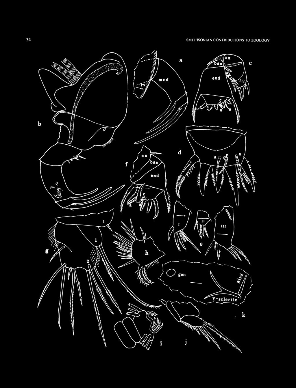
40 34 SMITHSONIAN CONTRIBUTIONS TO ZOOLOGY


: Single endite with 2 or 3 bristles; end joint with 11-13 bristles with short spines (spines not shown) separated by space from 2 posterior plumose bristles; posterior")
41 NUMBER FIGURE 17 (left). Eusarsiella tyanae, new species, adult female, holotype, USNM : a, view of right mandible protruding from carapace; b, right mandible, mv; c, left maxilla (endites I and II and 3 bristles of endite III not shown), lv; d, 2nd endopodial joint, right maxilla, mv; e, endites, left maxilla, mv;/ left maxilla (endites and exopodite not shown), mv; g, distal part of 5th limb; h. left 6th limb, lv; i, tip of 7th limb;y, right lamella and part of left lamella striated from right side; k, portion of posterior of body from left side. 2 spinous bristles; joints 2-5 fused, hirsute; 2nd joint with 3 bristles; fused 3rd to 5th joints with total of 6 bristles including 1 very short bristle. Sixth Limb (Figure 17 hi): Single endite with 2 or 3 bristles; end joint with bristles with short spines (spines not shown) separated by space from 2 posterior plumose bristles; posterior margin of limb hirsute. Seventh Limb (Figure 17/): Each limb with 8 bristles (2 proximal, 1 on each side, and 6 terminal, 3 on each side); each bristle with 3-7 bells and no marginal spines. Terminus with opposing combs each with 5 or 7 curved teeth. Furca (Figure 17/): Each lamella with 5 claws followed by few small spines; claw 1 continuous with lamella, others separated from lamella by suture; all claws with pointed tips and with teeth along posterior margins; 4 or 5 teeth on claw 1 much longer and stouter than others; slender medial spines or hairs present near base of claw 1; anterior edge of right lamella with few minute spines; right lamella anterior to left by width of base of claw 1. Bellonci Organ (Figure 16e): 1-jointed, short, with broadly rounded tip. Eyes: Lateral eye small, unpigmented, with 5 amber-colored divided ommatidia (Figure 16e); eye visible through shell (Figure 16a,b). Medial eye bare, larger than lateral eye, with small area of brown pigment (Figure \6e,h). Posterior of Body: Evenly rounded, bare. Upper Lip (Figure 16h): Helmet shaped, bare. Genitalia (Figure 17it): Oval ring anterior to Y-sclerite. Y-Sclerite (Figure 1 Ik): With usual ventral branch. Eggs (Figure 16a): USNM with 3 eggs in marsupium and smaller unextruded eggs. Length of 1 extruded egg 0.15 mm. DESCRIPTION OF ADULT MALE (Figures 18-20). Carapace more elongate than that of adult female and with incisur almost forming right angle (Figures 18, 19a). Surface with 2 lateral ribs similar to those of female except not connected posteriorly by undulating vertical rib. Curved vertical row of small spines connect anterior ends of horizontal ribs; 2 short parallel horizontal closely spaced rows of small spines connect the curved vertical row (at midpoint) with tip of rostrum. Ornamentation: Surface with shallow round fossae rimmed by minute spines similar to those of adult female (Figures 18, 19a). Long bristles present similar to those of adult female. Transparent gel-like substance visible filling space between bristles on ribs and valve edges. Infold: Anterior infold with small bristles near midheight well below rostrum. Infold of caudal process with 6 or 7 setose bristles forming row similar to that of adult female. Posterior infold with 2 setal bristles near midheight. Selvage: Lamellar prolongation of selvage extending well past tip of caudal process. Broad anterior and ventral lamellar prolongation with smooth outer edge. Central Adductor Muscle Attachments: Partly obscured, 13 shown in Figure 19b. FIGURE 18. Eusarsiella ryanae, new species, adult male, paratype, USNM , carapace from right side, length 0.71 mm.

; c, portion of")
42 36 SMITHSONIAN CONTRIBUTIONS TO ZOOLOGY a FIGURE 19. Eusarsiella ryanae, new species, adult male, paratype, USNM : a, complete specimen from right side, length 0.71 mm; b. central adductor muscle attachments of right valve and lower rib (from a); c, portion of anterior of body from right side; d, part of right 2nd antenna, mv; e, part of left mandible, mv;/ right mandible, Iv.
 0.81 mm.")
43 NUMBER Carapace Size (mm): USNM , length with caudal process 0.85, length without caudal process 0.71, height USNM , 4 adult males (length with caudal process): length 0.80, height 0.50; length 0.77, height 0.47; length 0.83, height 0.52; length 0.79, height Average length (including caudal process) 0.81 mm. First Antenna (Figure 19c): 2nd joint with bare dorsal bristle. 3rd and 4th joints fused; 3rd joint with 2 bristles (1 dorsal, 1 ventral short); 4th joint with 3 bristles (1 dorsal, 2 ventral). 5th joint small and wedged ventrally between 4th and 6th joints; sensory bristle with hour-glass shaped base with numerous long thin terminal filaments and stem with 3 short filaments. 6th joint with short bare medial bristle. 7th joint: a-bristle bare, short, about V3 length of f-bristle; b-bristle longer than a-bristle, bare; c-bristle long, with 4 marginal filaments. 8th joint: d- and e-bristles slightly shorter than c-bristle, bare with blunt tips; f-bristle shorter than c-bristle, with 2 marginal filaments; g-bristle same length as c-bristle, with 5 marginal filaments. Second Antenna: Protopodite bare (Figure \9c,d). Endopodite not reflexed, 2-jointed (Figure 19a*): 1st joint with 2 anterior ringed proximal bristles; tip of 2nd joint with 2 short ringed bristles. Exopodite: 1st joint with minute curved medial spine on distal margin; bristles of joints 2-4 with slender ventral spines and natatory hairs; bristles of joints 5-8 with natatory hairs; 9th joint with 2 bristles (1 small bare dorsal, other long with natatory hairs). Mandible (Figure 19e,/): Coxale endite consisting of short spine (Figure \9e). Basale: 6 bare bristles on or near ventral margin; 2 distal bare bristles on dorsal margin. Hirsute exopodite with base on lateral side of basale but then bends onto medial side of 1st endopodial joint. 1st endopodial joint with ringed ventral bristle (with long marginal hairs), small indistinct terminal bare dorsal bristle, and abundant medial hairs. 2nd endopodial joint with ringed bare terminal ventral bristle and long slender ringed bare dorsal bristle at midlength. 3rd endopodial joint with stout bare terminal claw, fairly long ringed bare ventral bristle, and 2 minute indistinct bare bristles (1 ventral, 1 dorsal). Maxilla (Figure 20a): Reduced. Coxale with short dorsal bristle. Exopodite with 2 ringed bristles. Endites, basale, and endopodite with indistinct weakly developed bristles (not all shown in illustration). Fifth Limb (Figure 20b,d): Reduced with weakly defined indistinct bristles (not all bristles shown in illustration). Sixth Limb (Figure 20c,d): Single endite with bristles. End joint with about 11 bristles (differs from end joint of female in having more bristles hirsute to tip and in not having a space anterior to the 2 posterior bristles). Illustrated limb with endite folded over end joint, and not in natural position. Seventh Limb (Figure 20d,e): Limb long with many welldefined segments. Terminal group with 3 or 4 bristles, 1 or 2 on each side, each with 5-7 distal bells and no marginal spines. Terminus convex and without combs. Furca (Figure 20a*): Similar to that of adult female except all claws with few stout teeth; those of claws 1 and 2 very large. Claw 1 with row of proximal long medial spines near base. All claws with spines along anterior margins; claws 5 and 6 also with few small proximal teeth on anterior margins (these visible under oil immersion lens (xloo objective, x20 ocular)). Bellonci Organ (Figure 20/): Similar to that of adult female. Eyes: Lateral eye slightly larger than that of adult female and with 5 or 6 ommatidia (Figures 19a,c, 20/). Medial eye similar to that of adult female (Figure 20/). Posterior of Body (Figure 20*/): Evenly rounded bare. Upper Lip: Similar to that of adult female. Genitalia (Figure 20d,g,h): Lobate copulatory organ on each side of body anterior to furca; distal lobe with sclerotized curved tooth; all lobes with few short bristles. Y-Sclerite (Figure 20c/): Similar to that of adult female. Gut Content: Without crustacean fragments observed in female gut. DESCRIPTION OF INSTAR I (sex unknown) (Figures 21,22). Carapace with numerous pointed spines and scattered long bristles, some with broad part either near base or near midlength. Ribs absent (Figure 21a). Transparent gel-like substance filling space between spines visible along edge of valve, not elsewhere. Tip of caudal process with 4 slender bristles (2 bristles in middle with a flair at base and each mounted on minute cylindrical protuberance) (Figures 210-0*, 22g). Infold: Caudal infold with 1 small double bristle near anteroventral corner; posterior with 1 setal bristle dorsal to caudal process (Figure 210*). Selvage: With lamellar prolongation extending past tip of caudal process similar to that of adult female (Figure 2 la*). Carapace Size (total length, height in mm): Sta : USNM ,0.49, USNM , 3 specimens: 0.47, 0.34; 0.47, 0.36; 0.50, Sta : USNM : 0.45, Average length 0.48 mm. First Antenna (Figures 21 ej, 22/): 1 st and 2nd joints bare. 3rd joint fused to 4th; 3rd joint with 2 bristles (1 ventral, 1 dorsal); 4th joint with few distal dorsal spines. Sensory bristle of long 5th joint without bristles. 6th joint minute, fused to 5th, with short medial bristle near dorsal margin. 7th joint: a-bristle bare, about twice length of bristle of 6th joint; b-bristle about same length as a-bristle, bare; c-bristle long, bare. 8th joint: d-, e-, and g-bristles long, bare; f-bristle shorter than g-bristle, bare. Second Antenna (Figure 21/g): Protopodite bare. Endopodite 1-jointed with minute terminal node with small bristle with spine at tip. Exopodite similar to that of adult female, 9th joint with 2 bristles. Mandible (Figure 21 h): Coxale endite comprising small spine. Basale: dorsal margin with minute subterminal bristle; ventral margin with 4 small bristles. Exopodite absent. 1st, 2nd, and 3rd endopodial joints each with stout terminal claw; 3rd joint with small ventral bristle at base of claw.
; c, 6th limb (endite folded); d, posterior of body from right side; e, tip of right 7th")
44 38 SMITHSONIAN CONTRIBUTIONS TO ZOOLOGY FIGURE 20. Eusarsiella ryanae, new species, adult male, paratype, USNM : a, right maxilla, Iv; b, 5th limb (nabs); c, 6th limb (endite folded); d, posterior of body from right side; e, tip of right 7th limb (only proximal parts of bristles shown);/ portion of anterior of body from right side; g.h, lateral view of left copulatory organ, and medial view of right organ.
, Iv; c,d, caudal process, left valve (surface spines not shown), mv; e, right 1st antenna and right lateral eye, Iv;/ portion of anterior of body")
45 NUMBER a '--- "^ bas FIGURE 21. Eusarsiella ryanae, new species, Instar I (sex unknown), paratype, USNM : a, complete specimen from right side, length 0.49 mm; b. caudal process, right valve (not all spines shown), Iv; c,d, caudal process, left valve (surface spines not shown), mv; e, right 1st antenna and right lateral eye, Iv;/ portion of anterior of body from left side; g, part of left 2nd antenna, mv; h, left mandible (nabs), Iv; ij, endites I and II, right maxilla, Iv; k, endites I III, left maxilla, Iv; /, right maxilla (nabs), Iv; m, left maxilla and Sth and 6th limbs drawn in place on body; n, left 6th limb, Iv; o,p, portions of anterior of body from left side.
, paratypes.")

46 40 SMITHSONIAN CONTRIBUTIONS TO ZOOLOGY FIGURE 22. Eusarsiella ryanae, new species, Instar I (sex unknown), paratypes. USNM : a, 5th limb; b, claw 1 of right lamella of furca, Iv; c, furcal claw 1; d, posterior of body from right side; e, right and left 6th limb; / portion of anterior of body, pv. USNM : g. complete specimen from left side, length 0.45 mm; h,i, portion of posterior of body from right side.

47 NUMBER Maxilla (Figures 21 i-n, 22/): Endites I III each with 3 to 5 bristles (Figure 21/-/:). Coxale with short dorsal bristle (Figure 21/). Basale with slender bristle near exopodite. Exopodite with 2 fairly long bristles. 1st endopodial joint with stout pectinate alpha- and beta-bristles. 2nd endopodial joint with 2 slender a-bristles and 3 stout pectinate end bristles. Fifth Limb (Figure 22a, h): Single endite with small bristle. 1st and 2nd endopodial joints each with 2 bristles. Joints 3-5 fused with total of 5 bristles: 3rd joint with 3 bristles (2 inner, 1 outer); joints 4 and 5 with total of 2 bristles. Sixth Limb (Figure 22e,h): Small with marginal hairs but no bristles. Seventh Limb (Figure 22h,i): Minute, bare, thumb-like. Furca (Figure 22b-d,h,i): 1st claw fused to lamella, with stout posterior tooth at midlength, smaller posterior teeth proximal, and slender minute closely spaced teeth distal; 2nd claw separated from lamella by suture, with proximal teeth along posterior margin; 3rd claw a stout pointed process fused to lamella and with spines along posterior margin; hairs along lamella following claws (hairs of left lamella longer); claw 1 of right lamella anterior to claw 1 of left lamella. Bellonci Organ and Eyes (Figures 2\a,e,f,o, 22g): Similar to those of adult female. Anterior of Body (Figure 21 o,p): Convex. Posterior of Body (Figure 22d): Evenly rounded with few spines at posterodorsal corner dorsal to intersection of girdle and posterior margin. Y-Sclerite (Figure 22d,h,i): Without clearly visible ventral branch. Gut Content: With brown unrecognizable particles. DESCRIPTION OF INSTAR II?FEMALE (Figure 23). Carapace similar in shape to carapace of Instar I (Figure 23a). Without ribs. Ornamentation (Figure 23a): Spines and bristles of carapace similar to those of Instar I (not all spines or bristles shown in Figure 23a). Transparent gel-like substance visible Infold: Infold of caudal process with 2 to 4 setose bristles forming row and 1 small double bristle in anteroventral corner near inner margin of infold (Figure 236); 2 setal bristles just dorsal to caudal process; left valve of USNM with small bristle just dorsal to setal bristles. Selvage: Similar to that of both Instar I and adult female. Central Adductor Muscle Attachments (Figure 23a): Consisting of numerous ovoid attachments. Carapace Size (total length, height in mm): USNM ,0.57,0.39. First Antenna (Figure 23c): Differs from that of Instar I in having a short dorsal bristle on 2nd joint and a terminal dorsal bristle on 4th joint. Also, sensory bristle of 5th joint and some long bristles of 7th and 8th joints with few minute filaments. Second Antenna (Figure 23c): Protopodite and exopodite similar to those of Instar I and adult female. Endopodite differs from that of Instar I in having a proximal anterior bristle. Mandible: Similar to that of adult female but not examined in detail. Maxilla: Similar to that of adult female but endite bristles not counted. 2nd endopodial joint differs from that of Instar I in having 5 instead of 3 end bristles. Fifth Limb: Similar to that of adult female but not examined in detail. Sixth Limb (Figure 23e): With single bristle. Seventh Limb (Figure 23d,e): Bare, thumb-like, unsegmented, longer than 7th limb of Instar I. Furca (Figure 23a*): Similar to that of adult female. Differs from that of Instar I furca in having 5 instead of 3 claws, having suture at base of claw 3, and having 3 instead of 2 large teeth on posterior edge of claw 1. Hairs not observed on edges of lamellae following claws. Bellonci Organ and Eyes (Figure 23c): Similar to those of both Instar I and adult female. Posterior of Body (Figure 23a*): Posterodorsal corner of body of USNM with triangular protuberance. (Protuberance probably resulting from inward bending of dorsal margin of body and not true posterodorsal process.) No spines observed on posterior of body. Y-Sclerite (Figure 23o»: Similar to that of adult female. Differs from that of Instar I in having ventral branch well developed. Gut Content: USNM with several large seemingly complete crustaceans in posterior half of gut. DESCRIPTION OF INSTAR III MALE (Figure 24). Carapace similar in shape and ornamentation to that of Instar II (Figure 24a,b,f,i). No ribs present. Transparent gel-like substance visible covering spines along edge. Infold (Figure 24g): Infold of caudal process with 5 setose bristles forming row; 2 setal bristles present just dorsal to caudal process. 1 double bristle present in anteroventral corner of caudal process. Selvage: Similar to that of adult female. Central Adductor Muscle Attachments (Figure 24a,bJ,i): Numerous ovoid attachments. Carapace Size (total length, height in mm): USNM , 0.68, 0.48; USNM , 0.66, 0.46; USNM , 0.66, 0.49; USNM , 0.65, 0.48; USNM , 0.67, 0.49; USNM , 0.68, Average total length 0.67 mm. First Antenna (Figure 24c): Differs from Instar II in having a terminal ventral bristle on 4th joint. Second Antenna (Figure 24c): Protopodite and exopodite similar to those of adult female. Endopodite 2-jointed: 1st joint with 2 proximal anterior bristles; 2nd joint triangular with 1 short terminal bristle. Mandible, Maxilla, and Fifth Limb: Similar to those of adult female but not studied in detail. Sixth Limb (Figure 24e): With single endite with bristle and end joint with many bristles.

, Bellonci Organ (Figure 24c), Eyes (Figure 24a-cJ,i), Posterior of Body (Figure 24d), and Y-Sclerite (Figure 2Ad,h,j\"): Similar to those of adult female.")
48 42 SMITHSONIAN CONTRIBUTIONS TO ZOOLOGY FIGURE 23. Eusarsiella ryanae, new species, Instar II?female, paratype, USNM : a, complete specimen from left side (not all spines shown), length 0.57 mm; b, caudal process of right valve, iv; c, portion of anterior of bodyfromright side; d,e, portion of posterior of body from left andright sides. Seventh Limb (Figure 24d,h, i): Elongate, bare, without segments, longer than 7th limb of Instar II. Furca (Figure 24J), Bellonci Organ (Figure 24c), Eyes (Figure 24a-cJ,i), Posterior of Body (Figure 24d), and Y-Sclerite (Figure 2Ad,h,j"): Similar to those of adult female. Gut Content: With almost complete skeletons of nematodes and Crustacea and crustacean fragments. (A nematode (Figure 24/) was identified by Duane Hope (S.I.) as a free-living type, and was placed into the S.I. worm collection.) REMARKS. Three of the six instar Ill's were not dissected, but the undissected specimens all appeared to have a 2nd joint on the endopodite of the 2nd antenna when viewed through the shell, and therefore were interpreted to be males. COMPARISONS. The new species, E. ryanae, differs from E. "carinata "(Kornicker, 1986a) in lacking scalloped anterior and ventral carapace margins. The endopodite of the 2nd antenna of the female E. ryanae differs from that of E. cornuta Poulsen, 1965, in having a ventral node with a short bristle. The 7th limb of E. ryanae bears two proximal bristles on the 7th joint compared to four on E. spicata Poulsen, The carapace of E. ryanae differs from that of E. capillaris (K.ornicker, 1958, 1986a) by not having nodes bearing finger-like

. bristles. The carapace of E. ryanae differs from that of E. paniculata Kornicker, 1986a, and E.")
49 NUMBER a FIGURE 24. Eusarsiella ryanae, new species, Instar III male, paratypes. USNM : a, complete specimen fromright side, length 0.68 mm; b. complete specimen from left side, length 0.68 mm; c, portion of anterior of body from left side; d, posterior of body from left side; e, left 6th limb, lv. USNM :/ complete specimen fromright side, length 0.66 mm; g, posterior of left valve, iv. USNM : h, right 7th limb and Y-sclerite (as seen through right valve). USNM : i. complete specimen from left side, length 0.67 mm;/ nematode in gut andrighty-sclerite (as seen through right valve). bristles. The carapace of E. ryanae differs from that of E. paniculata Kornicker, 1986a, and E. donabbotti (Cohen, 1989a) in lacking numerous spinous nodes. The Bellonci organ of E. bedoyai Baltanas, 1992, is much longer than that of E. ryanae. The carapace of E. ryanae, although smaller and with a narrower caudal process, outwardly strongly resembles that of E. styx Kornicker and Iliffe, 1989b; they differ in that the bristles of the infold of the caudal process of ryanae are setose and form a vertical row (Figure 25a), whereas those of styx are pointed and scattered (Figure 25b). Also, the adult female of

50 44 SMITHSONIAN CONTRIBUTIONS TO ZOOLOGY a FIGURE 25. Comparison of infold bristles of caudal processes of right valves of adult females of Eusarsiella ryanae, new species, and Eusarsiella styx Kornicker and Iliffe, 1989: a, E. ryanae, holotype, USNM b, E. styx, paratype, USNM Both illustrations drawn as seen through the right valve and at same magnification. styx lacks a ventral bristle on the endopodite of the 2nd antenna. The orientation of the backward-pointing caudal process and its considerable length distinguishes E. ryanae from many previously described species of Eusarsiella. The adult male of E. ryanae differs from the adult male of many species of Eusarsiella by having a well-developed 7th limb that bears bristles, and by not having a reflexed clasping-type endopodite on the 2nd antenna. Kornicker (1996, table 2) listed certain morphologic characters for adult males of 21 species of Eusarsiella. Only three species, E. styx, E. chessi Kornicker, 1991, and E. "carinata," have a 7th limb bearing bristles as well as a non-prehensile endopodite of the 2nd antenna. The male mandible of E. chessi differs from that of E. ryanae in having a claw on the 1st and 2nd endopodial joints. The endopodite of the 2nd antenna of the male E. ryanae differs from that of E. "carinata," but it is similar to that of E. styx, thus suggesting a close relationship between E. styx and E. ryanae. ONTOGENY. None of the specimens had within their appendages the appendages of the next stage. Although no instar IV is in the collection, it does contain four instar I's (sex unknown), one instar II (?female), six instar Ill's (male), one ovigerous adult female, and five adult males. The calculated growth factors of total carapace length between successive instars are I II, 1.19; II III, The adult male is about 12% shorter than the adult female. The ribs that are present on the adult carapace are absent in the first three instars. The morphologic changes that took place in the appendages during the first three stages are, in general, similar to those described by Hiruta (1977) for Sarsiella japonica Hiruta, The changes for E. ryanae include the following. 1 st antenna: no bristles on 2nd and 4th joints in instar I; a dorsal bristle is added to both the 2nd and 4th joints in instar II; and a ventral bristle is added to the 4th joint in instar III. Maxilla: 2nd endopodial joint with three end bristles in instar I and five in the following stages. 6th limb: without bristles in instar I, with one bristle in instar II, and with many bristles in the following stages. 7th limb: bare in instars I III, but the limb is longer in each succeeding instar (instar IV not present but in other sarsiellids it has many bristles on limb). Furca: instar I with three claws and having 1 st and 3rd claws fused to the lamella, and in following stages with five claws and having only claw 1 fused to the lamella. Hiruta (1977:58) stated that the maxilla of the 1st instar of Sarsiella japonica differed from that of Spinacopia sandersi Kornicker, 1969, in having on the 2nd endopodial joint three rather than five terminal bristles and no c-bristle. Hiruta understandably, but incorrectly, interpreted Kornicker's 1969:23 description of the mandible, maxilla, and 5th limbs of instar I of S. sandersi, which stated, "well developed, but not examined in detail; similar type as that of adult female" as meaning that the number of bristles on the 2nd endopodial joint of the maxillae of instar I and adult were the same. The number of bristles on the 1st instar maxilla in my study was one of the details not examined. The presence of only three end bristles and the lack of a c-bristle on the 2nd endopodial joint of the maxilla of instar I are probably constant in the Sarsiellidae, but the 1st instar of more species should be studied to confirm this. Unlike the 2nd antennae of members of the Cypridinidae, on which the number of bristles of the 9th exopodial joint increases during ontogeny (Kornicker, 1992, fig. 5), the 9th joint of E. ryanae has two bristles on the juveniles as well as on the adults. The number of ommatidia in the lateral eye of E. ryanae remains at five in both juveniles and adults, except some adult males have either five or six.
 FIGURE 26 Sarsiella costata Kornicker, 1958:251, figs. 47:4A,B, 80A-E, 81A-E, 88B,C,G. Eusarsiella costata. Poulsen, 1965:83. Kornicker, 1986a: 61, figs. 31-35.")
51 NUMBER The carapaces of juveniles as well as the adults of E. ryanae have a gel-like coating covering the short bristles on the valve margins. Eusarsiella costata (Kornicker, 1958) FIGURE 26 Sarsiella costata Kornicker, 1958:251, figs. 47:4A,B, 80A-E, 81A-E, 88B,C,G. Eusarsiella costata. Poulsen, 1965:83. Kornicker, 1986a: 61, figs HOLOTYPE. USNM , dry specimen, length 1.02 mm, (Kornicker, 1958:252, number 156-2). TYPE LOCALITY. Bimini Islands, Bahamas. MATERIAL. Exuma Sound: Sta , transect A A: USNM , adult male in alcohol. DISTRIBUTION. Bahamas: Bimini Islands, San Salvador Island, the Wall off Lee Stocking Island, Exuma Cays (depth 67 m). Known depth range m. SUPPLEMENTARY DESCRIPTION OF ADULT MALE (Figure 26). Carapace Size (length, height in mm): USNM , 0.91,0.68. Eusarsiella species indeterminate MATERIAL. Exuma Sound: Sta , transect AA: USNM , 3 juveniles; USNM , 3 juveniles. Sta , transect AB: USNM , 1 instar IV; USNM , 1 juvenile. All specimens in alcohol. (The material includes two species with lateral outlines similar to that of E. costata, but one species is without ribs, and the other has 2 poorly defined ribs (1 dorsal, 1 at midheight).) DISTRIBUTION. Exuma Sound at depths of m. FIGURE 26. Eusarsiella costata (Kornicker, 1958), adult male, USNM , complete specimen from right side, length of carapace 0.91 mm. Eurypylus Brady, 1869 Eurypylus Brady, 1869:141. Kornicker and McKenzie, 1976:348. Kornicker, 1986a:33. TYPE SPECIES. Eurypylus petrosus Brady, 1869, monotypy. COMPOSITION AND DISTRIBUTION. Including the two new species described herein, the genus contains 10 species (Kornicker, 1996). Members of the genus have been reported on the continental shelf off Georgia; a mangrove area in the vicinity of Tanzania, east Africa; the Cape Verde Islands off west Africa; shallow water in the vicinity of Singapore; in the vicinity of Mayotte and Glorioso Islands, Indian Ocean; shallow waters off Australia; and the Wall off Lee Stocking Island, Exuma Cays, Bahamas (Kornicker, 1996). The previous known depth range was shallow water to a depth of about 30 m (Kornicker, 1986a:33, 1996:57); the maximum known depth is increased to 142 m herein. Eurypylus hapax, new species FIGURES ETYMOLOGY. From the Greek hapax (once only). HOLOTYPE. USNM , Instar IV female on slide and in alcohol. TYPE LOCALITY. Sta , transect BA, Exuma Sound, Bahamas, depth 142 m. PARATYPES. None. DISTRIBUTION. Collected only at type locality. DESCRIPTION OF INSTARIV FEMALE (Figures 27-29). Carapace oval in lateral view with slightly projecting rostrum and short broad caudal process with straight posterior edge (Figure lla-c). Ornamentation: Surface without either lateral ribs, short spines, or gel-like coating. Posterior edge of caudal process with 7 bristles with bases either on lateral side or along edge (Figure 21a,c,e). Bristles, some with wide proximal part, present along valve edge and scattered on outer surface; all bristles with pointed tips (Figure 21a-e). Outer surface with minute triangular processes (on USNM processes restricted to edges of valves and best visible on posterior edge of caudal process (Figure 27e)). Infold: Anterior infold with small bristle dorsal to incisur (Figure 21d). Caudal process with scattered small pointed bristles (Figure 27e). Two setal bristles on infold just dorsal to caudal process. Selvage: Broad lamellar prolongation with smooth outer edge present along free margins and extending past posterior end of caudal process. Central Adductor Muscle Attachments: Consisting of numerous small oval attachments (Figure 27/). Carapace Size (total length, height in mm): USNM ,0.87,0.70.

.")
52 46 SMITHSONIAN CONTRIBUTIONS TO ZOOLOGY FIGURE 27. Eurypylus hapax, new species, Instar IV female, holotype, USNM : a, complete specimen from left side, length 0.87 mm; b,c, anterior and posterior of left valve, ov; d,e, anterior and posterior of left valve, iv;/ central adductor muscle attachments on left valve, ov. First Antenna (Figure 28a,b): 1 st joint bare. 2nd joint with dorsal bristle with indistinct spines. 3rd and 4th joints fused; 3rd joint with 1 dorsal bristle and no ventral bristle; 4th joint with ventral spines and 3 bristles (1 dorsal, 2 ventral). 5th joint with long ventral bristle with minute distal marginal spine or filament and terminal spine. 6th joint minute, fused to 5th joint,


53 NUMBER FIGURE 28. Eurypylus hapax, new species, Instar IV female, holotype, USNM : a, portion of anterior of body from left side; b, portion of anterior of body from left side; c, part of right 2nd antenna, mv; d, right mandible, mv; e, upper lip and part of left mandible, both in place on body and drawn from right side;/ endites I and II, left maxilla, mv; g, endite III, right maxilla, Iv; h, part of right maxilla, lv.


54 48 SMITHSONIAN CONTRIBUTIONS TO ZOOLOGY FIGURE 29. Eurypylus hapax, new species, Instar IV female, holotype, USNM : a, right 5th limb, lv; b, 6th limb; c,d, left 7th limb; e, right 7th limb viewed through right valve;/ left and right lamellae of fiirca (marginal teeth of claws not shown on left lamella), Iv; g, left lateral eye, medial eye, and Bellonci organ in place on body (stippling in medial eye represents brown pigment); h, views of portions of posterior of body from right and left sides. with short medial bristle. 7th and 8th joints fused. 7th joint: a-bristle about twice length of bristle of 6th joint; b-bristle medial and longer than a-bristle; c-bristle about same length as bristle of 5th joint, with small proximal filament and terminal spine. 8th joint: d-bristle about same length as bristle of 5th joint, bare with blunt tip; e-bristle about % length of d-bristle, bare with blunt tip; f-bristle shorter than c-bristle, with minute terminal spine; g-bristle about same length as bristle of 5th joint, with minute indistinct proximal filament and terminal spine. Second Antenna (Figure 2$a-c): Protopodite bare. Endopodite 1-jointed, with medial spines, proximal ringed anterior bristle, and minute spine or bristle adjacent to slight terminal protuberance (Figure 28c). Exopodite: 1st joint with
.")
55 NUMBER small terminal medial bristle with blunt tip; bristles of joints 1-8 long with slender ventral spines and distal natatory hairs; 9th joint with 2 bristles (dorsal short with few minute spines, ventral long with slender ventral spines and distal natatory hairs). Mandible (Figure 2$a,d,e): Coxale endite consisting of 1 or 2 short stout spines in proximal ventral corner; ventral margin of coxale with hairs, and surface near ventral margin with slender spines. Basale: ventral margin with 4 small medial and 2 small lateral bristles; dorsal margin with 3 small bristles (1 distal to midlength, 2 subterminal). Exopodite absent. 1st endopodial joint with medial, ventral, and dorsal spines, and stout ventral terminal claw with proximal ventral and dorsal spines. 2nd endopodial joint with stout ventral claw and small dorsal bristle. 3rd endopodial joint with stout terminal claw and 2 minute bristles (1 ventral, 1 dorsal). Maxilla: Endite I with 2 lobes with medial spines, proximal lobe with 1 unringed terminal bristle, distal lobe with S bristles (1 ringed, 4 claw-like) (Figure 28/); endite II with 4 bristles (Figure 28/); endite HI with 5 bristles (Figure 2$g,h). Coxale with short ringed dorsal bristle and long dorsal hairs (Figure 2Sh). Basale with bristle near exopodite (Figure 2Sh). Exopodite with 3 bristles (1 long, 2 short). 1st endopodial joint with few distal spines on anterior margin and pectinate alphaand beta-bristles. 2nd endopodial joint with 2 a-bristles, 1 c-bristle, and 5 pectinate end bristles (middle bristle shortest, anterior longest). Fifth Limb (Figure 29a): Single endite with short bristle. 1st exopodial joint with 2 spinous bristles; 2nd exopodial joint with 3 spinous terminal bristles; joints 3-5 fused with total of 7 bristles. Exopodial joints 2-5 hirsute. Sixth Limb (Figure 296): Single endite with 3 short bristles. End joint with 11 spinous anterior bristles followed by short space and 2 stout hirsute bristles. Seventh Limb (Figure 29c-e): Limb with 8 tapered bristles, 4 in proximal and 4 in terminal group, each with 2 or 3 bells. Terminus with opposing combs, each comb with about 3 minute teeth. Furca (Figure 29/): Each lamella with 5 claws decreasing in length along lamella; claws 1 and 2 nonarticulated, claws 3-5 articulated. All claws with teeth along posterior margin; few teeth on claws 1-3 slightly stouter than others; teeth of claw 5 indistinct. Few spines on lamellae following claws. Right lamella anterior to left by width of base of claw 1. Bellonci Organ (Figure 29g): Elongate, broadening near rounded tip. Eyes: Medial eye bare with line of brown pigment near midheight (Figure 29g). Lateral eye smaller than medial eye, with 5 divided amber-colored ommatidia (Figures 27a, 2%b, 29g). Upper Lip (Figure 28e): Projecting anteriorly, bare. Posterior of Body (Figure 29h): With few indistinct spines at posterodorsal corner dorsal to posterior end of girdle. Genitalia: None observed. Y-Sclerite (Figure 29h): With ventral branch typical for family. REMARKS CONCERNING 7TH LIMB. The holotype, USNM , is an instar IV female and the 7th limb has some juvenile characters. It is likely that the 7th limb of the adult female will have cylindrical rather than tapered bristles, 2 proximal anterior bristles rather than only 1, 6 rather than 4 terminal bristles, and more strongly developed opposing terminal combs. COMPARISONS. The absence of secondary claws on the furca of E. hapax places the species in the E. rousei group of Kornicker (1996, table 3). The only known species in that group having five furcal claws on each lamella is E. concentricostatus (Hartmann, 1974). The carapace off. hapax is without the concentric rib present on E. concentricostatus. CORRECTION. Kornicker (1996, table 3) incorrectly listed E. concentricus instead of E. concentricostatus. Eurypylus eagari, new species FIGURES ETYMOLOGY. Named for Dr. Stephan Eagar, ostracodologist. HOLOTYPE. USNM , ovigerous female on slide and in alcohol. TYPE LOCALITY. Sta , transect AA, Exuma Sound, Bahamas, depth 67 m. PARATYPES. None. DISTRIBUTION. Collected only at type locality. DESCRIPTION OF ADULT FEMALE (Figures 30-32). Carapace oval in lateral view with projecting caudal process, and lateral projection at valve midheight extending posteriorly just past valve edge (Figure 30). Location of incisur indicated by indentation in lateral surface of anterior margin of valve at about midheight, but no indentation present in valve edge. Ornamentation (Figure 30): Surface with lateral ribs both dorsal and ventral to valve midheight; a peripheral rib located well in from valve edge and with small riblets extending from rib to valve edge. Surface with numerous oval fossae with minute papillae on bottom. Bristles, some with broad base, numerous along valve edge and sparse on lateral surface. Ventral and anterior margins scalloped. Valves without gel-like coating. Infold: Small bristle near inner margin of anterior infold just ventral to incisure. Infold of caudal process with 9-11 small bristles and with few smaller bristles along inner edge of infold (Figure 32/). Two setal bristles present dorsal to caudal process at about VA valve height measured from caudal process. Selvage: Wide lamellar prolongation along anterodorsal, anterior, anteroventral, and ventral edges of valves; lamellar prolongation extending well past tip of caudal process; outer edge of anteroventral lamellar prolongation with fringe of closely spaced coarse spines; fringe along anterior and anterodorsal lamellar prolongation with slenderer spines; edge of ventral and posterior lamellar prolongation without spines. (Poulsen (1965:48) stated that the outer edge of the selvage of
56 50 SMITHSONIAN CONTRIBUTIONS TO ZOOLOGY FIGURE 30. Eurypylus eagari, new species, adult female, holotype, USNM : complete specimen from right side, length 0.64 mm. the Sarsiellidae is without a fringe of hairs that is characteristic of the Philomedinae and Rutidermatidae. Thus, the fringe of hairs along the outer edge of part of the lamellar prolongation of E. eagari is unusual, and has not previously been reported on other Sarsiellinae, but has been reported on species of Dantyinae by Kornicker and Cohen (1978:497).) Carapace Size (length, height in mm): USNM , 0.64, First Antenna (Figure 3 \c,d): 1 st joint bare. 2nd joint with dorsal spines and dorsal bristle with indistinct spines. 3rd and 4th joints fused; 3rd joint with 2 bristles (1 dorsal, 1 ventral (ventral bristle missing on right limb of USNM ; the bristle shown dashed on illustrated right limb (Figure 31c) is based on length of bristle of left limb)); 4th joint with 3 bristles (1 dorsal, 2 ventral). 5th joint with long ventral bristle with minute spine at midlength and 1 terminal spine. 6th joint minute, fused to 5th joint, with small terminal medial bristle. 7th and 8th joints fused. 7th joint: a-bristle short; b-bristle about V2 length of a-bristle; c-bristle long. 8th joint: d- and e-bristles equilength, slightly shorter than bristle of 5th limb, bare with blunt tips; f-bristle claw-like in proximal half with curved and oblique cross-sutures, and bristle-like in distal half with closely spaced rings (proximal end of distal half narrower than distal end of proximal half); g-bristle similar to f-bristle, but slightly longer. Second Antenna (Figure 3\c,e): Protopodite bare. Endopodite 2 jointed: 1st joint with 1 or 2 proximal ringed anterior bristles; 2nd joint with terminal bristle (with slender drawn-out tip) slightly longer than bristles of 1st joint (endopodite could be interpreted to be 1-jointed if terminal protuberance considered part of 1st joint). Exopodite: 1st joint with small recurved terminal medial bristle with blunt tip; bristles of joints 2 and 3 with slender proximal ventral spines and distal natatory hairs; bristles of joints 4-8 with natatory hairs, no spines; 9th joint with 2 bristles (dorsal short bare, ventral long with few proximal hair-like ventral spines and natatory hairs). Mandible (Figure 31/-/i): Coxale endite consisting of medial spine in proximal ventral corner; ventral margin of coxale with hairs; medial surface near ventral margin with few indistinct small spines. Basale: ventral margin with 4 small medial and 2 small lateral bristles; dorsal margin with 2 small FIGURE 31 (right). Eurypylus eagari, new species, adult female, holotype, USNM : a, complete specimen from right side, length 0.64 mm; b, adductor muscle attachments on right valve (not all shown), ov; c, portion of anterior of body from right side; d, tip of left 1st antenna (8th joint not shown), mv; e, part of right 2nd antenna, mv;/ right mandible, lv; g.h, proximal part and endopodite of right mandible, mv; /', furca (only part of left lamella shown, striated); j, right lateral eye, medial eye, and Bellonci organ; k, left lateral eye; /, portion of posterior of body from right side; m, genital organ.

57 NUMBER lft

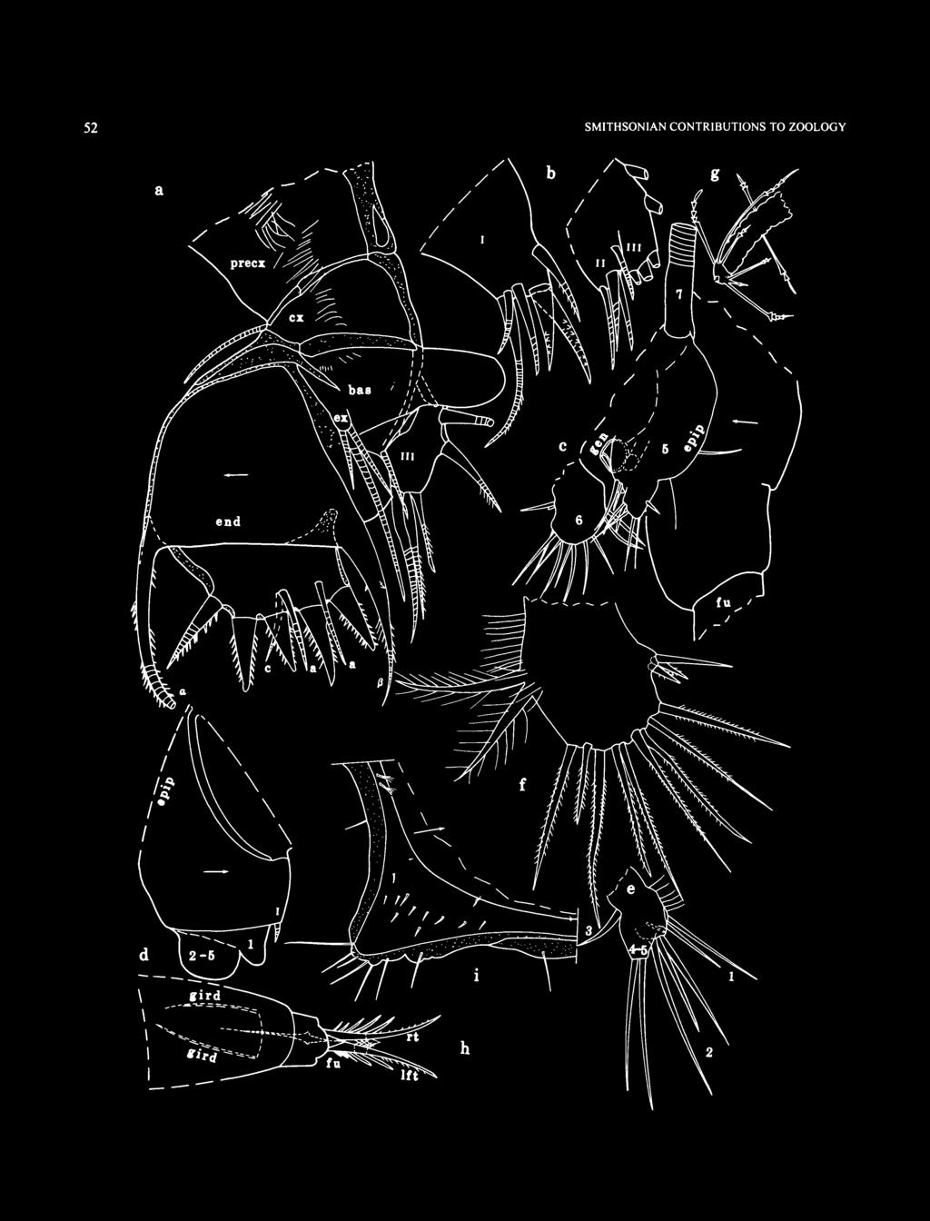
58 52 SMITHSONIAN CONTRIBUTIONS TO ZOOLOGY r

59 NUMBER FIGURE 32 (left). Eurypylus eagari, new species, adult female, holotype, USNM : a, left maxilla, lv; b, endites I III, right maxilla, mv; c, portion of posterior of body from left side; d. 5th limb (only bristle of endite I shown); e, distal 5th limb (folded on slide);/ right 6th limb, lv; g, left 7th limb; h. posterior of body, av; i, posteroventral infold of left valve, iv. indistinct bristles (1 near midlength, 1 subterminal). Exopodite absent. 1st endopodial joint with medial spines, a row of slender medial spines along distal edge near dorsal margin, a few indistinct terminal spines on dorsal margin, and stout bare ventral claw. 2nd endopodial joint with small dorsal bristle and stout bare ventral claw. 3rd endopodial joint with stout terminal claw and 2 minute bristles (1 ventral, 1 dorsal). Maxilla (Figure 32a,b): Precoxale and coxale with long hairs. Coxale with bare dorsal bristle. Endite I with 6 bristles (2 claw-like pectinate); endite II with 4 bristles; endite III with 5 bristles (2 proximal, 3 terminal). Transparent thumb-like flap projects from basale (or from near base of endites). Basale with bristle near exopodite. Exopodite with 3 bristles (1 long, 2 short). 1st endopodial joint with pectinate alpha- and beta-bristles. 2nd endopodial joint with 2 a-bristles, 1 c-bristle, and 5 pectinate end bristles (middle bristle shortest, anterior bristle ringed). Fifth Limb: Epipodite with about 22 bristles. Single endite with 1 small bristle (Figure 32*/). 1st exopodial joint with 2 bristles (Figure 32e); 2nd exopodial joint with 3 terminal bristles; 3rd exopodial joint with 1 bristle on outer lobe; fused 4th and 5th joints with 2 terminal bristles; exopodite with long hairs. (A small bristle illustrated by dashes on folded limb shown in Figure 32e could be either the endite bristle or a bristle on an exopodial joint.) Sixth Limb (Figure 32/): Single endite with 1 terminal bristle and 1 or 2 shorter medial bristles. End joint with 9 or 10 bristles with short spines and 2 broader plumose posterior bristles; medial surface and posterior margin of end joint with long hairs. Seventh Limb (Figure 32c,g): Limb with 8 cylindrical bristles: 2 in proximal group (1 on each side) each with 2 bells; 6 in terminal group (3 on each side) with either 1-4, or 2 4 bells; clappers of terminal bells unusually long. Terminus with opposing combs, each with 2 or 3 minute teeth. Furca (Figures 31/,/, 32c,h): Each lamella with 6 claws: claws 1 and 2 primary and nonarticulated; claws 3-6 secondary and articulated. Claws 1-5, and possibly 6, with teeth along posterior margin; a few teeth on claws 1 and 2 stouter than others. Long slender medial spines near base of claw 1; slender spines present along margins of lamellae following claws. Right lamella anterior to left by width of base of claw 1. Bellonci Organ (Figure 3 If): Elongate with suture proximal to midlength and rounded tip with minute node. Eyes: Medial eye bare unpigmented (Figure 31/). Lateral eye unpigmented, smaller than medial eye, with 5 amber-colored divided ommatidia (Figure 31/, A). Upper Lip: Indistinct, projecting anteriorly, bare. Posterior of Body (Figures 31/, 32c,h): With few spines dorsal to dorsal end of girdle. Genitalia (Figures 3\m, 32c): Rimmed oval on each side anterior to furca. Y-Sclerite (Figure 31/): With ventral branch typical for family. Eggs: USNM with 2 oval eggs in marsupium (1 shown in Figure 31a). Maximum lengths of each egg (mm): 0.15,0.17. COMPARISONS. The presence of secondary claws following the primary claws on the furca places E. eagari in the Eurypylus petrosus group of Kornicker (1996, table 3). The only species in that group having a total of six claws is E. darwinensis Kornicker, The caudal process of the female E. eagari projects farther posteriorly than that of E. darwinensis, and the endopodite of the female 2nd antenna of E. darwinensis is without a terminal bristle. Eurypylus eagari is the only species of the sarsiellinae reported to have spines along the outer edge of the lamellar prolongation of the selvage. Family RUTIDERMATIDAE Brady and Norman, 1896 This family contains two subfamilies: Rutidermatidinae Brady and Norman, 1896, and Metaschismatinae Kornicker, Subfamily RUTIDERMATIDINAE Brady and Norman, 1896 Rutiderma Brady and Norman, 1896 TYPE SPECIES. Rutiderma compressa Brady and Norman, COMPOSITION AND DISTRIBUTION. This genus is cosmopolitan between the latitudes of 45 N and 53 S, at depths from intertidal to 317 m (questionably to 1834 m), and contains about 31 species (Cohen and Kornicker, 1987; Kornicker, 1992, 1996). Rutiderma schroederi, new species FIGURES ETYMOLOGY. Named for Jack Schroeder, artist, who inked the camera lucida drawings of appendages for this and many other papers by the senior author. HOLOTYPE. USNM , adult female on slide and in alcohol. TYPE LOCALITY. Sta , transect AA, Exuma Sound, Bahamas, depth 67 m. PARATYPES. Sta , USNM , instar HI, in alcohol. Sta , USNM , undissected late instar in alcohol (length 1.16 mm, height 0.75 mm). DISTRIBUTION. Collected at Sta , off Lee Stocking Island, depth 67 m, and Sta , off Great Exuma Island, depth m.
; both valves with")
60 54 SMITHSONIAN CONTRIBUTIONS TO ZOOLOGY DESCRIPTION OF ADULT FEMALE (Figures 33-37). Carapace oval in lateral view with prominent rostrum and caudal process (Figure 33a). Ornamentation: Horizontal ribs present above and below central adductor muscle attachments form alar process on each valve that extend past posterior edge of valve (Figure 33a,c); both valves with small triangular process at anterodorsal corner (Figure 33a,c); surface of valves with abundant large fossa, each fossae lies within 5- or 6-sided polygon (polygons formed by indistinct intersecting thin sutures and not raised walls) (Figure 33a, b); tip of rostrum and anteroventral corner of valves extend well past valve edge (Figure 34a,b,e); long bristles, some with broad base, along anterior and ventral margins and scattered over valve surface. USNM with internal marginal pore canals along free margin (Figure 34c). Infold: Rostral infold with 17 bristles forming row parallel to valve edge, and with 2 paired bristles near inner end of incisur (Figure 34a); small bristles near inner margin of infold posterior to incisur (Figure 34a,e); anteroventral infold with about 11 short bristles and 6 or 7 parallel ridges (Figure 34e); infold of caudal process with list just within inner margin of infold at angle of 20 to 30 with ventral edge of valve (Figure 34d,f); list with 6 to 11 short bristles; infold of caudal process with 5 or 6 small scattered bristles between list and valve edge. Selvage: Wide lamellar prolongation with marginal fringe present along free margins; prolongation divided at inner end of incisur (Figure 34e) and probably also at tip of caudal process. Central Adductor Muscle Attachments (Figure 33e): Consisting of about 11 individual oval scars. Carapace Size (length, height in mm): USNM , 1.73, 1.4, height 66% of length. First Antenna (Figure 35a): 1 st joint bare. 2nd joint with spinous dorsal bristle near midlength. 3rd and 4th joints fused; 3rd joint short with 3 bristles (1 ventral, 2 dorsal); 4th joint with 3 spinous terminal bristles (2 ventral, 1 dorsal). 5th joint with row of minute lateral spines near base of sensory bristle; sensory bristle with small proximal filament and bifurcate tip. 6th joint minute, fused to 5th joint, with spinous medial bristle. 7th joint: a-bristle longer than bristle of 6th joint; b-bristle longer than a-bristle; c-bristle twice length of b-bristle. 8th joint: d- and e-bristles almost as long as c-bristle, bare with blunt tips (bristles not shown in Figure 35a); f-bristle almost as long as c-bristle, with 2 proximal filaments and bifurcate tip; g-bristle longer than c-bristle with 2 proximal filaments and bifurcate tip. (The 2nd joint of each limb of USNM is unusual in not having a lateral bristle; however, the 2nd joint of instar III, USNM , described below, does have a long lateral bristle indicating that the missing lateral bristle on the adult is either an aberrancy or was broken off during the dissection.) Second Antenna: Protopodite bare (Figure 356,c). Endopodite a single joint with 1 anterior and 4 posterior bristles (Figure 35b,c). Exopodite: 1st joint with small terminal medial bristle; bristle of 2nd joint reaching well past 9th joint, with small triangular ventral spines and hooked tip; bristles of joints 3-5 short, with triangular and rounded ventral spines; bristles of joints 6 to 8 long with natatory hairs, no spines; 9th joint with 5 bristles (3 long, 1 medium, 1 short dorsal) with natatory hairs. Mandible (Figure 35d,e): Coxale endite bifurcate (endite broken off right limb) (spines not observed on endite but present on endite of instar III described herein). Basale: dorsal margin with 3 distal bristles; ventral margin with 4 bristles in proximal corner (2 unringed pectinate, 2 ringed) and 3 or 4 distal bristles with bases medial (Figure 35e) (2 proximal bristles broken off in right limb (Figure 35a 1 )). 1st endopodial joint with 3 slender ventral bristles; medial surface with abundant spines. 2nd endopodial joint: dorsal margin with 5 proximal bristles (including 2 small bristles with bases medial); ventral margin with small a-bristle (a 2nd a-bristle may have been broken off left limb during dissection; both a-bristles broken off right limb; an instar III, USNM , described below has 2 a-bristles), small terminally ringed b-bristle on sclerotized base, and stout terminal claw-like c-bristle with ventral spines, proximal peg, and tapered tip (tip broken off c-bristle of one limb of adult USNM (Figure 35<?) but appears intact on other limb (Figure 35o*); however, tip of the c-bristle on instar III, USNM , more drawn-out than that of adult female, suggesting tip might have broken off both limbs of adult) with minute spines along distal inner margin, and 2 small d-bristles; medial side with abundant spines. 3rd endopodial joint with 3 slender medial a-bristles, long terminal b-bristle with minutely serrate ventral margin, and long terminal stout claw-like c-bristle with minutely serrate ventral margin and broadly rounded tip. Maxilla (Figure 36a-d): Precoxal and coxale with fringe of hairs (not shown); coxale with dorsal bristle. 3 endites each with 2 or 3 pectinate claw-like bristles in addition to a few slender bristles; endite III also with slender proximal bristle. Basale with 2 bristles (1 medial, 1 dorsal (dorsal bristle missing on right limb)). Exopodite with 3 bristles (2 long, 1 short). 1st endopodial joint with dorsal spines, 1 spinous alpha-bristle, and 2 spinous beta-bristles (beta-bristles missing on left limb). 2nd endopodial joint with 2 stout pectinate claws and 5 slender spinous or pectinate ringed bristles. Fifth Limb (Figures 36/ 37a- ): Endite I with 1-3 bristles; endite II with 5 bristles; endite HI with 5 or 6 bristles (Figure 31a,c)- 1st exopodial joint (Figure 37a»: with 2 anterior bristles at midwidth; inner edge of anterior side with 2 small cusps; minute spine-like bristle proximal to proximal cusp (arrow in Figure 31 e); main tooth composed of 4 prongs along inner edge; all but proximal prong with 1 to 4 cusps; small bristle adjacent to base of proximal prong. 2nd exopodial joint a large flat tooth with 3 smooth prongs along inner edge (Figure 31b,c); posterior side with spines, 2 adjacent distal bristles, 1 long proximal bristle near inner edge of tooth (not observed on left limb), and 1 minute ringed bristle near outer proximal corner (Figure 31b). 3rd exopodial joint with 3 bristles on inner lobe and 2 bare bristles on outer lobe. 4th and 5th exopodial

61 NUMBER a FIGURE 33. Rutiderma schroederi. new species, adult female, holotype, USNM : a. complete specimen from left side, length 1.73 mm; b, detail showing a bristle, fossae, and reticulations on valve surface; c.d. posterior of left valve, iv; e. central adductor muscle attachments on left valve, ov.

62 56 SMITHSONIAN CONTRIBUTIONS TO ZOOLOGY FIGURE 34. Rutiderma schroederi, new species, adult female, holotype, USNM : a-f, inside views of parts of right valve. Dashes in erepresentpore canals.


63 NUMBER a FIGURE 35. Rutiderma schroederi, new species, adult female, holotype, USNM : a, right 1st antenna, lv; b.c, parts of right and left 2nd antennae, mv; d, right mandible, mv; e, left mandible, lv;/ right lamella of furca, lv.

, mv; e.")
64 58 SMITHSONIAN CONTRIBUTIONS TO ZOOLOGY FIGURE 36 Rutiderma schmederi, new species, adult female, holotype, USNM : a, endites I III, left maxilla, mv; b, endites II and HI,rightmaxilla, mv; c, left maxilla (nabs), mv; d, right maxilla (nabs), mv; e. 7th limb;/ posterior of body from right side (nabs); g, right Y-sclerite.

, pv; c, part of right 5th limb (1st and 3rd exopodial joints not shown), av; d,e,")
65 NUMBER a FIGURE 37. Rutiderma schroederi, new species, adult female, holotype, USNM : a, endites, left 5th limb (as seen through 1 st and 2nd exopodial joints), pv; b, part of left 5th limb (1 st exopodial joint not shown), pv; c, part of right 5th limb (1st and 3rd exopodial joints not shown), av; d,e, right 5th limb, av;/ 6th limb; g, portion of anterior of body from right side.

: Each limb with 10 bristles (with small distal marginal spines), 4 proximal (2 on each side, each with 4 bells) and 6 terminal (3 on each side, each with 3-7 bells).")
66 60 SMITHSONIAN CONTRIBUTIONS TO ZOOLOGY joints fused, with total of 4 bristles (1 or 2 bristles missing on both limbs). Sixth Limb (Figure 37/): With 2 spinous epipodial bristles. Endites I, II, and IV each with 3 bristles; endite III with 4 bristles. End joint with 3 anterior bristles (with long proximal and short distal spines) on slight projection followed by 1 bristle with long proximal hairs and short distal spines, then 2 bristles plumose to tip. Seventh Limb (Figure 36e,/): Each limb with 10 bristles (with small distal marginal spines), 4 proximal (2 on each side, each with 4 bells) and 6 terminal (3 on each side, each with 3-7 bells). Terminus with comb of 5 alate teeth opposite comb with 2 alate teeth. Furca (Figures 35/ 36/): Each lamella with 4 primary claws followed by 2 secondary claws; primary claws with row of long medial spines near base; claw 1 with anterior spines and medial and lateral rows of slender spine-like teeth along posterior margin, teeth about same length except for few slightly longer than others; teeth along proximal half of claw slightly stouter than distal teeth (Figure 35/). Claws 2-4 with slender spine-like teeth along posterior margins. Secondary claws 5 and 6 spinous and with stout proximal anterior and posterior spines. Each lamella posterior to claws with long spines; right lamella anterior to left by width of base of claw 1. Bellonci Organ (Figure 37g): Long with broad proximal part and rounded tip bearing 2 minute spines. Eyes (Figure 37g): Medial eye elongate with area of brown pigment and slender dorsal filaments. Lateral eye small, unpigmented, with about 5 minute ommatidia (eye not visible through adult shell, but visible through shell of instar III, USNM ). Upper Lip: Simple, rounded. Genitalia: Small indistinct oval on each side anterior to furca. Anterior of Body: Not observed. Posterior of Body (Figure 36/): Part at midheight hirsute. Y-Sclerite (Figure 36/g): Branching distally; dorsal branch with bifurcate tip. Eggs: USNM with small unextruded eggs. Gut Content: USNM with many crustacean fragments. DESCRIPTION OF INSTAR III (Figure 38). Carapace similar in shape to that of adult female except less elongate (Figure 38a). a FIGURE 38. Rutiderma schroederi, new species, Instar III, paratype, USNM : a, complete specimen from right side (only few representative fossae shown), length 1.09 mm; b, right 1st antenna (nabs), Iv; c, part of right mandible as seen through right valve (nabs), lv; d, right 7th limb, lv; e, right lamella of furca, lv.
: USNM 194473, 1.09, 0.78, height 71.5% of length.")
67 NUMBER Ornamentation: Similar to that of adult female except with 3 low nodes, each bearing bristles, present between lower horizontal ridge and ventral margin of valve. Carapace Size (length, height in mm): USNM , 1.09, 0.78, height 71.5% of length. First Antenna: Illustrated in Figure 386 (bristles of 5th, 6th, and 8th joints not shown, and only a-bristle of 7th joint shown). Fifth Limb (Figure 38c): Not examined in detail, but c-bristle of 2nd exopodial joint with slightly drawn-out tip. Seventh Limb (Figure 38a 1 ): Elongate without bristles or terminal comb. Furca (Figure 38e): Similar to that of adult female. Bellonci Organ: Similar to that of adult female. Eyes: Lateral eye similar to that of adult female, but visible through shell. Medial eye also similar to that of adult female, with brown pigment and 4 dorsal filaments. Gut Content: Crustacean fragments. COMPARISONS. The carapace of/?, schroederi outwardly resembles that of/?, darbyi Kornicker, 1983:36. The infold of the caudal process of the left valve of/?, schroederi is without the pleated ruffle present on /?. darbyi (the ruffle is easily visible through the outside of the left valve). The carapace of R. schroederi also outwardly resembles /?. gyre Kornicker, 1983:54, but the infold of the caudal process of/?, schroederi is without the vertical crescent list present on /?. gyre. The endopodites of the 2nd antennae of /?. darbyi and /?. gyre are without the ringed posterior bristle present on /?. schroederi. The carapace of/?, schroederi also outwardly resembles that of/?, cohenae Kornicker, 1983:62, which was collected near San Salvador, Bahamas, and Key West, Florida, from subtidal to 4 m depth (Kornicker, 1983:62). The morphology of the infolds of the caudal processes of the two species are also similar. The rostral infold of/?, schroederi bears 17 bristles compared to 7 or 8 for /?. cohenae. The length of the carapace of the unique female of/?, schroederi is 1.73 mm, compared to a range of mm for two females of/?, cohenae (Kornicker, 1983:63). A major difference between the two species occurs in the mandible: the c-bristle of the 2nd exopodial joint of/?, cohenae has a prolonged finger-like tip that is absent on /?. schroederi. Slides of three type-specimens of/?, cohenae (USNM , juvenile female; USNM , ovigerous female; and USNM , ovigerous female) were reexamined during the present study, and all six limbs have the long finger-like tip on the c-bristle. The two specimens of /?. schroederi examined do not have the long finger-like tip, but the tip of one limb of the holotype is obviously broken. The endopodites of the 2nd antennae of/?, schroederi and /?. cohenae both share the unusual character of having a fairly long posterior bristle; the bristle is ringed in R. schroederi and unringed in /?. cohenae, but more specimens should be examined to determine whether this character might be variable. Superfamily CYLINDROLEBERIDOIDEA Muller, 1906 Family CYLINDROLEBERIDIDAE Muller, 1906 Subfamily CYLINDROLEBERIDINAE Muller, 1906 Diasterope Kornicker, 1975 TYPE SPECIES. Diasterope pilosa Poulsen, 1965; subsequent designation by Kornicker (1975:388). COMPOSITION AND DISTRIBUTION. Species of this genus are widespread at depths from m (Kornicker, 1986:84). Two species, D. tenuiseta Poulsen, 1965, and D. canina Poulsen, 1965, have been previously reported from the Virgin Islands, West Indies, to near the present study area. Both species are known only from juvenile males. The new species described herein from the Bahamas, D. procax, is known from an adult female as well as from juveniles. REMARKS. The genera Diasterope and Parasterope differ mainly on the presence or absence, respectively, of a short proximal bristle on the sensory bristle of the 1st antenna of the adult female. The juvenile male of some species of Parasterope has a short proximal bristle on the sensory bristle that is lacking on the adult female; therefore, it is possible that when adult females are known for the two species described by Poulsen from the Virgin Islands, they may have to be referred to Parasterope. Diasterope procax, new species FIGURES 39-44,52* ETYMOLOGY. From the Latin procax (bold, forward, impudent). HOLOTYPE. USNM , adult female on slide and in alcohol. TYPE LOCALITY. Sta , transect BB, Exuma Sound, Bahamas, depth 88 m. PARATYPES. Exuma Sound, off Lee Stocking Island: Sta , transect BB: USNM , A-2 male on slide and in alcohol. Sta , transect BA: USNM , A-4 instar in alcohol; USNM , A-4 instar in alcohol; USNM , A-l male in alcohol. Sta , transect BB: USNM , A-4 instar in alcohol; USNM , A-3 male in alcohol; USNM , A-3 female in alcohol; USNM , A-2 male in alcohol; USNM , A-2 male in alcohol. DISTRIBUTION. Exuma Sound at depths from m, collected with baited trap and grab sampler. DESCRIPTION OF ADULT FEMALE (Figures 39, 40,43a,/;). Carapace elongate with convex dorsal margin, fairly straight ventral margin, evenly rounded anterior margin, and posterodorsal corner of posterior margin more oblique than posteroventral corner (Figures 39a, 43a). Tip of ventral edge of rostrum projecting ventrally past inner straight part of ventral edge (Figure 39c).

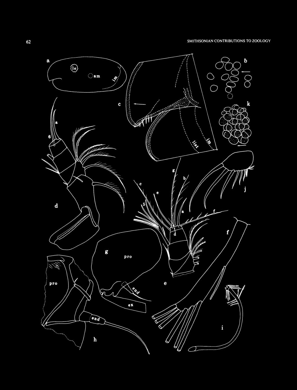
68 62 SMITHSONIAN CONTRIBUTIONS TO ZOOLOGY
, Iv; e, distal right 1st antenna,")
69 NUMBER FIGURE 39 (left). Diasterope procax, new species, adult female, holotype, USNM : a, complete specimen from left side, length 2.01 mm; b, central adductor muscle attachments on right valve, iv; c, anterior of left valve, ov; d, left 1st antenna (not all bristles of 7th and 8th joints shown), Iv; e, distal right 1st antenna, mv;/ part of sensory bristle, 5th joint, right 1st antenna, Iv; g, part of left 2nd antenna, Iv; h, part of left 2nd antenna, mv; i, right 7th limb, l\;j, left furcal lamella, Iv; k, right lateral eye, Iv. Infold: Rostral infold with about 125 long and short bristles. Anteroventral infold with about 115 long and short bristles. Ventral infold with about 28 bristles forming row ending posteriorly at point where list becomes broad. List beginning at inner margin of infold near posterior end of incisur, extending along ventral margin, and continuing on posterior infold where it broadens; list approaching valve edge at midlength. Broad posterior list with 25 broad transparent flap-like bristles and 43 slender bristles (1-3 bristles between each pair of flap-like bristles); 4 tubular processes forming row along midwith of posterior infold; about 15 long and 17 short bristles forming row between posterior list and posterior edge of valve (long bristles ventral to valve midheight). Tip of rostrum of each valve with minute indistinct hairs along inside edge (Figure 39c). Selvage: Short broad fringed lamellar prolongation along inner part of ventral edge of incisur. Posterodorsal curvature of right valve with short segment of fringed lamellar prolongation. Central Adductor Muscle Attachments (Figure 39a,b): Consisting of about 14 oval attachments. Carapace Size (length, height in mm): USNM , 2.01, 0.89, height 44% of length. First Antenna (Figure 39d-f ): 1 st and 2nd joints spinous (not shown) and with long spinous dorsal bristle and shorter distolateral bristle. Combined 3rd and 4th joints shorter than wide, separated by curved suture. 3rd joint with short ventral bristle; left limb of USNM with 5 dorsal bristles (3 single with long spines, followed by 2 paired bristles (lateral with long spines, medial with short spines)), right limb with 6 dorsal bristles (2 single bristles with long spines, 2 paired bristles (lateral with long spines, medial with short spines), followed by 2 paired bristles (lateral with long spines, medial with short spines)). 4th joint with 2 terminal ventral bristles and 1 terminal dorsal bristle with short spines. Sensory bristle of 5th joint with short slender proximal filament and 6 long terminal filaments (Figure 39e,f). Medial bristle of 6th joint long. 7th joint (Figure 39e): a-bristle claw-like, concave dorsally, bare; b-bristle with 3 long marginal filaments; c-bristle long with 7 short marginal filaments. 8th joint: d-bristle represented by minute peg; e-bristle reaching tip of sensory bristle of 5th joint, bare with blunt tip; f-bristle oriented dorsally, with 4 short proximal filaments; g-bristle long with 6 or 7 short marginal filaments. Filaments of sensory bristle of 5th joint and filaments of bristles of 7th and 8th joints, and also tips of b-, c-, f-, and g-bristles of 7th and 8th joints each with minute terminal papilla. Second Antenna: Protopodite with numerous medial spines mostly on distal dorsal half (Figure 39g,h), and small distomedial bristle (Figure 39h). Endopodite with 3 joints indicated by indistinct suture between 1st and 2nd joints and thinning of sclerotized outer layer between 2nd and 3rd joints (Figures 39g,h, 43/J); 3rd joint with long terminal filament. Exopodite: bristle of 2nd joint reaching just past 9th joint, with dense row of slender ventral spines, and dorsal margin either bare or with few slender spines; bristles of joints 3-6 with small ventral spines and natatory hairs; bristles of joints 7 and 8 with natatory hairs, no spines; 9th joint with 4 bristles (2 long with natatory hairs, 2 small bare); joints 3-8 with basal spines increasing in length on distal joints (spine of 8th joint about % length of 9th joint); 9th joint with either single spine or double lateral spines, about same length as spine of 8th joint; joints 2-8 with row of small spines along distal edges. Mandible (Figure 40a-c): Coxale endite: small bristle near base of ventral branch; ventral branch with spines forming 4 oblique rows, and with long slender tip; ventral margin of dorsal branch with proximal spines, 3 pairs of low pointed teeth followed by about 8 minute uneven teeth, short main spine, and without terminal spinous bristle; marginal spines present between main spine and tip; dorsal margin serrate distally and with slender bristle set well back from tip. Basale endite with 4 spinous end bristles (2 long, 2 about % length of long bristles), small glandular peg, 2 dwarf bristles (1 aboutvfe length of other), and 3 triaenid bristles with 5-7 pairs of spines proximal to terminal pair. Ventral margin bare with narrow U-shaped process set well back from valve edge; dorsal margin with backward-pointing bristle (with indistinct spines at midlength), and with 2 terminal bristles (lateral short, medial long); basale without spines. Exopodite about 62% length of dorsal margin of 1st endopodial joint (Figure 40c), hirsute distally, and with 2 short terminal bristles. 1st endopodial joint with 3 long ventral bristles (2 with long and short spines, 1 with short spines). 2nd endopodial joint: ventral margin with 3 long terminal bristles with short marginal spines; dorsal margin with a- to g-bristles (c-bristle slightly stouter than b- and d-bristles; d-bristle about same width as b-bristle, both stouter than a-bristle); 1 short spinous bristle proximal to a-bristle, and 1 short spinous (cleaning-type bristle) between a- and b-bristles; medial side with few rows of indistinct short spines, an oblique row of 2 or 3 spinous cleaning bristles between b- and c-bristles, and an oblique row of 4 or 5 spinous cleaning bristles between c- and d-bristles. 3rd endopodial joint with 5 spinous bristles and stout straight claw with ventral spines. Maxilla (Figure 40d): Epipodite triangular, hirsute. Endite I with 4 bristles (3 long, 1 short); endite II with 3 long bristles. Basale: dorsal margin with 2 short bristles with bases on medial side (1 proximal near base of epipodite, 1 distal); ventral margin with 1 backward-pointing proximal bristle, 1 minute distal bristle with base on lateral side, and 1 long spinous terminal bristle; lateral side with short proximal bristle near midheight; medial surface and dorsal margin spinous. 1st

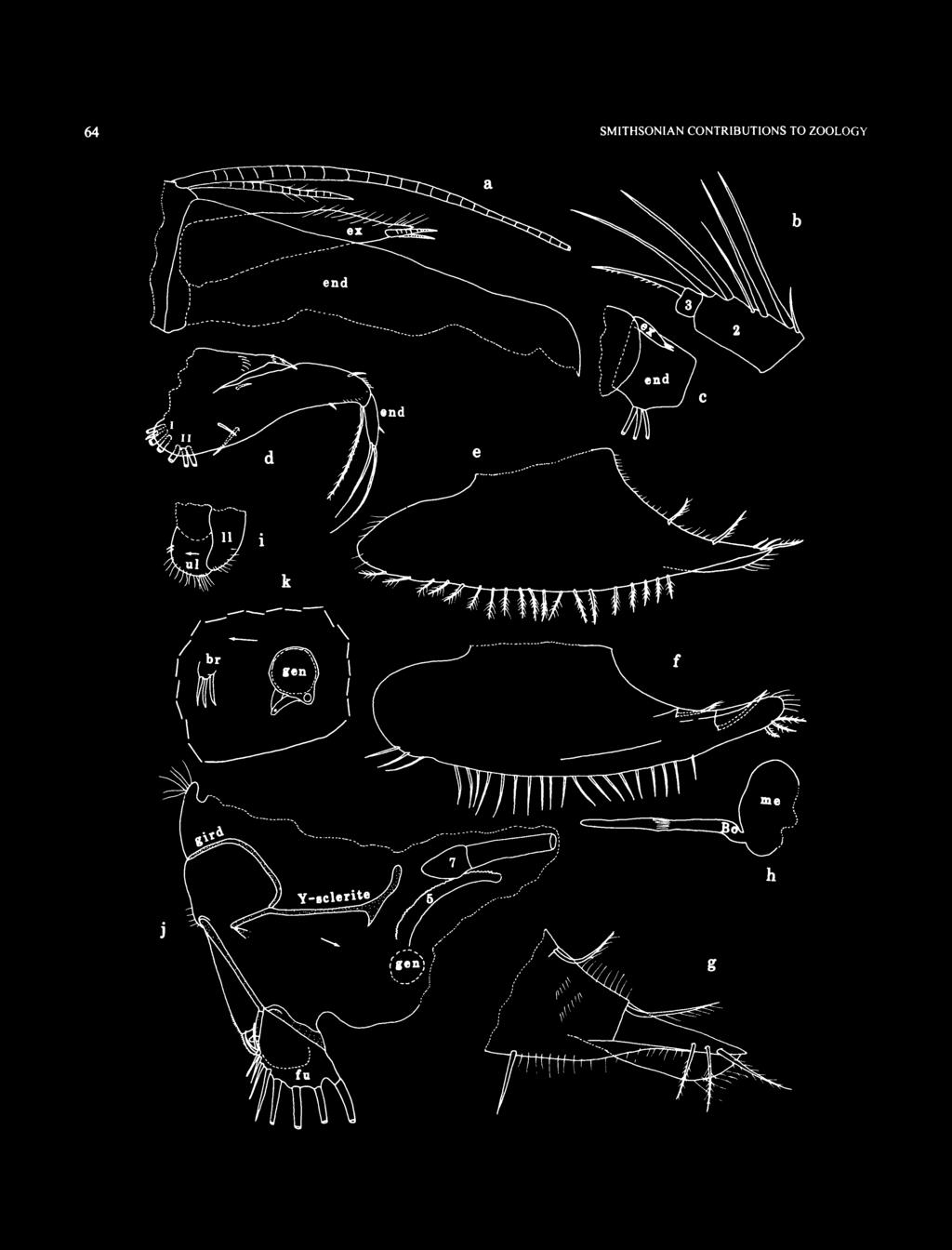
70 64 SMITHSONIAN CONTRIBUTIONS TO ZOOLOGY
, lv;")
71 NUMBER FIGURE 40 (left). Diasterope procax, new species, adult female, holotype, USNM : parts of left mandible: a, exopodite and part of basale and 1st endopodial joint, mv; b, 1st and 2nd endopodial joints (nabs), lv; c, exopodite and parts of basale and 1st endopodial joints, lv; d, right maxilla, lv; e, left 6th limb, mv;/ right 6th limb, lv; g, anterior tip of left 6th limb, mv; h, medial eye and Bellonci organ; i. lips from left side;/ posterior of body from right side; k, left genitalia and brush-like organ. endopodial joint with short bare alpha-bristle and long bare beta-bristle; 2nd joint with long terminal bare bristle reaching past beta-bristle. Fifth Limb: Ventral margin of comb with spinous bristles. Comb otherwise similar to that of A-2 male described below. Sixth Limb (Figure 40e-g): Ventral margin of skirt with bristles, otherwise similar to that of A-2 male described below. Seventh Limb (Figure 39/): Limb very long. Proximal group with 5 cylindrical bristles (3 on 1 side, 2 on other), each with 3-5 bells. Distal group with 6 cylindrical bristles, 3 on each side (4 on terminus, 2 on segment proximal to terminus), each with 3-6 bells. Terminus with opposing combs, each with 11 or 12 spinous teeth. Furca (Figure 39/): Each lamella with 10 claws; 4 posterior claws bristle-like but without rings (2 bent backwards); claws 1-6 with teeth along posterior edge, some longer than others; claws 1-5 with distal anterior spines; claws 9 and 10 also with distal anterior spines; right lamella anterior to left by width of base of claw 1. Lamellae following claws with small spines. Bellonci Organ (Figure 40h): Elongate with broadly rounded tip. Eyes: Medial eye bare, without pigment (Figure 40A). Lateral eye larger than medial eye, with 20 divided ommatidia and brown pigment (Figures 39a, k, 41a). Lips (Figure 40/): Similar to that of A-2 male described below. Genitalia (Figure 40j,k): Oval (with sclerotized tube along ventral edge) on each side of body anterior to furca. Brush-like Organ (Figure 40k): Several indistinct minute bristles on each side of body anterior to genitalia. Posterior of Body (Figure 40/): With small spinous dorsal process, and with few long hairs near midheight. Gills: Well developed. Y-Sclerite (Figures 40/, 52h): Without ventral branch. DESCRIPTION OF A-l MALE (Figure 43b,i,j). Carapace shape similar to that of adult female (Figure 43b). Carapace Size (length, height in mm): USNM , 1.95, 0.90, height 46% of length. First Antenna: Bristles similar in number and location to those of adult female, f-bristle of 8th joint with about 20 short proximal filaments. Bristles on joints similar in number to those of adult female. Second Antenna: Endopodite 3-jointed, larger than that of A-2 male, with 2 short bristles on 2nd joint and 1 long proximal bristle on 3rd joint; tip of 3rd joint tapers to point (Figure 43/,/). Exopodite with 4 bristles on 9th joint. Protopodite and exopodite similar to those of adult female. Mandible: Similar to that of adult female. 2nd endopodial joint with 2 rows of cleaning bristles between b- and c-bristles (2 in proximal row, 6 in distal row). Maxilla and 5th Limb: Not examined in detail but appearing to be similar to the same limbs on the adult female. Sixth Limb: End joint with 19 ventral bristles. Seventh Limb: Bristles cylindrical and same number as those of adult female. Each bristle with 3-5 bells. Furca: Each lamella with 10 claws, similar to those of adult female. Bellonci Organ: Similar to that of adult female. Eyes: Medial eye similar to that of adult female. Lateral eye with 20 ommatidia and brown pigment. Genitalia: A small lobe anterior to furca may be part of copulatory limb, but not identified as such with certainty. DESCRIPTION OF A-2 MALE (Figures 41,42,43c,A:). Carapace elongate with evenly rounded anterior and posterior margins, convex dorsal margin, and slightly convex ventral margin (Figures 41a, 43c); incisur slit-like and just ventral to valve midheight. Surface of valves with minute scattered pores. Infold: Rostral infold with about 50 bristles (Figure 41 b). Anteroventral infold with about 57 short and long bristles (Figure 416). Ventral infold with about 18 bristles forming row ending posteriorly at point where list becomes broad. List beginning at inner margin of infold near posterior end of incisur, extending along ventral margin, and continuing on posterior infold where it broadens; list approaching ventral edge of valve at midlength. Broad posterior list with broad transparent flap-like bristles and slender bristles (1 or 2 bristles between each pair of flap-like bristles) (Figure 41c); 4 tubular processes forming row along midwith of posterior infold; about 20 bristles forming row between posterior list and posterior edge of valve. Tip of rostrum of each valve with minute indistinct hairs along inside edge (possibly part of selvage) (detail in Figure 41 b). Selvage: Short broad fringed lamellar prolongation along inner part of ventral edge of incisur. Carapace Size (length, height in mm): USNM , 1.58, 0.80, height 50% of length; USNM , 1.67, 0.76, height 46% of length; USNM , 1.56, 0.73, height 47% of length. First Antenna (Figure 4ld,e): 1 st joint with medial and lateral spines and hairs. 2nd joint with medial, lateral, ventral, and dorsal spines, 1 spinous dorsal bristle, and 1 shorter spinous distolateral bristle. 3rd and 4th joints partly fused; 3rd joint with minute medial spines in ventral half, small ventral bristle, and 6 dorsal bristles (2 single with long spines, 2 paired bristles with long spines, followed by 2 paired bristles (lateral with long spines, medial with short spines)); 4th joint with few ven-

72 66 SMITHSONIAN CONTRIBUTIONS TO ZOOLOGY a FIGURE 41. Diasterope procax. new species, Instar A 2 male, paratype, USNM : a, complete specimen from left side, length 1.58 nun; b, anterior of left valve, iv; c, posterior of right valve, iv; d, right 1st antenna, Iv; e, part of left 1st antenna, mv;/ part ofright 2nd antenna, mv; g, comb of left Sth limb, mv; h, right 6th limb, mv; i, medial eye and Bellonci organfromright side;/ portion of posterior of body from right side.

73 NUMBER tral spines, 2 terminal ventral bristles, and 1 terminal dorsal bristle. Sensory bristle of 5th joint with small proximal filament and 6 long terminal filaments. Medial bristle of 6th joint long. 7th joint: a-bristle claw-like, concave dorsally, bare; b-bristle with 3 long marginal filaments; c-bristle long, but broken, with 7 short filaments on remaining part. 8th joint: d-bristle represented by minute peg; e-bristle reaching tip of sensory bristle of 5th joint, bare with blunt tip; f-bristle oriented dorsally, with 10 short proximal filaments and terminal papilla; g-bristle long with 7 marginal filaments and terminal papilla. Second Antenna: Protopodite with numerous medial spines mostly in distal dorsal half, and small distomedial bristle (Figure 41/). Endopodite 3-jointed (Figures 41/ 43k); 2nd joint with distal ventral bristle; 3rd joint with long filament at midlength and minute diaphanous terminal process. Exopodite: bristle of 2nd joint reaching 9th joint, with dense row of slender ventral spines and shorter dorsal spines; bristles of joints 3-7 with slender ventral spines and natatory hairs; bristle of joint 8 with natatory hairs, no spines observed; 9th joint with 4 bristles (2 long with natatory hairs, 2 short with short indistinct spines); joints 3-8 with basal spines increasing in length on distal joints (spine of 8th joint % length of 9th joint); 9th joint with lateral spine about same length as basal spine of 8th joint; joints 2-8 with row of small spines along distal edges. Mandible: Coxale endite (Figure 42a-c): small bristle near base of ventral branch; ventral branch with spines forming 4 oblique rows, and long slender tip with minute subterminal spine; ventral margin of dorsal branch with proximal spines, 3 pairs of low pointed teeth followed by 6-10 single minute uneven teeth, short main spine, and small terminal spine; marginal spines present between main spine and tip; dorsal margin serrate distally and with slender bristle set well back from tip. Basale endite with 4 (3 long, 1 short) spinous end bristles, small glandular peg, 2 dwarf bristles, and 3 triaenid bristles with 4 or 5 pairs of spines proximal to terminal pair. Ventral margin bare with U-shaped boss set well back from ventral edge; dorsal margin with backward-pointing bristle (with indistinct spines) at midlength, and 2 terminal bristles (lateral short, medial long); lateral and medial sides without spines. Exopodite about % length of dorsal margin of 1st endopodial joint, hirsute distally, with 2 short terminal bristles. 1st endopodial joint with 3 long ventral bristles (2 with long and short spines, 1 with short spines). 2nd endopodial joint: ventral margin with 3 spinous terminal bristles; dorsal margin with a- to g-bristles (c-bristle stouter than b- and d-bristles; d-bristle about same width as b-bristle), 1 short bristle proximal to a-bristle, and 1 short spinous bristle (cleaning-type bristle) between a- and b-bristles; medial side with few rows of indistinct spines, 2 spinous cleaning bristles between b- and c-bristles, and 4 or 5 spinous cleaning bristles forming oblique row between c- and d-bristles (Figure 42b). 3rd endopodial joint with stout straight claw with ventral teeth, and 5 spinous bristles (Figure 42c). Maxilla (Figure 42d): Epipodite triangular, hirsute. Endite I with 4 bristles (3 long, 1 short); endite II with 3 long bristles. Basale: dorsal margin with 2 short bristles with bases on medial side (1 proximal near base of epipodite, 1 distal); ventral margin with 1 backward-pointing proximal bristle, 1 minute distal bristle with base on lateral side, and 1 long spinous terminal bristle; lateral side with 1 short proximal bristle near midheight; medial surface and dorsal margin spinous. 1st endopodial joint with short alpha-bristle and long bare beta-bristle, 2nd joint with long terminal bristle (with indistinct spines) reaching past beta-bristle. Fifth Limb (Figure 4lg): Comb: lateral side with stout spinous exopodial bristle reaching past end of comb, 1 short slender bristle ventral to base of stout bristle, 2 pairs of bristles closer to ventral margin, and 1 bristle just proximal to proximal pair with base almost on ventral margin, and 1 proximal and 2 distal bristles with bases close to ventral margin. Ventral margin with 34 spinous bristles (4 anterior bristles longer than others; shorter bristles of ventral margin comprising row of alternating long and short bristles, all with short proximal spines and longer spines at tip; anterodorsal corner of comb with few long hairs. Sixth Limb (Figure 41 A): Small indistinct medial bristle near proximal anterior corner. Anterior margin with slender spinous bristle at upper endite and longer and stouter spinous bristle at lower endite. Skirt: anterior end with 3 hirsute ventral bristles and no bristles on lateral flap; ventral margin at midlength with bristles (either with long proximal and short distal spines or only long spines) followed by short space and 2 short bristles with long spines; posterior end of skirt broadly rounded; limb hirsute. Seventh Limb (Figure 42e): Proximal group with 5 tapered bristles (3 on one side, 2 on other), each with 2-4 bells. Distal group with 6 tapered bristles, 3 on each side (4 on terminus, 2 on segment proximal to terminus), each with 2-4 bells. Terminus with opposing combs each with 9 or 10 spinous teeth. Furca (Figure 42/): Each lamella with 9 claws; 2 or 3 posterior claws bristle-like but unringed (1 or 2 bent backwards); claws 1-6 with teeth along posterior edge, some longer than others; claws 1 and 2 with distal anterior spines; right lamella anterior to left by width of claw 1. Lamellae following claws with small spines. Bellonci Organ (Figure 41/): Elongate with broadly rounded tip. Eyes: Medial eye without pigment, bare (Figure 41/). Lateral eye missing from USNM (probably broken off), but well developed with 16 ommatidia and brown pigment on USNM Lips (Figure 42g): Upper lip comprising hirsute lobe on each side of saddle, with 2 minute anterior spines on each lobe. Lower lip comprising hirsute flap on each side of mouth. Genitalia: None observed. Posterior of Body (Figure 41/): With small spinous dorsal process, and with long marginal hairs near midheight.
, mv; c, distal right mandible, lv; d, right maxilla, mv; e, 7th limb;/ left lamella of furca, Iv; g, portion of anterior of body from left side")
74 68 SMITHSONIAN CONTRIBUTIONS TO ZOOLOGY FIGURE 42. Diasterope procax, new species, Instar A 2 male, paratype, USNM : a, coxale endite, left mandible, mv; b. left mandible (nabs), mv; c, distal right mandible, lv; d, right maxilla, mv; e, 7th limb;/ left lamella of furca, Iv; g, portion of anterior of body from left side (mandible removed).
. Carapace Size (length, height in mm): USNM 194466, 1.36, 0.63, height 46% of length. First Antenna: 3rd joint with 6 dorsal bristles.")
75 NUMBER Y-Sclerite: Typically unbranched (only proximal part shown in Figure 4\h). Gills: Well developed. DESCRIPTION OF A-3 FEMALE (Figure 43e,l). Carapace similar in shape to that of adult female (Figure 43e). Carapace Size (length, height in mm): USNM , 1.36, 0.63, height 46% of length. First Antenna: 3rd joint with 6 dorsal bristles. Sensory bristle of 5th joint of right limb, but not left limb of USNM , with small proximal filament. 8th joint: f-bristle with 4 proximal filaments. Second Antenna: Endopodite similar to that of adult female. Exopodite: 9th joint with 3 bristles. Mandible: 2nd endopodial joint with e-bristle, and short bristle proximal to a-bristle (Figure 43/). Maxilla and 5th Limb: Not examined in detail but, in general, similar to adult female. Sixth Limb: Skirt with 15 ventral bristles. Seventh Limb: With same number of bristles as on adult female; each bristle with 2-3 bells. Furca: Each lamella with 8 claws. Bellonci Organ: Similar to that of adult female. Eyes: Medial eye similar to that of adult female. Lateral eye with 18 ommatidia and brown pigment. Y-Sclerite: Similar to that of adult female. Posterior of Body: With short spinous thumb-like process similar to that of adult female. DESCRIPTION OF A-3 MALE (Figure 43d,m). Carapace similar in shape to that of A-2 male (Figure 43d). Carapace Size (length, height in mm): USNM , length 1.36, height 0.64, height 47% of length. First Antenna: Differs from that of adult female in not having short proximal filament on sensory bristle of 5th joint. 8th joint: f-bristle with 7 short filaments. Second Antenna: Endopodite weakly 3-jointed with short distal bristle on 2nd joint and long subterminal filament on 3rd joint (Figure 43m). Exopodite: 9th joint with 3 bristles. Mandible: 2nd exopodial joint with e-bristle. Maxilla and Fifth Limb: Not examined in detail but, in general, similar to those of adult female. Sixth Limb: Similar to that of adult female except with only 14 ventral bristles on skirt. Anterior tip of skirt with 3 bristles. Seventh Limb: With same number of bristles as on adult female; each bristle with 1-3 bells; bristles slightly tapered. Furca: Each limb with 8 claws. Bellonci Organ: Similar to that of adult female. Eyes: Medial eye similar to that of adult female. Lateral eye large with 15 ommatidia and brown pigment. Y-Sclerite: Similar to that of adult female. Gills: Each side with 7 well-developed gills. Posterior of Body: With short spinous thumb-like process similar to that of adult female. DESCRIPTION OF A-4 INSTAR (sex unknown) (Figure 43f,g,n,6). Carapace similar in shape to that of adult female (Figure 43/g). Carapace Size (length, height in mm): USNM , 1.03, 0.49, height 48% of length; USNM , 1.02, 0.50, height 49% of length; USNM , 1.10, 0.52, height 47% of length. First Antenna: Dorsal margin of 3rd joint with 4 or 5 bristles; sensory bristle of 5th joint with 6 terminal filaments but without short proximal filament; limb otherwise similar to those of adult female, but filaments of bristles of joints 7 and 8 not counted, except f-bristle with 3 or 4 proximal filaments. Second Antenna: Endopodite similar to that of adult female (Figure 43n,o). Exopodite: 9th joint with 3 bristles. Mandible: 2nd endopodial joint without e-bristle, without dorsal bristle proximal to a-bristle, and with fewer cleaning bristles than on adult. Maxilla and Fifth Limb: Not examined in detail, but in general similar to those of adult female. Sixth Limb: Similar to that of adult female except with fewer ventral bristles (7-12) on skirt. Seventh Limb: Proximal group with either 5 bristles (3 on one side, 2 on other) or 6 bristles (3 on each side). Distal group with 4 bristles, 2 on each side. All bristles with 1 or 2 bells and bristles tapered (juvenile character). Furca: With 7 claws on each lamella. Bellonci Organ: Similar to that of adult female. Eyes: Medial eye similar to that of adult female. Lateral eye with about 14 ommatidia and brown pigment. Y-Sclerite: Similar to that of adult female. Gills: Each side with 7 well-developed slender gills reaching past posterior of body. Posterior of Body: With short spinous thumb-like process similar to that of adult female. COMPARISONS. This species differs from D. canina in not having a terminal claw-like dorsal bristle on the basale of the mandible, and from D. tenuiseta in having one of the terminal bristles on the dorsal margin of the basale being much shorter than the other, in having 1 dorsal midbristle on the mandibular basale, and in having 2 instead of 4 dorsal bristles on the basale of the maxilla. Diasterope procax differs from D. pilosa in the maxilla having 2 instead of 3 dorsal bristles on the basale and in not having a short bristle just distal to endite II. Diasterope procax differs from D. bisetosa Poulsen, 1965, in not having 2 finger-like anterior processes on the upper lip. Disaterope procax differs from D. grisea (Brady, 1898), and D. schmitti Kornicker, 1975, in having 1 instead of 3 or 5 dorsal midbristles on the mandibular basale. ONTOGENY. Recognition of Stages: It is helpful in identifying stages to examine specimens in which ecdysis has proceeded to where the appendages of the next stage are visible within the appendages of the present stage. Unfortunately, no specimens were at this stage in the 10 specimens of D. procax in the present collection. The adult male of the Cylindroleberidinae is easily recognized by its 3-jointed reflexed endopodite of the 2nd antenna, and by a brush-like sensory bristle on the 5th joint of the 1st
: a, adult female, holotype, USNM 194460, right valve, length 2.")

76 70 SMITHSONIAN CONTRIBUTIONS TO ZOOLOGY FIGURE 43. Diasterope procax, new species: a-g,, carapaces (Figures a-f drawn at same magnification): a, adult female, holotype, USNM , right valve, length 2.01 mm, iv; b, Instar A-l male, paratype, USNM , complete specimen from right side, length 1.9S mm; c, Instar A-2 male, paratype, USNM , complete specimen from left side, length 1.67 mm; d, Instar A-3 male, paratype, USNM , complete specimen from left side, length 1.36 mm; e, Instar A-3 female, paratype, USNM , complete specimen from left side, length 1.36 mm;/g, Instar A-4 (sex unknown), USNM , complete specimen from right and left sides at different magnifications, length 1.10 mm. h-o, endopodites of 2nd antennae (drawn at same magnification): h, adult female, holotype, USNM , right limb, mv; i, Instar A-l male, paratype, USNM , left limb, mv;y, Instar A-l male, paratype, USNM , right limb, mv; *, Instar A-2 male, paratype, USNM , right limb, mv; /, Instar A-3 female, paratype, USNM , left limb, lv; m, Instar A-3 male, paratype, USNM , left limb, lv; n, Instar A-4 (sex unknown), paratype, USNM , right limb, lv; o, Instar A-4 (sex unknown), paratype, USNM , one limb.
 was identified by having genitalia and also by a brush-like organ anterior to the genitalia.")

77 NUMBER 606 antenna. No adult males of D. procax were in the present collection. Adult females are most easily recognized if the marsupium of the carapace contains eggs. No ovigerous females were in the present collection. One adult female in the collection (USNM ) was identified by having genitalia and also by a brush-like organ anterior to the genitalia. An A-l and an A-2 male were identified as male by the large size of the endopodite of the 2nd antenna and by having a characteristic long proximal filament on an elongate 3rd joint. The growth stage of the A-l male was identified by having a carapace almost as large as the adult female, which is a common relationship among species of the Cylindroleberidinae. The growth stage of the A-2 male was identified by comparing the development of the endopodite of its 2nd antenna with that of the A-l male, which has a larger endopodite with a longer 3rd joint. The endopodite of the 2nd antenna of the A-3 male differs from that of the female in being slightly larger and in having the long filament of the 3rd joint in a subterminal rather than a terminal location. The f-bristle of the 8th joint of the 1st antenna of the A-3 male of D. procax differs from that of the A 3 female in having 7 rather than 4 short proximal filaments. Recognition of the growth stage of the A-3 female cannot be determined with certainty because the appendages are fairly similar to those of later female instars. In the present collection, the stage of the A-3 female was estimated by comparing it with the A-3 male: (1) both have the same number of claws on the furca; (2) both have the same maximum number of bells on the bristles of the 7th limb; and (3) both have a carapace of similar length. Also, if a growth factor of 1.23 is assumed, the A-3 female, USNM , should produce an adult female of approximately the size of the adult female USNM (No A-2 females were in the collection.) It is difficult to identify the sex of A-4 instars with certainty, so the sexes of the three A-4 instars in the present collection were left unidentified. The endopodite of the 2nd antenna of USNM appears slightly larger and more strongly developed than those of the other two A-4 instars, which suggests that it is a male and that the other two are females. If the sex of the A-4 instars were known with certainty, the degree of development of the endopodite of the 2nd antenna could be used to separate the A-4 instars from the male A-3 instar. Without knowing the sex of the A-4 instars and, also, the variability of the appendages at either stage, it is difficult to separate the A-3 and A-4 instars with certainty. The A-4 instar stage is estimated by comparing it with the A-3 instar: (1) the carapaces of the A-4 instars are smaller and (2) there are fewer appendage bristles, as well as fewer filaments or bells on the bristles, on the A-4 instars (Table 5). 71 TABLE 5. Selected morphological changes during ontogeny of Diasteropeprocax. (d = dorsal; no. = number; v = ventral; = absent; + = present.) Feature No. of specimens Sex Carapace length (mm)* First Antenna 3rd joint, d bristle 4th joint, v/d bristles 5th joint, sensory bristle short proximal filament 8th joint, f-bristle proximal filaments Second Antenna Exopodite 9th joint bristles Mandible 2nd endopodite joint e-bristle Sixth Limb v bristles Seventh Limb distal bristles proximal bristles no. of bells on bristle Furca, claws Lateral eye no. of ommatidiat A-4 3? / A-3 1 F / Instars A-3 1 M / A-2 3 M / A-l 1 M / Adult 1 F / * Lengths of specimens averaged, t Approximate.


78 72 SMITHSONIAN CONTRIBUTIONS TO ZOOLOGY Although the number of specimens are too few for statistical analysis, it is interesting to note that two A-4 instars have 6 proximal bristles on both 7th limbs, and one A-4 instar has 5 on one limb and 6 on other, compared to a total of 5n nine later instars and adults. The number of bristles on the 7th limb was not determined on a tenth later instar in the collection, USNM (A-l male). A decrease in the number of proximal bristles on the 7th limb in later developmental stages has not been previously reported. Determination of the Number of Instars: The collection of D. procax on hand is interpreted to contain the last three instars and the adult female (Figure 44). Because of the absence of younger stages, it is not possible to be certain of the total number of instars in the species. Both Kornicker (1969:3) and Hiruta (1983:673), however, presented a key to early myodocopid instars in which instar III is without bristles on the 7th limb, whereas later instars have bristles on the 7th limb. If this were to follow in D. procax, the A-4 instar would be at least instar IV, because the 7th limb bears bristles. If that is so, D. procax would have at least seven instars. An alternative interpretation is that instar III of D. procax differs from most other myodocopids in having bristles on the 7th limb. With that interpretation, D. procax would have six instars. Final determination must await the collection of the earlier instars. Two previously studied species of the Cylindroleberidinae (Bathyleberis yamadai Hiruta, 1979, and Synasterope calyx Kornicker, 1981) have only five instars. The number of instars of D. procax is more similar to some members of the Cyclasteropinae (Cyclasteropini and Cycloleberidini), which have six instars (Hiruta, 1983:675). Synasterope Kornicker, 1975 TYPE SPECIES. Synasterope implumis Poulsen, 1965; subsequent designation by Kornicker (1975:440). COMPOSITION AND DISTRIBUTION. Species of the genus are widespread between latitudes of about 47.5 N and 73 S and depths from m (Kornicker, 1992:194). Only one species of Synasterope (S. setisparsa Kornicker, 1958) has been reported previously from the Bahamas. Three species of Synasterope (S. implumis Poulsen, 1965; S. longiseta Poulsen, 1965; and 5. serrata Poulsen, 1965) have been reported from the Virgin Islands, West Indies. Four additional species (one left in open nomenclature) have been reported from the continental shelves and bays of North America (Atlantic Ocean and Gulf of Mexico) (Kornicker, 1986b). An additional species, collected at the Belize Barrier Reef near Carrie Bow Cay, was listed by Cohen (1989b) as Synasterope new species A. Synasterope browni, new species FIGURES 45-47,52g ETYMOLOGY. The species is named in honor of Roland Brown, Smithsonian Institution, who has been of considerable assistance in facilitating research in the Department of Invertebrate Zoology. HOLOTYPE. USNM , adult female on slide and in alcohol. TYPE LOCALITY. Sta , transect BB, Exuma Sound, Bahamas, depth 96 m. PARATYPES. None. DISTRIBUTION. Collected only at type locality, in baited trap A-2 Adult # o A " A-4 A-3 O Female Male % Sex unknown 0.5 -* * n A 1 i 1 1 l Length (mm) FIGURE 44. Graph of carapace length to carapace height for specimens of Diastervpe prolixa in the collection.
.")
79 NUMBER DESCRIPTION OF ADULT FEMALE (Figures 45-47). Carapace oval with evenly rounded anterior and posterior margins; dorsal margin more convex than ventral margin (Figure 45a). Infold: Obscure on slides, but appearing to have relatively few bristles on infold of rostrum (Figure 45c/). Approximate number of flap-like bristles and small bristles on broad posteroventral list shown in Figure 4Se. No processes observed between broad posteroventral list and valve edge. Each valve with 2 large?glands containing closely spaced light amber-colored globules present in space between inner surfaces of anteroventral infold and outer shell (Figure 45b, c). Selvage: Posterodorsal curvature of right valve with short segment of fringed lamellar prolongation (Figure 45e). Central Adductor Muscle Attachments (Figure 45a,/): Comprising about 8 ovoid attachments. Carapace Size (length, height in mm): USNM , 0.80,0.50, height 63% of length. First Antenna (Figure 45g,h): 1st joint with long lateral spines. 2nd joint with row of short lateral spines near distal margin, spinous dorsal bristle, and without lateral bristle. 3rd and 4th joints combined shorter than wide; 3rd joint with small ventral bristle and 6 dorsal bristles (2 single bristles with long spines, 2 paired bristles (lateral with short spines, medial with long spines), and 2 paired bristles with short spines); 4th joint with 1 terminal ventral bristle and 1 terminal dorsal bristle with short spines; suture separating 4th and 5th joints convex. Sensory bristle of 5th joint with 6 terminal filaments. Medial bristle of 6th joint long with short spines. 7th joint: a-bristle claw-like, concave dorsally, with proximal dorsal spines; b-bristle with 3 long dorsal filaments; c-bristle long with 4 or 5 filaments. 8th joint: d-bristle represented by minute peg; e-bristle reaching tip of sensory bristle of 5th joint, bare with blunt tip; f-bristle oriented dorsally, with 3 marginal filaments; g-bristle long with 4 marginal filaments. Filaments of sensory bristle of 5th joint and filaments of 7th and 8th joints each with minute terminal papilla. Second Antenna (Figure 45/,/): Protopodite with small distomedial bristle, and with few rows of medial spines in distodorsal corner. Endopodite 3-jointed with long terminal filament. Exopodite: bristle of 2nd joint reaching 8th joint, with slender ventral spines; bristles of joints 3-8 with slender ventral spines and natatory hairs; 9th joint small, with 2 bristles (1 about 1 /2 length of bristle of 8th joint and with natatory hairs, 1 short with small spines); joints 2 and 3 with small spines along distal medial margin; lateral side of exopodite obscured, but some joints with minute spines along distal margins (spines not shown), basal spines not observed (obscured), but not large if present. Mandible (Figure 46a, b): Coxale endite broken off both limbs of USNM Basale endite with 3 spinous end bristles, glandular peg, 2 dwarf bristles (one % length of other), and 2 triaenid bristles (one close to base) with 3 pairs of spines proximal to terminal pair (terminal pair well developed, about V3 length of bristle). Basale: ventral margin with U-shaped boss near midlength; dorsal margin with 2 subequal terminal bristles with short spines. Exopodite aboutvfe length of dorsal margin of 1 st endopodial joint, with rounded hirsute tip and 2 short subterminal bristles (exopodite of right limb of USNM larger than that of left limb (compare Figures 46a and 46b). 1st endopodial joint with 3 long ventral bristles (2 with long spines, 1 with short spines). 2nd endopodial joint: ventral margin with 3 terminal bristles with short marginal spines; dorsal margin with a-, b-, c-, d-, f-, and g-bristles (c-bristle with slightly stouter base than b- and d-bristles), and with 1 short bristle proximal to a-bristle; medial surface of joint with 4 cleaning bristles in row between b- and c-bristles but closer to c-bristle. 3rd endopodial joint with slightly curved claw with ventral and dorsal spines, and with 5 spinous bristles. Maxilla (Figure 46c,d): Epipodite triangular, short, hirsute at tip. Endite I with 4 bristles (3 long, 1 short); endite II with 3 long bristles. Basale: medial surface with 1 short proximal bristle near ventral margin, and 2 short bristles (proximal near midwidth, distal closer to dorsal margin); lateral surface with 1 short proximal bristle near midwidth; ventral margin with long stout spinous terminal bristle; medial surface and dorsal margin spinous. 1st endopodial joint with short bare alpha-bristle and long bare beta-bristle; 2nd joint with long terminal bare bristle reaching past tip of beta-bristle. Fifth Limb (Figure 46e): Comb (right limb USNM ): lateral side with stout spinous exopodial bristle reaching past end of comb, 1 short slender bifurcate bristle ventral to base of stout bristle (tip of both branches broken off on illustrated limb), 1 pair of bristles and 1 single bristle closer to ventral margin, 1 single bristle proximal to paired bristles and almost on ventral margin, and 1 proximal and 2 distal bristles almost on ventral margin; anterodorsal tip of comb with long hairs. Sixth Limb (Figure 47a): Small indistinct medial bristle near proximal anterior corner. Anterior margin with slender bristle at upper endite and longer spinous bristle at lower endite. Skirt: anterior end with 3 spinous ventral bristles; lateral flap with slender hirsute anterior bristle; ventral margin posterior to midlength with 9 spinous bristles; posterior edge of skirt slightly concave. Seventh Limb (Figure 41b): Proximal group with 5 or 6 bristles (2 or 3 on each side), each with 2-4 bells; distal group with 6 bristles (3 on each side), each with 2-4 bells. Terminus with opposing combs, each with 8 spinous teeth. Furca (Figures 46/ 47c): Each lamella with 9 claws; posterior 4 claws bristle-like but not ringed (2 bent backward); claws 1-5 with teeth along posterior edge (not shown), some longer than others; claw 1 with distal anterior spines; right lamella anterior to left by width of base of claw 1. Bellonci Organ (Figure 46g): Elongate, broad near midlength, with rounded tip. Eyes: Medial eye without pigment, bare (Figures 46g, 41 d). Lateral eye well developed with brown pigment and 15 ommatidia (Figure 45a,j,k).


80 74 SMITHSONIAN CONTRIBUTIONS TO ZOOLOGY FIGURE 45. Synasterope browni. new species, adult female, holotype, USNM : a, complete specimen from left side, length 0.80 mm; b,c, anterior of right and left valves (bristles not shown), iv; d, anterior of left valve, iv; e, posterior of right valve, iv;/ central adductor muscle attachments on left valve, ov; g,h. parts of left 1st antenna, Iv; i, part of right 2nd antenna, mv;y, left lateral eye and part of left 2nd antenna, Iv; k, left lateral eye, Iv; /, 3 eggs in marsupium.

81 NUMBER a FIGURE 46. Synastempe browni, new species, adult female, holotype, USNM : a, left mandible, mv; b, exopodite of right mandible and dorsal edge of 1st endopodial joint, mv; c, right maxilla (limb torn apart during dissection as shown), mv; d, part of left maxilla (nabs), mv; e, comb of right 5th limb, lv;/ right lamella of furca, lv; g, medial eye and Bellonci organ from left side.

82 76 SMITHSONIAN CONTRIBUTIONS TO ZOOLOGY FIGURE 47. Synasterope browni, new species, adult female, holotype, USNM : a, right 6th limb, rav; b, 7th limb; c, portion of posterior of body from left side showing left genitalia and left lamella of furca; d, anterior view of body (Bellonci organ broken off medial eye and not shown); e, lips;/ posterior of body from right side (not all furcal claws shown).
. Lower lip a hirsute flap on each side of mouth. Genitalia (Figures 45a, 47c,/): Oval with sclerotized peripheral rim, present on each side anterior to furca.")
83 NUMBER Lips (Figure 47d,e): Upper lip comprising 2 hirsute lobes on each side of saddle; minute anterior spine on each lobe; saddle not visible in lateral view (Figure 47e) but observed in anterior view (Figure 47d). Lower lip a hirsute flap on each side of mouth. Genitalia (Figures 45a, 47c,/): Oval with sclerotized peripheral rim, present on each side anterior to furca. Brush-like Organ: Not observed, but could have been torn off during dissection. Posterior of Body (Figure 47/): Posterodorsal corner spinous and forming right angle; posterior margin with few long hairs at midheight. Gills: Seven on each side reaching just past posterior of body (2 shown in Figure 47/). V-Sclerite (Figures 47/ 52g): Without ventral branch. Eggs: USNM with small unextruded eggs (3 shown in Figure 45/; 1 shown in Figure 47/); diameter of 3 eggs 0.07 mm, 0.10 mm, 0.10 mm. COMPARISONS. The new species, S. browni, differs from S. setisparsa (Kornicker, 1958), the only other species of the genus reported in the Bahamas, in having many more ventral and posteroventral bristles on the 6th limb (nine compared to at most two or three for S. setisparsa), and also in having a much shorter bristle at the anterior tip of the 6th limb. Synastrope browni differs from 5. cushmani Kornicker, 1974, S. williamsae Kornicker, 1986, and S. psitticina (Darby, 1965) in having well-developed lateral eyes. The 1st antenna of S. browni differs from that of S. serrata Poulsen, 1965, in lacking a lateral bristle on the 1st joint. The 6th limb of S. browni differs from that of S. longiseta Poulsen, 1965, in not having two long bristles at the anterior tip of the skirt and also differs by having many more bristles along the ventral and posteroventral edge (nine compared to four or five for 5. longiseta). The stem proximal to the terminal filaments of the sensory bristle of the 5th joint of the 1st antenna is much longer in S. longiseta and S. implumis Poulsen, 1965, than it is in S. browni. Parasterope Kornicker, 1975 TYPE SPECIES. Asterope Mulleri Skogsberg, 1920; subsequent designation by Kornicker (1975:401). COMPOSITION AND DISTRIBUTION. Members of the genus are widespread between latitudes of about 55 N to 65 S with a depth range from intertidal to 4303 m (Kornicker and Caraion, 1974:7). Two species of Parasterope, P. muelleri (Skogsberg, 1920) and P. extrachelata Kornicker, 1958, have been reported previously from the Bahamas. Four additional species of the genus have been reported from the eastern Atlantic: P. longungues Poulsen, 1965, from about 900 m off St. Croix, U.S. Virgin Islands; P. pollex Kornicker in Bowman and Kornicker, 1967, which is widespread along the North American coast of the Atlantic and Gulf of Mexico; P. zeta Kornicker, 1986b, on the continental shelf of Texas; and P. hulingsi Baker, 1978, on the continental shelf off North Carolina and Georgia (Kornicker, 1986b). Parasterope muelleri (Skogsberg, 1920) FIGURES 48-51, S2a-f Asterope Mulleri Skogsberg, 1920:483. Parasterope muelleri (Skogsberg). Kornicker, 1986b:20 [synonymy]. Maddocks and Kornicker, 1986:284, fig. 90 [in part]. Kornicker and Iliffe, 1989b:45, fig. 27. LECTOTYPE. Adult female on slides in Swedish State Museum, Stockholm. TYPE LOCALITY. English Channel, off Salcombe, coast of England. MATERIAL. Exuma Sound: Sta , transect AA: USNM , adult female on slide and in alcohol. Bermuda: USNM , adult female on slide and in alcohol. Biscayne Bay, Florida: USNM , ,2 adult females each on slide and in alcohol. Mauritania: Sta X070, USNM , adult female on 2 slides and in alcohol. DISTRIBUTION. Exuma Sound at depth of 67 m; also, widespread in North Atlantic Ocean (Kornicker and Iliffe, 1989b:45). DISCUSSION. This species was originally described from the English Channel (Skogsberg, 1920:483). It was subsequently reported in the western Atlantic extending from Bermuda to the West Indies (Kornicker, 1986b:20), and in the eastern Atlantic off Mauritania (Kornicker and Caraion, 1974:8). Because this wide distribution is unusual in benthic myodocopids, an adult female from the Wall was studied in considerable detail to determine whether a character might have been overlooked that could be used to separate the eastern and western Atlantic specimens. The Wall specimen is described below. The 5th joint of the 1st antenna and the Y-sclerite of specimens from four localities are compared in the Figures. The 1st antenna of the Wall female differs from that of the holotype described and illustrated by Skogsberg (1920:484, fig. 89.4) in having a distal row of minute spines just within the dorsal margin of the 5th joint (Figure 48e/). Could Skogsberg have overlooked such spines in the holotype? The lectotype was not reexamined herein, but a female specimen in the Smithsonian collection (USNM ) that had been collected in the eastern Atlantic off Mauritania (depth m) and identified as Parasterope muelleri by Kornicker and Caraion (1974:8) was examined; this specimen is not from the type locality, but it is at least from the same side of the Atlantic. The 1st antenna of the Mauritanian female bears a row of lateral distal spines near the dorsal margin (Figure 52d). These had been overlooked by Kornicker and Caraion (1974:16), who reported no spines on the 5th joint. Unlike the spines on the Bahamian female, this row of spines is slightly curved ventrally at its distal end. Kornicker (1986b:8) reported P. muelleri from eastern Florida and the Florida Keys, but did not mention spines on the 5th joint of the 1st antenna. The 1st antennae of two Florida females (USNM ) from Biscayne Bay, which had been identified by Kornicker in 1975 as P. muelleri, were reexamined. Both limbs of the specimens have distal spines along the

84 78 SMITHSONIAN CONTRIBUTIONS TO ZOOLOGY dorsal margin of the 5th joint (Figure 52b,c). These differ from both the Bahamian and Mauritanian females in projecting past the dorsal edge of the joint and in being more strongly developed; the spines on the Florida specimens are visible using an x40 objective, whereas the spines on the Bahamian and Mauritanian specimens are clearly visible only at a higher magnification (objective: xloo, oil immersion). The row of spines of the Florida specimens also differs from the Mauritanian specimen in not curving ventrally at its distal end. Kornicker (1981:9,fig. 7c) reported that the 5th joint of the 1st antenna of an adult female of P. muelleri from Bermuda has short spines forming a row along the distal dorsal margin. A 1st antenna from one of the Bermudian specimens in that collection (USNM ) was reexamined. The row of spines project past the dorsal edge of the joint but are only clearly visible using an oil immersion objective (xloo) (Figure 52a). The row of spines differs from that of the Mauritanian female in not bending ventrally at its distal end. The spines are less strongly developed than those of the female from Florida, and they project farther past the dorsal edge of the 5th joint then they do in the female from the Bahamas. An insufficient number of specimens were studied at the four localities to determine the variability at each locality in either distribution or the degree of development of the spines on the 5th joint of the female 1st antenna. If future examination of additional specimens also reveals small differences in the distribution and development of the spines of specimens from these different localities, it would suggest that populations of P. muelleri differ in the East and West Atlantic, and that they also differ within the eastern Atlantic in Bermuda, the Bahamas, and Biscayne Bay, Florida. Because spines are present on all specimens examined herein from various localities, it is hypothesized that the spines were overlooked in the original description of the species by Skogsberg (1920:483). No differences were observed in the morphology of the carapace and appendages of the specimens from the four localities studied to warrant the proposal of a new species. The Y-sclerites of the females from the four localities studied are similar (Figure 52e-h). DESCRIPTION OF ADULT FEMALE FROM THE WALL (Figures 48-51, 52g). Carapace tumid in lateral view with dorsal margin more convex than ventral margin; incisur ventral to midheight (Figure 48a). Infold (Figure 486,c): Rostrum and anterodorsal infold with 42 bristles; list of rostrum restricted to tip. Anteroventral infold with about 30 small bristles; about 10 bristles present along ventral infold to point opposite lowermost hyaline flap-like bristle on posterior list. List beginning near inner margin of anterior infold, extending along ventral infold, and continuing on postero-ventral and posterior infold where it broadens; broad postero-ventral and posterior list with 18 or 19 hyaline flap-like transparent bristles and about 12 minute bristles, generally only 1 minute bristle between adjacent flap-like bristles, and no more than 2; 8 long bristles along posteroventral infold between broad list and valve edge, all confined to ventral half of posterior infold. No processes on posterior infold between list and valve edge. Central Adductor Muscle Attachments (Figure 48a,d): Consisting of about 12 ovoid attachments. Vestment (Figure 486): Cluster of few indistinct hairs on vestment just proximal to anterodorsal infold. Carapace Size (length, height in mm): USNM , 1.05,0.70, height 67% of length. First Antenna (Figure 48e/): 1 st joint with few distal lateral and medial spines. 2nd joint with ventral and dorsal spines, 1 spinous dorsal bristle, and 1 shorter lateral bristle with few indistinct spines. 3rd joint with 6 dorsal bristles (2 single with long spines, 1 single bristle with long spines and base slightly lateral, 1 single bristle with long spines, and 2 paired bristles (medial with short spines, lateral with long spines)) and 1 minute ventral bristle. 4th joint with 1 long dorsal bristle with short spines and 2 terminal ventral bristles (longest with short spines). 5th joint with distal row of minute pointed spines along dorsal margin (base of spines on lateral side of joint) (spines seen best using oil immersion objective, and better developed on some limbs than on others); sensory bristle long slender and with 6 terminal filaments. 6th joint with long spinous medial bristle. 7th joint: a-claw with indistinct dorsal spines; b-bristle with 4 marginal filaments; c-bristle longer than sensory bristle of 5th joint, with 5 marginal filaments. 8th joint: d-bristle represented by indistinct minute peg; e-bristle long bare with blunt tip; f-bristle bent dorsally, with 3 marginal filaments and bifurcate tip; g-bristle similar to c-bristle of 7th joint but with 4 marginal filaments. Second Antenna (Figure 49a-c): Protopodite with small distal medial bristle and rows of indistinct distomedial spines (medial bristle missing on right limb of USNM ). Endopodite distinctly 3-jointed with long terminal filament. Exopodite: 1st joint with few distal dorsal spines, but without minute medial terminal bristle; bristle of 2nd joint reaching 9th joint, with abundant slender ventral spines; bristles of joints 3-6 with slender ventral spines and natatory hairs; bristles of joints 7 and 8 with natatory hairs, no spines; 9th joint with 3 bristles with natatory hairs (1 short dorsal, 1 long ventral, 1 medium at joint midwidth); basal spines obscured but observed on joints 5-8; spine of 8th joint about Vb length of 9th joint; lateral spine of 9th joint about % length of 9th joint; joints 2-8 with row of minute spines along distal edges. Mandible (Figure 49*/-/): Coxale endite with slender medial bristle at base of ventral branch; ventral branch with 4 rows of spines and prolonged tip with 2 minute teeth; ventral margin of dorsal branch with 3 small paired nodes, very small main spine, and slender drawn-out tip; dorsal margin of dorsal branch without distal bristle but with minute spines at location of base of missing bristle (bristle missing from both limbs of USNM , but whether this is normal for the species, or whether they are broken off this specimen is not known). Basale with 4 end-type bristles (only 1 shown in Figure 49e) and 3 triaenid bristles with 4 or 5 pairs of marginal spines in addition to small terminal pair, 1 fairly long dwarf bristle, and

85 NUMBER FIGURE 48. Parasterope muelleri (Skogsberg, 1920), adult female, USNM : a, complete specimen from right side, length 1.05 mm; b.c, anterior and posterior of right valve, iv; d, adductor muscle attachments on right valve, iv; ej, parts of right 1st antenna, Iv.

86 80 SMITHSONIAN CONTRIBUTIONS TO ZOOLOGY FIGURE 49. Parasterope muelleri (Skogsberg, 1920), adult female, USNM : a, right lateral eye and right 2nd antenna (nabs), Iv; b. part of left 2nd antenna, mv; c, sclentes attached to protopodite of left 2nd antenna, mv; d, coxale endite, left mandible, mv; e, part of right mandible, mv;/ part of right mandible, Iv; g, right lateral eye, Iv.

87 NUMBER glandular peg; ventral margin with U-shaped boss near base of endite; dorsal margin with 2 long bristles with short spines. Exopodite about same length as dorsal margin of 1st endopodial joint, with 2 small subterminal bristles and hirsute tip. 1st endopodial joint with 3 ventral bristles (2 long with long spines, 1 shorter with short spines). 2nd endopodial joint: dorsal margin with 1 short proximal bristle, long a-, b-, c-, d-bristles, and short cleaning-type bristle between b- and c-bristles; base of c-bristle very slightly stouter than bases of b- and d-bristles; lateral surface with long e- and g-bristles; medial surface with 1 small cleaning bristle and oblique row of 5 cleaning bristles between b- and c-bristles, 1 long g-bristle close to base of d-bristle, and rows of spines; ventral margin with 3 bristles with short spines. 3rd endopodial joint with slightly curved claw with few minute dorsal teeth near tip, and with 5 slender bristles. Maxilla (Figure 50a,b): Endite I with 4 bristles (3 long spinous, 1 short); endite II with 3 long spinous bristles. Epipodite with drawn-out hirsute tip reaching to about midlength of dorsal margin of basale. Basale: medial surface near dorsal margin with bristles (1 proximal, 1 distal); lateral surface with 1 proximal bristle near base of epipodite; ventral margin with 1 proximal backward-oriented bristle and 1 long spinous terminal bristle; dorsal margin with indistinct hairs. 1st endopodial joint with short distal alpha-bristle and long bare beta-bristle about % length of long bare terminal bristle of small 2nd endopodial joint. Fifth Limb (Figure 50c): Comb with spinous exopodial bristle, 1 slender bristle just ventral to base of exopodial bristle (this area somewhat obscured on both limbs of USNM and the possibility that 2 bristles are present cannot be ruled out), and 5 bristles at midlength of comb and almost on ventral margin. Sixth Limb (Figure 50d): Medial side with minute bristle in proximal anterior corner and a bristle at each endite; bristle of lower endite longer and set back slightly from anterior margin. Skirt: anterior tip with 2 bristles (longer bristle anterior and spinous, other bristle bare); ventral margin with 12 or 13 spinous bristles in posterior % (bristles at posteroventral corner longer); lateral anterior flap with hairs but no bristles. Seventh Limb: 6 bristles in proximal group, 3 on each side, same number and distribution in distal group, each bristle with 2-4 bells; terminus consisting of opposing combs, each with 8 spinous teeth. Furca (Figure 50e): Each lamella with nine claws; 2 posterior claws bent backward, bristle-like, ringed, bare; claws 1-6 with teeth along posterior edges, some slightly longer than others; claws 1 and 2 with distal anterior spines. Bellonci Organ (Figure 50/): Elongate, broadening near midlength, with rounded tip. Eyes: Lateral eye well developed with 16 ommatidia and brown pigment (Figures 48a, e, 49a,g). Medial eye about same size as lateral eye, unpigmented (Figure 50/) (whether or not medial eye bears hairs not determined). Upper Lip (Figure 50g): Consisting of 2 hirsute lobes, each with indistinct minute anterior spine, one on each side of saddle bearing minute indistinct anterior spine. Lower lips consisting of hirsute lateral flap on each side of mouth. Genitalia (Figures 50/J, 5\a,e): Small oval ring on each side of body anterior to furca. Brush-like Organ (Figure 50h): 4 minute bristles on each side of body near genitalia. Posterior of Body (Figure 5 la.b.c): Posterodorsal margin with 3 low lobes; ventral of these with long spines; 3 spinous crescents on each side proximal to margin; long hairs along middle of posterior margin. Y-Sclerite (Figures 51a,/ 52g): Without ventral branch; anterior attached to upward curving sclerite. Gills (Figure 510,0*): 7 on each side with small terminal lobe on ventral edge. Eggs (Figure 5\d): USNM with small round unextruded eggs; diameter of 1 egg 0.16 mm. Genus and Species Indeterminate MATERIAL. Exuma Sound: Sta , transect AB, 1 partly dissected juvenile plus 3 early instars, all in alcohol. Sta , transect BB, 1 early instar in alcohol. Sta , transect BB, 2 early instars in alcohol. DISCUSSION. The specimens are early instars and were not identified further. Subfamily ASTEROPTERONINAE Kornicker, 1981 Actinoseta Kornicker, 1958 TYPE SPECIES. Actinoseta chelisparsa Kornicker, COMPOSITION AND DISTRIBUTION. The type species has previously been reported from the Bahamas, Florida, West Indies, and from off Venezuela (Kornicker, 1981). Another species, A. hummelincki Kornicker, 1981, has been reported from the Florida Keys, West Indies, and off Argentina (Kornicker, 1981). Two additional species have been described in this genus: A.jonesi Kornicker, 1981, from off Argentina, and A. nodosa Kornicker, 1981, from the Indian Ocean. Actinoseta chelisparsa Kornicker, 1958 Actinoseta chelisparsa Komicker, 1958:244, figs. 43a-l, 46:10a,b, 69a-f, 70a-i, 89hj,p,q; 1981:192, figs. 9o, lie, 12c, 16d, 17e, 72-77, pis HOLOTYPE. Ovigerous female (lost). TYPE LOCALITY. Bimini, Bahamas. MATERIAL. Exuma Sound: Sta , transect AA, USNM , 2 specimens in alcohol. DISTRIBUTION. Wall off Lee Stocking Island, Exuma Cays, Sta , depth 67 m. REMARKS. The two specimens in the collection were not dissected.
, adult female, USNM 194470: a.")

88 82 SMITHSONIAN CONTRIBUTIONS TO ZOOLOGY FIGURE SO. Parasterope muelleri (Skogsberg, 1920), adult female, USNM : a.b, parts of right maxilla, mv; c. comb of right 5th limb (nabs), mv; a\ right 6th limb, mv; e. right lamella of furca, lv;/ medial eye and Bellonci organ from left side; g, upper and lower lips (distorted); h, genitalia and brush-like organ on right side of body.
, adult female, USNM 194470: a, posterior of body from right side (not all furcal claws shown); b, posterior of body from left side (gills and spinous crescents")
89 NUMBER \ \ \ \ FIGURE 51. Parasterope muelleri (Skogsberg, 1920), adult female, USNM : a, posterior of body from right side (not all furcal claws shown); b, posterior of body from left side (gills and spinous crescents not shown); c, posterior of body from right side (gills not shown); d, posterior of body from left side showing 4 gills and 1 egg); e, genitalia on left side of body;/ right Y-sclerite and girdle of posterior of body from right side.
, adult females: a-d, 5th joints of 1st antennae: a, right limb, Iv, Bermuda, USNM 158293; b, right limb, mv; c.")
90 84 SMITHSONIAN CONTRIBUTIONS TO ZOOLOGY a FIGURE 52. Parasterope muelleri (Skogsbeig, 1920), adult females: a-d, 5th joints of 1st antennae: a, right limb, Iv, Bermuda, USNM ; b, right limb, mv; c. left limb, Iv, Florida, USNM ; d, right limb, lv, Mauritania, USNM e-h, left Y-sclerites and girdles of adult females: e, Bermuda, P. muelleri, USNM ;/ Mauritania, P. muelleri, USNM ; g, Bahamas, Synasterope browni, holotype, USNM ; h, Bahamas, Diasterope procax, holotype, USNM

91 NUMBER Asteropella Kornicker, 1975 TYPE SPECIES. Asteropella mortenseni Poulsen, 1965; subsequent designation by Kornicker (1975:557). COMPOSITION AND DISTRIBUTION This genus contains 12 named species from the Atlantic and Pacific oceans in the vicinity of North and South America between latitudes of about 37 30'N and 41 48'S from depths of intertidal to 57 m (Kornicker, 1981, 1986b). Asteropella species indeterminate MATERIAL. USNM , undissected juvenile, length 0.75 mm, height 0.55 mm. DISTRIBUTION. Sta , Wall off Lee Stocking Island, Exuma Cays, Bahamas, depth 67 m. Order HALOCYPRIDA Dana, 1853 Suborder HALOCYPRIDINA Dana, 1853 The Halocypridina contains the superfamilies Halocypridoidea Dana, 1853, and Thaumatocypridoidea Muller, Both are represented in the present collection. Superfamily HALOCYPRIDOIDEA Dana, 1853 Family HALOCYPRIDIDAE Dana, 1853 Subfamily DEEVEYINAE Kornicker and Iliffe, 1985 Spelaeoecia Angel and Iliffe, 1987 TYPE SPECIES. Spelaeoecia bermudensis Angel and Iliffe, COMPOSITION AND DISTRIBUTION. The genus contains eight species from anchialine caves in Bermuda, Bahamas, Jamaica, Cuba, and Mexico (Kornicker and Yager, 1996; Kornicker and Iliffe, 1998). Spelaeoecia bermudensis Angel and Iliffe, 1987 Spelaeoecia bermudensis Angel and Iliffe, 1987:545, figs Kornicker and Iliffe, 1989b:46, fig. 29. Kornicker, 1989:313, figs HOLOTYPE. USNM , adult female on five slides. TYPE LOCALITY. Green Bay Cave, Bermuda. MATERIAL. Bermuda: Church Cave, sta : USNM , 25 specimens including an adult male. Bitumen Cave, sta : USNM , 8 specimens. DISTRIBUTION. Anchialine caves in Bermuda. HOLOTYPE. USNM , empty carapace in alcohol (sex and age unknown). TYPE LOCALITY. Alfonso Dean Blue Hole, Long Island, Great Bahama Bank. MATERIAL. Exuma Cays: Oven Rock Cave, Great Guana Cay, Sta : USNM , adult male; USNM , 3 specimens; USNM , 1 A-4 instar, 9 adult males, 9 adult females, 24 specimens. Sta (all specimens undissected and in alcohol): USNM , 1 A-3 instar (sex unknown); USNM , 5 adult males; USNM , 5 adult females; USNM , 3 A-l instars (sex unknown); USNM , 2 A-l instars (sex unknown); USNM , 3 A-2 instars (sex unknown). Norman's Pond Cave, Norman's Pond Cay, Sta : USNM ,1 adult female. DISTRIBUTION. Great Bahama Bank: Alphonso Dean Blue Hole, Long Island (type locality), at depth of 13 m, salinity about 20 ppt. Oven Rock Cave, Great Guana Cay, Exuma Cays, at depths of 0-20 m, salinity ppt. Norman's Pond Cave, Norman's Pond Cay, Exuma Cays, at depths of 0-50 m. SUPPLEMENTARY DESCRIPTION OF ADULT FEMALE FROM OVEN ROCK CAVE. Surface of all carapaces with distinct striations. Carapace Size (length, height in mm): USNM , 5 specimens: A, 3.03, 1.42; B, 3.05,1.45; C, 2.87, 1.40; D, 2.71, -1.16; E, 2.73, SUPPLEMENTARY DESCRIPTION OF ADULT MALE FROM OVEN ROCK CAVE. Surface of all carapaces with distinct striations. Carapace Size (length, height in mm): USNM , 5 specimens: A (length only), 2.74; B, 2.78, 1.30; C, -2.66, -1.21; D, 2.82,1.23; E, -2.90, SUPPLEMENTARY DESCRIPTION OF A-l INSTAR (Instar VI?, sex unknown) FROM OVEN ROCK CAVE. Surface of all carapaces with distinct striations. Carapace Size (length, height in mm): USNM , 3 specimens: A, 2.08, 0.94; B, 2.21, 1.02; C, 2.20, USNM ,2 specimens: 1.97,0.81; 2.05,0.85. SUPPLEMENTARY DESCRIPTION OF A-2 INSTAR (Instar V?, sex unknown) FROM OVEN ROCK CAVE. Surface of all specimens with distinct striations. Carapace Size (length, height in mm): USNM , 3 specimens: A, 1.52, 0.69; B (length only), 1.48; C (length only), SUPPLEMENTARY DESCRIPTION OF A-3 INSTAR (Instar IV?, sex unknown) FROM OVEN ROCK CAVE. Surface of specimen with distinct striations. Carapace Size (length, height in mm): USNM , 1.13,0.50. Spelaeoecia capax Kornicker in Kornicker et al., 1990 Spelaeoecia capax Kornicker in Kornicker et al., 1990:23, fig. 14. Komicker and Iliffe, 1998:10, figs. 3a, Spelaeoecia styx Kornicker in Kornicker et al., 1990 Spelaeoecia styx Komicker in Kornicker et al., 1990:6, figs Komicker and Iliffe, 1998:26, figs

92 86 SMITHSONIAN CONTRIBUTIONS TO ZOOLOGY HOLOTYPE. USNM , undissected adult male in alcohol. TYPE LOCALITY. El Dorado Cave, South Andros Island, Great Bahama Bank. MATERIAL. Exuma Cays: Norman's Pond Cave, Norman's Pond Cay: Sta : USNM , 1 specimen in alcohol. Oven Rock Cave, Great Guana Cay, sta : USNM , 1 adult female without carapace, 1 adult male, both in alcohol. DISTRIBUTION. Great Bahama Bank: Eldorado Cave, South Andros Island; Norman's Pond Cave, Norman's Pond Cay, Exuma Cays; Oven Rock Cave, Great Guana Cay, Exuma Cays. SUPPLEMENTARY DESCRIPTION OF ADULT MALE FROM NORMAN'S POND CAVE. Valves decalcified and without visible surface ornamentation. Carapace Size (length, height in mm): USNM , 0.91,0.54. Spelaeoecia mayan Kornicker and Iliffe, 1998 Spelaeoecia mayan Kornicker and Iliffe, 1998:43, figs HOLOTYPE. USNM , undissected adult male in alcohol. TYPE LOCALITY. Cenote Maya Blue, Tulum, Quintana Roo, Mexico. MATERIAL. Cenote 27 Steps, Sta : USNM , 4 specimens. DISTRIBUTION. Quintana Roo, Mexico: Cenote Maya Blue (type locality) and Cenote 27 Steps. Superfamily THAUMATOCYPRIDOIDEA Muller, 1906 Family THAUMATOCYPRIDIDAE Muller, 1906 This family includes two fossil genera known only from the Permian and Mesozoic, and three known from the Holocene {Thaumatocypris Muller, 1906; Thaumatoconcha Kornicker and Sohn, 1976; and Danielopolina Kornicker and Sohn, 1976). Danielopolina Kornicker and Sohn, 1976 TYPE SPECIES. Danielopolina carolynae Kornicker and Sohn, COMPOSITION AND DISTRIBUTION. Including the new species described herein, the genus contains 10 species (see Kornicker and Iliffe, 1998, for key to nine species) from anchialine habitats in the Bahamas, Canary Islands, Cuba, Galapagos Islands, Jamaica, Mexico (Yucatan Peninsula), and Australia, and in the South Atlantic (3459 m depth). Danielopolina mexicana Kornicker and Iliffe, 1989 Danielopolina mexicana Komicker and Iliffe, 1989a: 15, figs. 1, 7, 8; 1998:62, figs HOLOTYPE. USNM , adult female on slide and in alcohol. TYPE LOCALITY. Cenote Maya Blue, Tulum, Quintana Roo, Mexico. MATERIAL. Cenote Ponderosa: Sta : 3 specimens in alcohol; Sta , 2 specimens in alcohol. Cenote 27 Steps: Sta : 3 specimens in alcohol; Sta : 1 specimen in alcohol. All above specimens returned to Iliffe for DNA analysis. Cenote 27 Steps: Sta : USNM , 1 specimen in alcohol. DISTRIBUTION. Quintana Roo Province, Yucatan Peninsula, Mexico: Cenote Maya Blue, Cenote Ponderosa, Cenote 27 Steps. Danielopolina exuma Kornicker and Iliffe, 1998 Danielopolina exuma Kornicker and Iliffe, 1998:70, figs HOLOTYPE. USNM , undissected adult female in alcohol. TYPE LOCALITY. Norman's Pond Cave, Norman's Pond Cay, Exuma Cays, Great Bahama Bank. MATERIAL. Exuma Cays: Oven Rock Cave, Great Guana Cay, Sta : USNM , undissected adult male in alcohol. DISTRIBUTION. Norman's Pond Cave, Norman's Pond Cay, and Oven Rock Cave, Great Guana Cay, Exuma Cays, Bahamas. SUPPLEMENTARY DESCRIPTION OF ADULT MALE. Carapace Size (length, height in mm): (including small anterior process), Danielopolina kakuki, new species FIGURES ETYMOLOGY. This species is named in honor of Brian Kakuk, an exceptional cave diver, and Diving Officer at the Caribbean Marine Research Center, who carried out exploration and biological collections from Oven Rock and other caves in the Bahamas. HOLOTYPE. USNM ,1 juvenile male on slide and in alcohol. TYPE LOCALITY. Sta , Oven Rock Cave, Great Guana Cay, Exuma Cays, Bahamas. PARATYPES. None. DISTRIBUTION. Collected only at type locality. DESCRIPTION (Figures 53-56). Carapace slightly elongate with fairly straight dorsal margin in vicinity of hinge and also straight margin between anterior and anteroventral processes (Figures 53, 54a). Ventral and posterior margins evenly

: Surface reticulate with small spine at intersections of walls forming reticulations. Spines also present on 3 processes.")

93 NUMBER FIGURE 53. Danielopolina kakuki. new species, juvenile male, holotype, USNM 194S34, length (including processes) 0.63 mm, lateral and dorsal views. rounded; valves at greatest width at about midlength and midheight, in vicinity of adductor muscle attachments. Fairly long anterior, anteroventral, and posterodorsal processes with bases just lateral to valve edge, and in similar location on each valve. Ornamentation (Figures 53, 54a): Surface reticulate with small spine at intersections of walls forming reticulations. Spines also present on 3 processes. Walls of reticulations generally broader in vicinity of intersections then tapering to point midway between intersections so that walls appear as 3-pointed star with spine at center. Reticulations mostly hexagonal but few with 4 or 5 sides. Right valve with few long bristles along dorsal and posterodorsal margins (Figure 54a); both valves with divided bristle on anteroventral margin just ventral to anteroventral process (Figure 54a). Muscle Attachments: Adductor muscle attachments central in location (Figure 54a); subround with greatest diameter trending towards posterior end of hinge; consisting of several obscure segments. Oval auxiliary muscle scar located anteroventral to adductor muscle attachments (Figure 54a; not shown in Figure 53). Carapace Size: USNM , length including anterodorsal and posterodorsal processes 0.63 mm; length excluding processes 0.56 mm; height including anteroventral process 0.49 mm; height excluding anteroventral process mm. First Antenna (Figure 546): 1 st joint with 2 ringed bristles (1 lateral, 1 dorsal). 2nd joint with distal medial spines and 1 ringed dorsal bristle. 3rd and 4th joints fused, but place of fusion indicated by thinning in sclerotized dorsal margin and

94 88 SMITHSONIAN CONTRIBUTIONS TO ZOOLOGY

95 NUMBER FIGURE 54 (left). Danielopolina kakuki, new species, juvenile male, holotype, USNM : a, inside view of right valve showing representative reticulations as seen through shell; b, right 1st antenna, lv; c, left 2nd antenna, lv; d, right furcal lamella and unpaired bristle on endopodite of right 2nd antenna, mv; e, proximal left 5th limb, lv;/ left 5th limb, lv; g, right 6th limb, lv. slight decrease in joint width shown by minute step near midlength of ventral margin; 4th joint with short unringed filament-like ventral terminal bristle (bristle broad except for narrow tip). 5th joint with 2 filament-like bristles (1 long (tip missing on illustrated limb), 1 small indistinct). 6th joint bare. 7th joint with 3 bristles (1 short ringed a-bristle, 2 long filament-like b- and c-bristles). 8th joint with 3 terminal bristles (slender dorsal ringed spinous d-bristle, 1 long filament-like e-bristle with widely separated marginal spines, and 1 long filament-like medial f-bristle narrower than e-bristle (tips of e- and f-bristles missing on illustrated limb). Filament-like bristles weakly ringed proximally. Second Antenna: Protopodite with few long hairs at proximal ventral corner (Figure 54c). Endopodite 3-jointed (Figures 54c, 55): 1st joint with 2 ringed a- and b-bristles; 2nd joint with short ringed dorsal bristle with base near midwidth of lateral side and 3 filament-like ventral bristles (2 long, 1 short); 3rd joint of left limb long slender with 3 bristles or processes at tip (ventral process with 2 or 3 minute terminal papillae; dorsal process short unringed; middle process longer than others and somewhat bristle-like, but at most weakly ringed) (Figures 54c, 55b)\ 3rd joint of right limb with terminal processes broken off; stem shorter than on left limb (Figure 55a). Exopodite 8-jointed (Figure 54c): 1st joint divided into long proximal and short distal parts; bristles of joints 2-7 long, with natatory hairs; 8th joint with 2 bristles (1 long with natatory hairs, 1 shorter and narrower with small marginal spines); joints 3-7 with indistinct minute spines along distal dorsal margins; only proximal parts shown of long bristles; natatory hairs not shown on long exopodial bristles. Mandible: Coxale endite with proximal and distal sets of teeth separated by a space (Figure 56a, b); proximal set comprising 4 cusps plus a triangular tooth close to distal set of teeth; surface between cusps and medial and lateral surfaces just proximal to cusps with slender spines; 2 spinous bristles with bases just proximal and another bristle with base just distal to triangular tooth; distal set of teeth consisting of 2 flat teeth, each having 3-5 cusps; 1 slender bristle with base proximal to distal set of teeth. Basale (Figure 56a, c): tooth of endite with 5 triangular cusps (anterior 4 with indistinct minute marginal teeth); posterior edge of endite spinous, with 2 short ringed bristles (distal of these tubular); anterior margin of endite with 1 long ringed bristle near midlength; lateral side of endite with 5 ringed bristles (4 long, 1 short) near midlength and 1 distal bristle ringed except near tip; medial side of basale FIGURE 55. Danielopolina kakuki. new species, juvenile male, holotype, USNM : a, endopodite, right 2nd antenna, mv; b, endopodite, left 2nd antenna, lv.

96 90 SMITHSONIAN CONTRIBUTIONS TO ZOOLOGY b

97 NUMBER FIGURE 56 (left). Danielopolina kakuki, new species, juvenile male, holotype, USNM : a, left mandible, lv; b, distal coxale, left mandible, lv; c, distal basale, left mandible, lv; d, distal 3rd endopodial joint, left mandible, lv; ef, parts of maxilla (limb B); g,h, parts of maxilla (limb A); /', portion of posterior of bodyfromright side showing 7th limb and oval containing round brown cells (stippled);/ left lower lip from left side; k. posterior of bodyfromright side showing limbs in place on body (nabs). near dorsal margin with 2 stout ringed bristles; dorsal margin of basale with cluster of spines at apex. Endopodite 3-jointed: 1st joint with lateral spines and dorsal bristle near midlength; 2nd joint spinous with 1 ringed terminal ventral bristle, and 2 ringed dorsal bristles; 3rd joint with dorsal and medial spines and 6 bristles (middle lateral bristle long claw-like) (Figure 56a,d). Rings not shown on all bristles. Maxilla (Figure 56e-h,k): Endites I to III each with 5 to 8 obscured bristles. Coxale without usual dorsal bristle (probably broken off). Basale with 2 bristles (1 ventral with widely separated long marginal hairs, 1 medial at midwidth). Endopodite: 1st joint with long proximal anterior spines, lateral spines at midlength, 3 long ringed distal anterior bristles, and 3 distal bristles near posterior margin; 2nd joint with 1 anterior stout straight nonarticulated terminal bare claw, 3 slender ringed bristles with bases lateral, and 1 stout proximal claw-like bristle with distal rings and indistinct short marginal spines. Fifth Limb (Figures 54e^ 56k): Protopodite with 5 bristles (1 tubular). Basale with 6 bristles (1 short claw-like, 2 long plumose, 2 ventral tubular, 1 slender medial proximal). Endopodite with 6 bristles (1 small medial tooth-like, 2 long sclerotized ventral claw-like, 1 ventral tubular, and 2 long plumose). Exopodite 3-jointed: 1st joint with 6 bristles; 2nd joint with 2 ventral bristles at midlength; 3rd joint with 3 bristles (middle bristle about 58% and smallest bristle about 34% of longest bristle). Epipodite obscured. Sixth Limb (Figures 54g, 56k): Epipodite with 3 groups of plumose bristles (dorsal and ventral group each with 5 bristles, middle group with 4). Precoxale and coxale each with 2 bristles. Basale with 4 bristles. Small endopodite with 2 bristles. Exopodite: joints 1 and 2 fused, with 2 ventral bristles at midlength; 3rd joint with 1 long bristle and 1 small obscured bristle. With appendages in place, tip of 6th limb extends posteriorly slightly past tip of 5th limb. Seventh Limb: A 7th limb with 2 bristles was observed prior to dissection of specimen (Figure 56i,k), but the limb was not found after specimen was dissected. Whether the limb was lost during the dissection, or was mistakenly observed on the whole specimen prior to dissection, is not known with certainty. Furca (Figure 54d): Each lamella with 4 claws. Claws 1 and 2 anterior, articulated, and weakly ringed, claw 3 on anteroventral corner, nonarticulated, slight recurved, unringed; claw 4 separated by space from claw 3, nonarticulated, not recurved, triangular, unringed; claws 3 and 4 with minute teeth along posterior edges. Each lamella with medial rows of minute spines, some extending ventrally past edge of lamellae. Stout unpaired process on posterior of body just proximal to lamellae. Bellonci Organ: Not observed on specimen but could have been obscured. Lips: Lower lip sclerotized and triangular (Figure 56/). Upper lip with small sclerotized triangular process on each side of anterior of body dorsal to upper lip. Copulatory Organ: Not observed. Ganglion (Figure 56/): Oval on each side containing round brown cells (stippled) present posterior to 2nd antenna. DISCUSSION. This unique specimen bears an elongate 3rd joint on the endopodite of the 2nd antenna; this indicates that the specimen is a male. Because of the absence of a copulatory organ, the specimen is interpreted to be a late instar rather than an adult. Because known A-l males in the Thaumatocyprididae usually have a fairly well-developed copulatory organ (Kornicker and Sohn, 1976, table 4), the specimen may be younger than A-l, but it certainly is not younger than A-2 because of the well-developed 3rd joint of the endopodite of the 2nd antenna. COMPARISONS. The new species, D. kakuki, differs from D. exuma Kornicker and Iliffe, 1998, in having an a-bristle on the 7th joint of the 1st antenna. Also, the reticulations on the carapace of D. kakuki have continuous walls, whereas the walls of reticulations of D. exuma are formed by rows of papillae. Danielopolina exuma is reportedfromoven Rock Cave for the first time herein. The carapace of D. kakuki resembles that of D. mexicana Kornicker and Iliffe, 1989a, in having numerous spines, but the surface of D. kakuki is more strongly reticulated. Each lamella of the furca of the specimen of D. kakuki in the present collection, which is probably an A-2 male, bears only two unarticulated ventral claws, whereas each lamella of the furca of the A-2 instar of D. mexicana probably bears four unarticulated ventral claws (extrapolated from Kornicker and Iliffe, 1998). A specimen interpreted to be an instar II and left in open nomenclature as Danielopolina species A, has been reported previously from Oven Rock Cave (Kornicker and Iliffe, 1998). The carapace of that specimen is without the numerous spines present on the carapace of D. kakuki and each valve is without a posterodorsal process. The 1st antenna of that specimen has an a-bristle on the 7th joint similar to that on the 1st antenna of D. kakuki. The carapace differences between D. kakuki and D. species A do not permit them to be synonymized at this time. Oven Rock Cave is the second cave reported to have more than one species of Danielopolina (Kornicker and Iliffe, 1995).

98 Appendix Station Data with Specimens Collected (in chronological order) Submarine Escarpment All stations were from the escarpment off Lee Stocking Island, Exuma Cays, Bahamas, with the exception of Sta , which was collected in a suction sample off Great Exuma Island by the submersible Clelia. For these stations, the tube trap samples and grab samples were made from the submersible Nekton Gamma, plankton net samples were collected by scuba divers, and a suction sample was made from the submersible Clelia. Two listed species of Jimmorinia are deliberate nomen nuda (being described by Cohen et al., in press), which are included herein to present all myodocopids collected on the escarpment. Open-ocean salinities of 34.9 g/ g/1 are assumed to be present at all stations on the escarpment, based on profiles obtained in September Sta , 7 May 1993, Transect BB Buoy; collected with tube trap baited with sandwich meat and left for 48 hours on a sandy ledge near possible cave entrance at 88 m depth. In addition to ostracodes, the sample contained copepods and nebaliaceans. Diasterope procax: USNM , A-2 male. Jimmorinia gunnari: USNM , Instar I; USNM , adult female; USNM , Instar III; USNM , Instar V. Sta , 19 May 1994, Transect AA Buoy; collected with plankton net from sandy sediments on ledges at 67 m depth of the upper slope. In addition to ostracodes, the sample contained copepods, cumaceans, tanaidaceans, and polychaetes. Actinoseta chelisparsa: USNM , 2 specimens. Asteropella sp. indet.: USNM , 1 juvenile. Eurypylus eagari: USNM , ovigerous female. Eusarsiella costata: USNM , adult male. Eusarsiella ryanae: USNM , ovigerous female; USNM , adult male; USNM , 4 adult males; USNM , Instar I; USNM , 3 Instar I; USNM , , , , 4 Instar HI males. Eusarsiella sp. indet.: USNM , 3 juveniles; USNM , 3 juveniles. Parasterope muelleri: USNM , adult female. Rutiderma schroederi: USNM , adult female; USNM , Instar III. Sta , 22 May 1994, Transect AB Buoy; collected with plankton net from sandy sediments on ledges at 62 m depth of upper slope. In addition to ostracodes, the sample contained copepods and cumaceans. Eusarsiella ryanae: USNM , Instar I; USNM , Instar II?female; USNM , , 2 Instar III males. Eusarsiella sp. indet.: USNM , Instar IV; USNM ,1 juvenile. Vargula exuma: USNM , Instar V female. Vargula sp. indet.: USNM , Instar I. Cylindroleberidinae: 4 juveniles. Sta , 12 May 1995, Transect AA Buoy, collected with grab sampler from sandy slope at 240 m depth. This was from the deepest depth sampled and was from a site where a few boulders projecting from the sand had probably fallen from the rocky escarpment above. The sample contained amphipods, copepods, tanaidaceans, and polychaetes. Myodocopid ostracodes: none. Sta , 12 May 1995, Transect BA Buoy; collected with grab sampler from sandy sediment on ledge beneath rock outcropping at 142 m depth. In addition to ostracodes, the sample contained amphipods, copepods, cumaceans, isopods, nebaliaceans, and tanaidaceans. Diasterope procax: USNM , A-4 instar; USNM , A-4 Instar; USNM , A-l male. Eurypylus hapax: USNM , Instar IV female. Sta , 15 May 1995, Transect BB Buoy; collected with tube trap baited with lobster legs and set on a sandy ledge near possible cave entrance at 99 m depth for 24 hours. In addition to ostracodes, the sample contained amphipods, copepods, and fish. Jimmorinia gunnari: USNM , adult female; USNM , ,5 adult males. Cylindroleberidinae: 1 juvenile. Sta , 15 May 1995, Transect BB Buoy; collected with tube trap baited with lobster legs and set on bare rock ledge near possible cave entrance at 96 m depth for 24 hours. In addition to ostracodes, the sample contained amphipods, copepods, and polychaetes. Jimmorinia gunnari: USNM , 2 specimens. Skogsbergia lerneri: USNM ,4 specimens. Synasterope browni: USNM , adult female. Cylindroleberidinae: 2 juveniles. Sta , 14 May 1995, Transect BB Buoy; collected with tube trap baited with lobster legs and set on a sandy ledge 92

99 NUMBER near possible cave entrance at 105 m depth for 24 hours. In addition to ostracodes, the sample contained amphipods, copepods, isopods, and nebaliaceans. Diasterope procax: USNM , adult female; USNM , A-2 male; USNM , A-2 male; USNM , A-3 female; USNM , A-3 male; USNM , A-4instar. Jimmorinia gamma: USNM , adult female. Jimmorinia gunnari: USNM , adult female; USNM , 2 juveniles; USNM , 8 specimens. Skogsbergia lerneri: USNM , Instar IV. Sta , 15 May 1995, within 50 m of Transect BB; collected with tube trap baited with sandwich meat and set on ledge at 93 m depth on 7 May Due to inclement weather it could not be recovered in two days as planned. The next opportunity to collect this trap did not occur until 15 May At that time, the trap appeared undisturbed, but it was partially encrusted with sponges. All bait had long disappeared from this trap; therefore, it was functioning primarily as a protective habitat and not as a feeding attractant. If ostracodes were initially present, attracted by the original bait, they had since been eaten or had decomposed and had not bred in the trap. The trap contained amphipods, copepods, isopods, tanaidaceans, and fish (2 blennies). Myodocopid ostracodes: none. Sta , 14 Sep 1996, submarine escarpment off George Town, Great Exuma Island, Exuma Cays, Bahamas (about 50 km southeast of the other stations); collected with a suction sampler from sandy ledges at m depth. Eusarsiella ryanae: USNM , 1 juvenile. Jimmorinia gunnari: USNM , 1 specimen (length 1.04 mm, height 0.73 mm). Rutiderma schroederi: USNM , 1 late instar. Anchialine Caves Sta , 22 May 1995, Oven Rock Cave, Great Guana Cay, Exuma Cays, Bahamas; salinity 35 ppt; collected with 93 im mesh plankton net and suction bottle from water column at 1-22 m depths. Kornicker and Iliffe (1998) reported 41 specimens of Spelaeoecia capax and 7 specimens of 5". styx from this sample. Danielopolina kakuki: USNM , 1 juvenile male. Sta , 25 Jul 1995, Cenote Ponderosa, Puerta Aventuras, Quintana Roo, Mexico; salinity 35 ppt; collected with 93 )i.m mesh plankton net in the passage to the Chapel at m depths, which was at or below halocline. Danielopolina mexicana: 3 specimens. Sta , 27 Jul 1995, Cenote 27 Steps, Akumal, Quintana Roo, Mexico; salinity 35 ppt; collected with 93 urn mesh plankton net from water column at m depths. Danielopolina mexicana: 3 specimens. Sta , 15 Dec 1995, Church Cave, Bermuda; salinity 35 ppt; collected with 93 (im mesh plankton net from water column at 1-20 m depths. Spelaeoecia bermudensis: USNM , 25 specimens (including adult male). Sta , 16 Dec 1995, Bitumen Cave, Bermuda; salinity 35 ppt; collected with 93 \im mesh plankton net from water column at 1-25 m depths. Spelaeoecia bermudensis: USNM , 8 specimens. Sta , 11 Jan 1996, Cenote 27 Steps, Akumal, Quintana Roo, Mexico; salinity 35 ppt; collected with 93 \im plankton net from water column at m depths. Danielopolina mexicana: 1 specimen. Spelaeoecia mayan: USNM , 4 specimens. Sta , 2 Jan 1996, Cenote Ponderosa, Puerto Aventuras, Quintana Roo, Mexico; salinity 35 ppt; collected with 93 (im mesh plankton net from water column at m depths, which was at or below halocline. Danielopolina mexicana: 2 specimens. Sta , 17 Mar 1996, Cenote 27 Steps, Akumal, Quintana Roo, Mexico; collected with 93 \im mesh plankton net from water column at m depths, with halocline at 12 m depth. (Salinity on 1 Jan 1996 was 35 ppt.) Danielopolina mexicana: USNM , 1 specimen. Sta , 10 Sep 1996, Norman's Pond Cave, Norman's Pond Cay, Exuma Cays, Bahamas, collected with 93 ^im mesh plankton net and vials from water column at m and 50 m depths. Spelaeoecia capax: USNM , 1 adult female. Spelaeoecia styx: USNM , 1 specimen. Sta , 13 Sep 1996, Oven Rock Cave, Great Guana Cay, Exuma Cays, Bahamas, collected with 93 im mesh plankton net from water column at 0-18 m depths. Danielopolina exuma: USNM , 1 adult male. Spelaeoecia capax: USNM , , : 47 specimens. Spelaeoecia styx: USNM , 1 adult male, 1 adult female. Sta , 18 Aug 1996, Oven Rock Cave, Great Guana Cay, Exuma Cays, Bahamas, collected with suction bottle from water column at 0-18 m depths. Spelaeoecia capax: USNM : 19 specimens. Tidal Blue Holes The tidal Blue Holes have strong tidal currents. Typical open-water species can be swept for considerable distances into these caves and likewise, troglobitic species, if present, could be washed out. No troglobitic ostracodes were collected in these caves. The species in blue holes may be described in a later paper.
100 94 SMITHSONIAN CONTRIBUTIONS TO ZOOLOGY Sta , 5 Aug 1995, Sugar Cay Blue Hole, Sugar Cay, Exuma Cays, Bahamas; collected with 93 [im mesh plankton net from surface of silt mound on wall of cave passage at 31 m depth. Harbansus sp.: 3 specimens. Sarsiellinae: 10 specimens. Sta , 9 Sep 1996, Mystery Cave, Stocking Island, Exuma Cays, Bahamas; collected with 93 jxm mesh plankton net from sandy bottom and walls at 50 m depth. Cylindroleberidinae: 3 specimens. Sta , 9 Sep 1996, Master Harbour Cave, Great Exuma Island, Exuma Cays, Bahamas; collected with 93 (im mesh plankton net from ceiling at m depths. Cypridininae: 2 specimens. Cylindroleberidinae: 1 specimen. Sta , 6 Apr 1996, Conch Sound Blue Hole, Andros Island, Bahamas; collected with 93 (im mesh plankton net from surface of silt mound at 22 m depth. Harbansus sp.: 11 specimens. Sarsiellinae: 1 specimen.
:541-553, figures 1-7, tables 1-3. Baird, W. 1850. The Natural History of the British Entomostraca. 364 pages, 36 plates. London. [Printed for the Ray Society.")
![] Baker, J.H. 1978. Two New Species of Parasterope (Myodocopina, Ostracoda) from Southern California. Crustaceana, 35(2):139-141, figures 1-4. Baltanas, A. 1992.](/docs-images/96/127785147/images/101-2.jpg "Eusarsiella bedoyai (Myodocopida, Sarsiellidae), a New Ostracode Species from a Marine Lava Cave in the Canary Islands. Bijdragen tot de Dierkunde, 61(4):251-255, figures 1-6. Barr, T.C., Jr., and J.")
101 Literature Cited Angel, M.V., and T.M. Iliffe Spelaeocia bermudensis New Genus, New Species, a Halocyprid Ostracod from Marine Caves in Bermuda. Journal of Crustacean Biology, 7(3): , figures 1-7, tables 1-3. Baird, W The Natural History of the British Entomostraca. 364 pages, 36 plates. London. [Printed for the Ray Society.] Baker, J.H Two New Species of Parasterope (Myodocopina, Ostracoda) from Southern California. Crustaceana, 35(2): , figures 1-4. Baltanas, A Eusarsiella bedoyai (Myodocopida, Sarsiellidae), a New Ostracode Species from a Marine Lava Cave in the Canary Islands. Bijdragen tot de Dierkunde, 61(4): , figures 1-6. Barr, T.C., Jr., and J.R. Holsinger Speciation in Cave Faunas. Annual Review of Ecology and Systematics, 16: , figure 1. Bowman, T.E., and L.S. Kornicker Two New Crustaceans: The Parasitic Copepod Sphaeronellopsis monothrix (Choniostomatidae) and Its Myodocopid Ostracod Host Parasterope pollex (Cylindroleberidae) from the Southern New England Coast. Proceedings of the United States National Museum, 123(3613): 29 pages, 7 figures, 1 plate. Bowman, T.E., S.P. Gamer, R.R. Hessler, T.M. Iliffe, and H.L. Sanders Mictacea, a New Order of Crustacea Peracarida. Journal of Crustacean Biology, 5:74-78, figure 1. Brady, G.S Entomostracgs. In A.G.L. de Folin and L. Perier, Les Fonds de la Mer, 1 (supplement): On New or Imperfectly Known Species of Ostracoda, Chiefly from New Zealand. Transactions of the Zoological Society of London, 14(part B): , plates Brady, G.S., and A.M. Norman A Monograph of the Marine and Freshwater Ostracoda of the North Atlantic and of Northwestern Europe. Scientific Transactions of the Royal Dublin Society, series 2, 5: Camacho, A.I., E. Bello, J.M. Becerra, and N. Vaticon A Natural History of the Subterranean Environment and Its Associated Fauna. In Ana Isabel Camacho, editor, The Natural History of Biospeleology, pages , 7 figures, 4 diagrams. Madrid: Museo Nacional de Ciencias Naturales. Christiansen, K Biological Processes in Space and Time: Cave Life in the Light of Modern Evolutionary Theory. In Ana Isabel Camacho, editor, The Natural History of Biospeleology. pages , table 1. Madrid: Museo Nacional de Ciencias Naturales. Cohen, A.C Rearing and Postembryonic Development of the Myodocopid Ostracode Skogsbergia lerneri from Coral Reefs of Belize and the Bahamas. Journal of Crustacean Biology, 3(2): , figures 1-10, tables Myodocopid Ostracodes on the Coral Barrier Reef at Carrie Bow Cay, Belize: Systematics, Juvenile Stages, Life History, and Distribution Two volumes, xvi pages, 128 figures, 26 tables. Doctoral dissertation, George Washington University, Graduate School of Arts and Sciences, Washington, D.C. 1989a. Eusarsiella donabbotti. New Ostracode Species (Sarsiellidae) from the Belize Barrier Reef. Bulletin of Marine Science, 45(2): b. Comparison of Myodocopid Ostracodes in Two Zones of the Belize Barrier Reef Near Carrie Bow Cay with Changes in Distribution Bulletin of Marine Science, 45(2): Cohen, A.C, and L.S. Kornicker Taxonomic Indexes to Ostracoda (Suborder Myodocopina) in Skogsberg (1920) and Poulsen (1962,1965). Smithsonian Contributions to Zoology, 204:29 pages Catalog of the Rutidermatidae (Crustacea: Ostracoda). Smithsonian Contributions to Zoology, 449: 11 pages. Cohen, A.C, L.S. Kornicker, and T.M. Iliffe In press. Jimmorinia, a New Genus of Myodocopid Ostracoda (Cypridinidae) with Two New Species from the Bahamas, Jamaica, Honduras, and San Bias Islands, Panama. Smithsonian Contributions to Zoology, 605. Cohen, A.C, and J.G. Morin Three New Luminescent Ostracodes of the Genus Vargula from the San Bias Region of Panama. Contributions in Science, Natural History Museum of Los Angeles County, 373: 23 pages, 8 figures Six New Luminescent Ostracodes of the Genus Vargula (Myodocopida, Cypridinidae) from the San Bias Region of Panama. Journal of Crustacean Biology, 9(2): ,14 figures, 1 table Morphological Relationships of Bioluminescent Caribbean Species of Vargula (Myodocopa). In R. Whatley and C. Maybury, editors, Ostracoda and Global Events, pages New York: Chapman and Hall The Cypridinid Copulatory Limb and a New Genus Kornickeria (Ostracoda: Myodocopida) with Four New Species of Bioluminescent Ostracods from the Caribbean. Zoological Journal of the Linnean Society, 108:23-84, figures Courbon, P., C. Chabert, P. Bosted, and K. Lindsley Atlas of the Great Caves of the World. 369 pages. St. Louis, Missouri: Cave Books. Dana, J.D Tribe III: Cyproidea = Ostracoda. In Crustacea. In United States Exploring Expedition during the Years , 1840, 1841, 1842, under the Command of Charles Wilkes, U.S.N., with Atlas of 96 plates, 13(2): , plates 90, 91. Philadelphia: C. Sherman. Danielopol, D.L The Origin of the Anchialine Cave Fauna the "Deep Sea" Versus the "Shallow Water" Hypothesis Tested Against the Empirical Evidence of the Thaumatocyprididae (Ostracoda). Bijdragen tot de Dierkunde, 60(3/4): , figure 1. Darby, D.G Ecology and Taxonomy of Ostracoda in the Vicinity of Sapelo Island, Georgia. In R.V. Kesling, editor, Four Reports of Ostracod Investigations, 2:1-77, 11 figures, 33 plates. Ann Arbor, Michigan: University of Michigan. Fairbanks, R.G A 17,000-Year Glacic-Eustatic Sea Level Record: Influence of Glacial Melting Rates on the Younger Dryas Event and Deep-Ocean Circulation. Nature, 342 (7 December 1989): , figures 1-6. Fosshagen, A., and T.M. Iliffe Two New Genera of Calanoida and a New Order of Copepoda, Platycopioida, from Marine Caves on Bermuda. Sarsia, 70: Hart, C.W., Jr., and R.B. Manning Two New Shrimps (Procarididae and Agostocarididae, New Family) from Marine Caves of the Western North Atlantic. Journal of Crustacean Biology, 6(3):408-^16, figures

102 96 SMITHSONIAN CONTRIBUTIONS TO ZOOLOGY Hart, C.W., Jr., R.B. Manning, and T.M. Iliffe The Fauna of Atlantic Marine Caves: Evidence of Dispersal by Sea Floor Spreading while Maintaining Ties to Deep Waters. Proceedings of the Biological Society of Washington, 98: Hartmann, G Zur Kenntnis des Eulitorals der afrikanischen Westkuste zwischen Angola und Kap der Guten Hoffnung und der afrikanischen Ostkuste von Sudafrika und Mozambique unter besonderer Berucksichtigung der Polychaeten und Ostracoden, 3: Die Ostracoden des Untersuchungsgebiets. Mitteilungen aus dem Hamburgischen Zoologischen Museum und Institut, 69: , plates Hiruta, Shinlchi A New Species of the Genus Sarsiella Norman from Hokkaido, with Reference to the Larval Stages (Ostracoda: Myodocopina). Journal of the Faculty of Science, Hokkaido University, series 6 (Zoology), 21(l):44-60, figures 1-12, plate A New Species of Bathyleberis Kornicker from Hokkaido, with Reference to the Larval Stages (Ostracoda: Myodocopina). Journal of the Faculty of Science, Hokkaido University, 22(1): Post-Embryonic Development of Myodocopid Ostracoda. In Rosalie F. Maddocks, editor, Applications of Ostracoda, pages '. University Park, Houston: Department of Geosciences, University of Houston. Humphreys, W.F Stygofauna in Semi-Arid Tropical Western Australia: A Tethyan Connection? Memoires de Biospeologie, 20: Humphreys, W.F., and M. Adams The Subterranean Aquatic Fauna of the North West Cape Peninsula, Western Australia. Records of the Western Australia Museum, 15: Huys, R Rotundiclipeidae, a New Family (Copepoda: Harpacticoida) from an Anchihaline Cave on Tenerife, Canary Islands. Stygologia, 4(l):42-63, figures 1-11, table 1. Iliffe, T.M The Zonation Model for the Evolution of Aquatic Faunas in Anchialine Waters. Stygologia, 2:2-9, table Crevicular Dispersal of Marine Cave Faunas. Memoires de Biospeologie, 17: Anchialine Cave Fauna of the Galapagos Islands. In Matthew J. James, editor, Galapagos Marine Invertebrates, , table 1, figures 1-8. New York: Plenum Press Fauna Troglobia acuatica de la peninsula de Yucatan. In Sergio I. Salazar-Vallejo and Norma Emilia Gonzalez, editors, Biodiversidad marina y costera de Mexico, 865 pages. Quintana Roo, Mexico: Comision Nacional para el Conocimiento y Aprovechamiento de la Biodiversidad, Centra de Investigaciones de Quintana Roo Bermuda. In V. Decu and C. Juberthie, editors, Encyclopaeida Biospeologica, 1: , figures 1-4, tables 1-2. Paris: Society of Biospeleology. Iliffe, T.M., C.W. Hart, Jr., and R.B. Manning Biogeography and the Caves of Bermuda. Nature, 302: , figure 1. Iliffe, T.M., H. Wilkens, J. Parzefall, and D. Williams Marine Lava Cave Fauna: Composition, Biogeography, and Origins. Science, 255: Jaume, D Presence of Troglobitized Janiridae (Isopoda: Asellota: Janiroidea) in Anchihaline Caves of the Balearic Islands (Mediterranean); Description of Trogloianiropsis lloberai n. gen., n. sp. Contributions to Zoology, 65(3): , figures Jaume, D., and G.A. Boxshall Stygocyclopia balearica, a New Genus and Species of Calanoid Copepod (Pseudocylopiidae) from Anchihaline Caves in the Balearic islands (Mediterranean). Sarsia, 80: , figures a. A New Genus and Two New Species of Cave-Dwelling Misophrioid Copepods from the Balearic Islands (Mediterranean). Journal of Natural History, 30: , figures b. Two New Genera of Cyclopinid Copepods (Crustacea) from Anchihaline Caves on Western Mediterranean and Eastern Atlantic Islands. Zoological Journal of the Linnean Society, 117: , figures 1-9. Juberthie, C, and T.M. Iliffe Bahama Islands. In V. Decu and C. Juberthie, editors, Encyclopaeida Biospeologica, 1: , figures 1-8, table 1. Paris: Society of Biospeleology. Kendall, C.G. St. C, R.F. Dill, and E.A. Shinn, editors Guidebook to the Marine Geology and Tropical Environments of Lee Stocking Island, the Southern Exumas, Bahamas. 74 pages, plates San Diego, California: KenDill Publishers. King, C.E., and L.S. Kornicker Ostracoda in Texas Bays and Lagoons: An Ecologic Study. Smithsonian Contributions to Zoology, 24: 92 pages, 15 figures, 21 plates, 19 tables. Kornicker, L.S Ecology and Taxonomy of Recent Marine Ostracodes in the Bimini Area, Great Bahama Bank. Publications of the Institute of Marine Science (The University of Texas), 5: , 89 figures, 1 map, 4 tables Morphology, Ontogeny, and Intraspecific Variation of Spinacopia, a New Genus of Myodocopid Ostracod (Sarsiellidae). Smithsonian Contributions to Zoology, 8: 50 pages, 26 figures, 6 plates, 7 tables Revision of the Cypridinacea of the Gulf of Naples (Ostracoda). Smithsonian Contributions to Zoology, 178: 64 pages, 26 figures, 2 tables Antarctic Ostracoda (Myodocopina), Parts 1 and 2. Smithsonian Contributions to Zoology, 163: 720 pages, 432 figures, 9 plates, 21 tables Revision, Distribution, Ecology, and Ontogeny of the Ostracode Subfamily Cyclasteropinae (Myodocopina: Cylindroleberididae). Smithsonian Contributions to Zoology, 319: 548 pages, 174 figures, 185 plates, 23 tables Rutidermatidae of the Continental Shelf of Southeastern North America and the Gulf of Mexico (Ostracoda: Myodocopina). Smithsonian Contributions to Zoology, 371: 89 pages, 51 figures, 3 plates, 1 table Cypridinidae of the Continental Shelves of Southeastern North America, the Northern Gulf of Mexico, and the West Indies (Ostracoda: Myodocopina). Smithsonian Contributions to Zoology, 401: 37 pages, 17 figures, 2 maps, 1 table. 1986a. Sarsiellidae of the Western Atlantic and Northern Gulf of Mexico, and Revision of the Sarsiellinae (Ostracoda: Myodocopina). Smithsonian Contributions to Zoology, 415: 217 pages, 113 figures, 34 plates, 7 tables. 1986b. Cylindroleberididae of the Western North Atlantic and the Northern Gulf of Mexico, and Zoogeography of the Myodocopina (Ostracoda). Smithsonian Contributions to Zoology, 425: 139 pages, 63 figures, 6 tables The Adult Male of the Troglobitic Ostracode Spelaeocia bermudensis Angel and Iliffe, 1989, from an Anchialine Cave in Bermuda (Crustacea: Ostracoda: Halocypridoidea). Proceedings of the Biological Society of Washington, 102(2): , figures Myodocopid Ostracoda of Enewetak and Bikini Atolls. Smithsonian Contributions to Zoology, 505: 140 pages, 71 figures Myodocopid Ostracoda of the Benthedi Expedition, 1977, to the NE Mozambique Channel, Indian Ocean. Smithsonian Contributions to Zoology, 531: 243 pages, 109 figures, 4 tables Ostracoda (Myodocopina) of the SE Australian Continental Slope, Part 1. Smithsonian Contributions to Zoology, 553: 200 pages, 111 figures, 4 tables.
.")
.")
103 NUMBER Ostracoda (Myodocopina) from Shallow Waters of the Northern Territories and Queensland, Australia. Smithsonian Contributions to Zoology, 578: 97 pages, 64 figures, 4 tables. Kornicker, L.S., and J.H. Baker Vargula tsujii, a New Species of Luminescent Ostracoda from Lower and Southern California (Myodocopa: Cypridininae). Proceedings of the Biological Society of Washington, 9O(2): , figures 1-6. Kornicker, L.S., and D.J. Barr Anchialine Ostracoda (Halocyprididae) from San Salvador, Bahamas. Smithsonian Contributions to Zoology, 588: 20 pages, 11 figures, 3 tables. Kornicker, L.S., and F.E. Caraion West African Myodocopid Ostracoda (Cylindroleberididae). Smithsonian Contributions to Zoology, 179: 78 pages, 43 figures, 1 table. Kornicker, L.S., and A.C. Cohen Dantyinae, a New Subfamily of Ostracoda (Myodocopina: Sarsiellidae). Proceedings of the Biological Society of Washington, 91(2): , figures 1-5, plates 1-7. Kornicker, L.S., and T.M. Iliffe Deeveyinae, a New Subfamily of Ostracoda (Halocyprididae) from a Marine Cave on the Turks and Caicos Islands. Proceedings of the Biological Society of Washington, 98(2): , figures a. New Ostracoda (Halocyprida: Thaumatocyprididae and Halocyprididae) from Anchialine Caves in the Bahamas, Palau, and Mexico. Smithsonian Contributions to Zoology, 470: 47 pages, 22 figures, 8 tables. 1989b. Ostracoda (Myodocopina, Cladocopina, Halocypridina) Mainly from Anchialine Caves in Bermuda. Smithsonian Contributions to Zoology. 475: 88 pages, 49 figures, 22 tables. 1989c. Troglobitic Ostracoda (Myodocopa: Cypridinidae, Thaumatocyprididae) from Anchialine Pools on Santa Cruz Island, Galapagos Islands. Smithsonian Contributions to Zoology, 483: 38 pages, 17 figures, 15 tables Ostracoda (Halocypridina, Cladocopina) from an Anchialine Lava Tube in Lanzarote, Canary Islands. Smithsonian Contributions to Zoology, 568: 32 pages, 16 figures, 1 table Myodocopid Ostracoda (Halocypridina, Cladocopina) from Anchialine Caves in the Bahamas, Canary Islands, and Mexico. Smithsonian Contributions to Zoology, 599: 93 pages, 64 figures, 2 maps, 9 tables. Komicker, L.S., and K.G. McKenzie Redescription of Eurypylus petrosus Brady, 1869, and a Key to the Genera of Sarsiellidae (Myodocopina: Ostracoda). Proceedings of the Biological Society of Washington, 89(27): , 1 figure The Adult Male of the Myodocopid Ostracode Philomedes cubitum Komicker, 1975, from the Strait of Magellan (Crustacea: Ostracoda: Myodocopina). Proceedings of the Biological Society of Washington, 109(3): , figures 1-7. Kornicker, L.S., and R.J. Palmer Deeveya bransoni, a New Species of Troglobitic Halocyprid Ostracode from Anchialine Caves on South Andros Island, Bahamas (Crustacea: Ostracoda). Proceedings of the Biological Society of Washington, 100(3): Kornicker, L.S., and G.C.B. Poore Ostracoda (Myodocopina) of the SE Australian Continental Slope, Part 3. Smithsonian Contributions to Zoology, 573: 186 pages, 102 figures, 17 tables. Komicker, L.S., and I.G. Sohn Phytogeny, Ontogeny, and Morphology of Living and Fossil Thaumatocypridacea (Myodocopa: Ostracoda). Smithsonian Contributions to Zoology, 219: 124 pages, 93 figures. Komicker, L.S., and J. Yager The Troglobitic Halocyprid Ostracoda of Anchialine Caves in Cuba. Smithsonian Contributions to Zoology, 580: 16 pages, 9 figures. Komicker, L.S., J. Yager, and D. Williams Ostracoda (Halocyprididae) from Anchialine Caves in the Bahamas. Smithsonian Contributions to Zoology, 495: 51 pages, 30 figures, 4 tables. Maddocks, R.F., and L.S. Komicker Class Ostracoda (Mussel Shrimps). In W. Sterrer, editor, Marine Fauna and Flora of Bermuda, pages New York: John Wiley and Sons. Morin, J.G., and A.C. Cohen Two New Luminescent Ostracodes of the Genus Vargula (Myodocopida: Cypridinidae) from the San Bias Region of Panama. Journal of Crustacean Biology, 8(4): , figures 1-7, table 1. Muller, G.W Ostracoda. In Wissenschaftliche Ergnebnisse der Deutsche Tiefsee-Expedition , 8(2): 154 pages, 31 plates. Mylroie, J.E., J.L. Carew, and H.L. Vacher Karst Development in the Bahamas and Bermuda. In H.A. Curran and B. White, editors, Terrestrial and Shallow Marine Geology of the Bahamas and Bermuda. Geological Society of America, Special Paper, 300: , figures 1-9. Palmer, R Life in a Sunless Sea. Sea Frontiers, 32(4): , photographs 2-9, map 1. Poulsen, E.M Ostracoda Myodocopa, 1: Cypridiniformes Cypridinidae. Dana Report, 57:1-414,181 figures. Copenhagen: Carlsberg Foundation Ostracoda Myodocopa, 2: Cypridiniformes Rutidermatidae, Sarsiellidae and Asteropidae. Dana Report, 65:1-484, 156 figures. Copenhagen: Carlsberg Foundation. Reddell, J.R A Preliminary Survey of the Caves of the Yucatan Peninsula. In J.R. Reddell, editor, Studies of the Caves and Cave Fauna of the Yucatan Peninsula, pages Association for Mexican Cave Studies, bulletin 6. Austin, Texas: Speleo Press. Rocha, C.E.F. da, and T.M. Iliffe Speleoithonidae, a New Family of Copepoda Cyclopoida, from Anchialine Caves on the Bahama Islands. Sarsia, 76: , figures Sars, GO ("1865"). Oversigt af Norges marine Ostracoder. Forhandlinger i Videnskabs-Selskabet I Christiania. 8: [Preprint, 1865.] Scott, A Report on the Ostracoda Collected by Professor Herdmann at Ceylon in Supplementary Reports, Ceylon Pearl Fisheries, 22: , figures 1,2. London. Sealey, N.E Bahamian Landscapes: An Introduction to the Geography of the Bahamas. Second edition, 128 pages, 142 figures. Nassau, Bahamas: Media Publishing. Sket, B Atlantasellus cavernicolus, n. gen. n. sp. (Isopoda Asellota, Atlantasellidae n. fam.) from Bermuda. Bioloski Vestnik (Ljubljana), 27(2): Ecology of the Mixohaline Hypogean Fauna along the Yugoslav Coasts. Stygologia, 2(4): The Anchihaline Habitats, a Dispersed "Center" of Biotic Diversity. In Ira D. Sasowsky, Daniel W. Fong, and Elizabeth L. White, editors, Conservation and Protection of the Biota of Karst, 125 pages. Charles Town, West Virginia: Karst Waters Institute, Special Publication 3.

104 98 SMITHSONIAN CONTRIBUTIONS TO ZOOLOGY Sket,B., and T.M. Iliffe Cave Fauna of Bermuda. Internationale Revue der Gesamten Hydrobiologie, 65: , figure 1, tables 1-3. Skogsberg, T Studies on Marine Ostracods, I: Cypridinids, Halocyprids, and Polycopids. Zoologiska Bidragfr&n Uppsala, supplement 1: Stock, J.H Deep Sea Origin of Cave Faunas, an Unlikely Supposition. Stygolo- #ia, 2: Suirez-Morales, E., and T.M. Iliffe New Superfamily of Calanoida (Copepoda) from an Anchialine Cave in the Bahamas. Journal of Crustacean Biology, 16(4): , figures 1-4, table 1. Tressler, W.L Marine Ostracoda from Tortugas, Florida. Journal of the Washington Academy of Sciences, 39(10): ,25 figures. Warner, G.F., and C.A.M. Moore Ecological Studies in the Marine Blue Holes of Andros Island, Bahamas. Cave Science, 1 l(l):30-44, figures 1,2, plates 1-9, tables 1-8. Wilkens, H., J. Parzefall, and T.M. Iliffe Origin and Age of the Marine Stygofauna of Lanzarote, Canary Islands. Mitteilungen aus dent Hamburgischen Zoologischen Museum undlnstitut, 83: , figures 1-3. Wilson, C.W., Jr Crustacean Parasites of West Indian Fishes and Land Crabs, with Descriptions of New Genera and Species. Proceedings of the United States National Museum, 44(1950): , plates Yager, J Remipedia, a New Class of Crustacea from a Marine Cave in the Bahamas. Journal of Crustacean Biology, 1(3):


, with 1 1 /4\" margins, submitted as ribbon copy (not carbon or xerox), in")
105 REQUIREMENTS FOR SMITHSONIAN SERIES PUBLICATION Manuscripts intended for series publication receive substantive review (conducted by their originating Smithsonian museums or offices) and are submitted to the Smithsonian Institution Press with Form SI-36, which must show the approval of the appropriate authority designated by the sponsoring organizational unit. Requests for special treatment use of color, foldouts, case-bound covers, etc. require, on the same form, the added approval of the sponsoring authority. Review of manuscripts and art by the Press for requirements of series format and style, completeness and clarity of copy, and arrangement of all material, as outlined below, will govern, within the judgment of the Press, acceptance or rejection of manuscripts and art. Copy must be prepared on typewriter or word processor, double-spaced, on one side of standard white bond paper (not erasable), with 1 1 /4" margins, submitted as ribbon copy (not carbon or xerox), in loose sheets (not stapled or bound), and accompanied by original art. Minimum acceptable length is 30 pages. Front matter (preceding the text) should include: title page with only title and author and no other information; abstract page with author, title, series, etc., following the established format; table of contents with indents reflecting the hierarchy of heads in the paper; also, foreword and/or preface, if appropriate. First page of text should carry the title and author at the top of the page; second page should have only the author's name and professional mailing address, to be used as an unnumbered footnote on the first page of printed text. Center heads of whatever level should be typed with initial caps of major words, with extra space above and below the head, but no other preparation (such as all caps or underline, except for the underline necessary for generic and specific epithets). Run-in paragraph heads should use period/dashes or colons as necessary. Tabulations within text (lists of data, often in parallel columns) can be typed on the text page where they occur, but they should not contain rules or numbered table captions. Formal tables (numbered, with captions, boxheads, stubs, rules) should be submitted as carefully typed, double-spaced copy separate from the text; they will be typeset unless otherwise requested. If camera-copy use is anticipated, do not draw rules on manuscript copy. Taxonomic keys in natural history papers should use the aligned-couplet form for zoology and may use the multi-level indent form for botany. If cross referencing is required between key and text, do not include page references within the key, but number the keyed-out taxa, using the same numbers with their corresponding heads in the text. Synonymy in zoology must use the short form (taxon, author, yearpage), with full reference at the end of the paper under "Literature Cited." For botany, the long form (taxon, author, abbreviated journal or book title, volume, page, year, with no reference in "Literature Cited") is optional. Text-reference system (author, yearpage used within the text, with full citation in "Literature Cited" at the end of the text) must be used in place of bibliographic footnotes in all Contributions Series and is strongly recommended in the Studies Series: "(Jones, 1910:122)" or "...Jones (1910:122)." If bibliographic footnotes are required, use the short form (author, brief title, page) with the full citation in the bibliography. Footnotes, when few in number, whether annotative or bibliographic, should be typed on separate sheets and inserted immediately after the text pages on which the references occur. Extensive notes must be gathered together and placed at the end of the text in a notes section. Bibliography, depending upon use, is termed "Literature Cited," "References," or "Bibliography." Spell out titles of books, articles, journals, and monographic series. For book and article titles use sentence-style capitalization according to the rules of the language employed (exception: capitalize all major words in English). For journal and series titles, capitalize the initial word and all subsequent words except articles, conjunctions, and prepositions. Transliterate languages that use a non-roman alphabet according to the Library of Congress system. Underline (for italics) titles of journals and series and titles of books that are not part of a series. Use the parentheses/colon system for volume (number):pagination: "10(2):5-9." For alignment and arrangement of elements, follow the format of recent publications in the series for which the manuscript is intended. Guidelines for preparing bibliography may be secured from Series Section, SI Press. Legends for illustrations must be submitted at the end of the manuscript, with as many legends typed, double-spaced, to a page as convenient. Illustrations must be submitted as original art (not copies) accompanying, but separate from, the manuscript. Guidelines for preparing art may be secured from the Series Section, SI Press. All types of illustrations (photographs, line drawings, maps, etc.) may be intermixed throughout the printed text. They should be termed Figures and should be numbered consecutively as they will appear in the monograph. If several illustrations are treated as components of a single composite figure, they should be designated by lowercase italic letters on the illustration; also, in the legend and in text references the italic letters (underlined in copy) should be used: "Figure 9b." Illustrations that are intended to follow the printed text may be termed Plates, and any components should be similarly lettered and referenced: "Plate 9b." Keys to any symbols within an illustation should appear on the art rather than in the legend. Some points of style: Do not use periods after such abbreviations as "mm, ft, USNM, NNE." Spell out numbers "one" through "nine" in expository text, but use digits in all other cases if possible. Use of the metric system of measurement is preferable; where use of the English system is unavoidable, supply metric equivalents in parentheses. Use the decimal system for precise measurements and relationships, common fractions for approximations. Use day/month/ year sequence for dates: "9 April 1976." For months in tabular listings or data sections, use three-letter abbreviations with no periods: "Jan, Mar, Jun," etc. Omit space between initials of a personal name: "J.B. Jones." Arrange and paginate sequentially every sheet of manuscript in the following order: (1) title page, (2) abstract, (3) contents, (4) foreword and/or preface, (5) text, (6) appendices, (7) notes section, (8) glossary, (9) bibliography, (10) legends, (11) tables. Index copy may be submitted at page proof stage, but plans for an index should be indicated when the manuscript is submitted.


106 i
Ostracoda of Moss Town Blue Hole, Great Exuma Island, Great Bahama Bank (Crustacea: Ostracoda: Myodocopa)
") PROCEEDINGS OF THE BIOLOGICAL SOCIETY OF WASHINGTON 121(2):237 268. 2008. Ostracoda of Moss Town Blue Hole, Great Exuma Island, Great Bahama Bank (Crustacea: Ostracoda: Myodocopa) Louis S. Kornicker*,
PROCEEDINGS OF THE BIOLOGICAL SOCIETY OF WASHINGTON 121(2):237 268. 2008. Ostracoda of Moss Town Blue Hole, Great Exuma Island, Great Bahama Bank (Crustacea: Ostracoda: Myodocopa) Louis S. Kornicker*,
ZOOTAXA. Ostracoda (Myodocopa) from Anchialine Caves and Ocean Blue Holes LOUIS S. KORNICKER, THOMAS M. ILIFFE & ELIZABETH HARRISON-NELSON
 from Anchialine Caves and Ocean Blue Holes LOUIS S. KORNICKER, THOMAS M. ILIFFE & ELIZABETH HARRISON-NELSON") ZOOTAXA 1565 Ostracoda (Myodocopa) from Anchialine Caves and Ocean Blue Holes LOUIS S. KORNICKER, THOMAS M. ILIFFE & ELIZABETH HARRISON-NELSON Magnolia Press Auckland, New Zealand Louis S. Kornicker, Thomas
ZOOTAXA 1565 Ostracoda (Myodocopa) from Anchialine Caves and Ocean Blue Holes LOUIS S. KORNICKER, THOMAS M. ILIFFE & ELIZABETH HARRISON-NELSON Magnolia Press Auckland, New Zealand Louis S. Kornicker, Thomas
Lecture Benthic Ecology
 Lecture Benthic Ecology Benthos Unlike the drifting plankton and swimming nekton, benthic organisms commonly referred to as benthos live on or near the ocean bottom A benthic habitat may be shallow or
Focus Benthic habitats found at Gray s Reef National Marine Sanctuary
 NOAA Gray s Reef National Marine Sanctuary Acoustic Fish Tagging Project Activity: Discover Gray s Reef Habitats Grades 6-8 Developed by C.J. Carroll and Cathy Sakas 9/3/2009 Focus Benthic habitats found
Blue Holes: Some of the Least Explored Areas on Earth
 30 November 2011 voaspecialenglish.com Blue Holes: Some of the Least Explored Areas on Earth Diving deep in a blue hole ngs.org (You can download an MP3 of this story at voaspecialenglish.com) BARBARA
Overview. What are Corals?
 Overview Coral Reefs extend back over 500 m.y. Grow in tropical seas with normal salinity Support a great variety of plant and animal life Cover less than 0.2% of sea floor Contain about 25% of marine
Marine Ecosystems. Aquatic Ecosystems Section 2
 Marine Ecosystems Marine ecosystems are located mainly in coastal areas and in the open ocean. Organisms that live in coastal areas adapt to changes in water level and salinity. Organisms that live in
SALINITY. It's the amount of dissolved salts the water contains.
 AQUATIC ECOSYSTEMS SALINITY The types of organisms in an aquatic ecosystem depend upon the water s Salinity. It's the amount of dissolved salts the water contains. Salinity determines the two main aquatic
Coasts. 1. Coastal Processes. 1.1 Coastal erosion. 1.2 Sediment transport. Coastal Processes and Landforms. i. Hydraulic action
 Coasts Coastal Processes and Landforms 1. Coastal Processes 1.1 Coastal erosion i. Hydraulic action When waves strike against a rock surface, the waves trap air in the rock joints. This air is compressed
Monitoring the Recruitment of Postlarval Caribbean Spiny Lobster (Panulirus argus) Using Witham Artificial Collectors
 Using Witham Artificial Collectors") Monitoring the Recruitment of Postlarval Caribbean Spiny Lobster (Panulirus argus) Using Witham Artificial Collectors Mark Butler Department of Biological Sciences Old Dominion University Norfolk, Va 23529-0266
Monitoring the Recruitment of Postlarval Caribbean Spiny Lobster (Panulirus argus) Using Witham Artificial Collectors Mark Butler Department of Biological Sciences Old Dominion University Norfolk, Va 23529-0266
Figure 4, Photo mosaic taken on February 14 about an hour before sunset near low tide.
 The Impact on Great South Bay of the Breach at Old Inlet Charles N. Flagg and Roger Flood School of Marine and Atmospheric Sciences, Stony Brook University Since the last report was issued on January 31
The 'Search by keywords' box is temporarily out of action. If you use 'Website Index' you will find every page on this site. Search by keyword(s)...
...") ( au-> wa-> cape_range ) Home Cave Diving Wreck Diving Sit U d t Toolbar loading... The 'Search by keywords' box is temporarily out of action. If you use 'Website Index' you will find every page on this
( au-> wa-> cape_range ) Home Cave Diving Wreck Diving Sit U d t Toolbar loading... The 'Search by keywords' box is temporarily out of action. If you use 'Website Index' you will find every page on this
Chapter 7. Marine Animals Without a Backbone
 Chapter 7 Marine Animals Without a Backbone Molluscs Characteristics of Phylum: More than 200,000 species Name means "soft body" Basic body plan head, muscular foot and visceral mass in most species Mantle-
TITLE: COASTAL EROSION AND LANDFORMS.
 TITLE: COASTAL EROSION AND LANDFORMS. AIM: What are the factors that influence the formation of coastal features along Fishing Pond beach Trinidad? LOCATION OF FIELD STUDY: The study was carried out in
NATIONAL BIORESOURCE DEVELOPMENT BOARD Dept. of Biotechnology Government of India, New Delhi
 NATIONAL BIORESOURCE DEVELOPMENT BOARD Dept. of Biotechnology Government of India, New Delhi For office use: MARINE BIORESOURCES FORMS DATA ENTRY: Form- 1(general ) Ref. No.: (please answer only relevant
The evolution of beachrock morphology and its influence on beach morphodynamics
 The evolution of beachrock morphology and its influence on beach morphodynamics Robert J. Turner Division of Natural Sciences, Southampton College, 239 Montauk Highway, Southampton, NY 11968, U.S.A. email:
Focus on New Sites for Caves and Reefs Issues Identified. Dr. Leyla Knittweis-Mifsud Department of Biology, Faculty of Science University of Malta
 Focus on New Sites for Caves and Reefs Issues Identified Dr. Leyla Knittweis-Mifsud Department of Biology, Faculty of Science University of Malta Presentation Overview Areas of conservation potential within
COASTAL SYSTEMS WAVE ENERGY
 WAVE ENERGY The energy of a wave determines its ability to erode and transport material on the coast Wave energy depends on the fetch, the distance the wind has blown the wave Wind strength and wind duration
Reef Watch Guidelines
 Reef Watch Guidelines What is Reef Watch? Reef watch is a simple one-dive-one-survey method to record the health of coral reefs. The method is easily learnt and it is even easier to participate in the
1.5 Understand how ocean waves and currents change the face of coastal areas. (Chapter 3)
") 1.5 Understand how ocean waves and currents change the face of coastal areas. (Chapter 3) 1. Hydraulic Action/Pressure - air being forced into crack in rocks. 2. Corrosion (Solution) Minerals such as calcium
1.5 Understand how ocean waves and currents change the face of coastal areas. (Chapter 3) 1. Hydraulic Action/Pressure - air being forced into crack in rocks. 2. Corrosion (Solution) Minerals such as calcium
SCI-5 MES_Lamb_Oceans Exam not valid for Paper Pencil Test Sessions
 SCI-5 MES_Lamb_Oceans Exam not valid for Paper Pencil Test Sessions [Exam ID:53S2JF 1 Two children are swimming and jumping in the waves at the beach. On which part of the ocean are they standing? A Abyssal
Chapter 10 Lecture Outline. The Restless Oceans
 Chapter 10 Lecture Outline The Restless Oceans Focus Question 10.1 How does the Coriolis effect influence ocean currents? The Ocean s Surface Circulation Ocean currents Masses of water that flow from one
NATIONAL BIORESOURCE DEVELOPMENT BOARD Dept. of Biotechnology Government of India, New Delhi
 NATIONAL BIORESOURCE DEVELOPMENT BOARD Dept. of Biotechnology Government of India, New Delhi For office use: MARINE BIORESOURCES FORMS DATA ENTRY: Form- 1(general ) Ref. No.: (please answer only relevant
EcoLogic Memorandum. TO: Ben Brezell; EDR FROM: Mark Arrigo RE: Possible Impacts of Dredging Snooks Pond DATE: 6/4/07
 EcoLogic Memorandum TO: Ben Brezell; EDR FROM: Mark Arrigo RE: Possible Impacts of Dredging Snooks Pond DATE: 6/4/07 Background EDR P.C. (EDR) has been contracted by a landowner to assess the feasibility
Temperature Main thermocline is present in the mesopelagic Relatively constant at the ocean depths, below 1000m About 35 F = 2 C
 Number of species Marine ecosystems 6: Deep Sea Delimited by the photic zone (200m) and the bottom of the ocean Mesopelagic Ocean depths 200-1000m Below 1000m Unique features Lack of primary production
NATIONAL BIORESOURCE DEVELOPMENT BOARD Dept. of Biotechnology Government of India, New Delhi
 NATIONAL BIORESOURCE DEVELOPMENT BOARD Dept. of Biotechnology Government of India, New Delhi For office use: MARINE BIORESOURCES FORMS DATA ENTRY: Form- 1(general ) (please answer only relevant fields;add
PHYLUM: PLATHYHELMINTHES
 PHYLUM: PLATHYHELMINTHES FLATWORMS Free living (living independently i.e. not attached) Bilaterally symmetrical with a definite front and back, and with left and right sides. Mobile creeping animals. Concentration
Examples of estuaries include bays, sounds, salt marshes, mangrove forests, mud flats, swamps, inlets, and sloughs.
 An estuary is a partially enclosed body of water where incoming seawater is mixed with fresh water coming from the land. Examples of estuaries include bays, sounds, salt marshes, mangrove forests, mud
CHAPTER 11.1 THE WORLD OCEAN MARINE BIOMES NOTES
 CHAPTER 11.1 THE WORLD OCEAN MARINE BIOMES NOTES 1. The world ocean can be divided into zones both and. The,, and zones that apply to large lakes also apply to the ocean. 2. In addition to the depth zones,
DEEVEYINAE, A NEW SUBFAMILY OF OSTRACODA (HALOCYPRIDIDAE) FROM A MARINE CAVE ON THE TURKS AND CAICOS ISLANDS
 FROM A MARINE CAVE ON THE TURKS AND CAICOS ISLANDS") 16 May 1985 PROC. BIOL. SOC. WASH. 98(2), 1985, pp. 476493 DEEVEYINAE, A NEW SUBFAMILY OF OSTRACODA (HALOCYPRIDIDAE) FROM A MARINE CAVE ON THE TURKS AND CAICOS ISLANDS Louis S. Kornicker and Thomas M.
16 May 1985 PROC. BIOL. SOC. WASH. 98(2), 1985, pp. 476493 DEEVEYINAE, A NEW SUBFAMILY OF OSTRACODA (HALOCYPRIDIDAE) FROM A MARINE CAVE ON THE TURKS AND CAICOS ISLANDS Louis S. Kornicker and Thomas M.
page - Laboratory Exercise #5 Shoreline Processes
 page - Laboratory Exercise #5 Shoreline Processes Section A Shoreline Processes: Overview of Waves The ocean s surface is influenced by three types of motion (waves, tides and surface currents). Shorelines
LAB: WHERE S THE BEACH
 Name: LAB: WHERE S THE BEACH Introduction When you build a sandcastle on the beach, you don't expect it to last forever. You spread out your towel to sunbathe, but you know you can't stay in the same spot
Overview. Beach Features. Coastal Regions. Other Beach Profile Features. CHAPTER 10 The Coast: Beaches and Shoreline Processes.
 Overview CHAPTER 10 The Coast: Beaches and Shoreline Processes Coastal regions constantly change. The beach is a dominant coastal feature. Wave activity continually modifies the beach and coastal areas.
MARINE SCIENCE. Monday 16 Apr 2018
 MARINE SCIENCE Monday 16 Apr 2018 Guest Teacher Grab a copy of the How Scientists Classify Marine Life article & question worksheet from the front counter. Using the article Read the information. Answer
170 points. 38 points In your textbook, read about modern oceanography. For each item write the word that meets the description.
 Ch 15 Earth s Oceans SECTION 15.1 An Overview of Oceans 38 points In your textbook, read about modern oceanography. For each item write the word that meets the description. (5 points) 1. German research
Diadema antillarum (Long-spined Black Urchin)
") Diadema antillarum (Long-spined Black Urchin) Order: Diadematoida (Hollow-spined Sea Urchins) Class: Echinoidea (Sea Urchins) Phylum: Echinodermata (Starfish, Sea Urchins and Sea Cucumbers) Fig. 1. Long-spined
Diadema antillarum (Long-spined Black Urchin) Order: Diadematoida (Hollow-spined Sea Urchins) Class: Echinoidea (Sea Urchins) Phylum: Echinodermata (Starfish, Sea Urchins and Sea Cucumbers) Fig. 1. Long-spined
NATIONAL BIORESOURCE DEVELOPMENT BOARD Dept. of Biotechnology Government of India, New Delhi
 NATIONAL BIORESOURCE DEVELOPMENT BOARD Dept. of Biotechnology Government of India, New Delhi MARINE BIORESOURCES FORMS DATA ENTRY: Form- 1(general ) Ref. No.: (please answer only relevant fields;add additional
Keywords: marine habitat recovery, derelict fishing gear
 Marine habitat recovery after derelict fishing net removal. Jeff June*, Kyle Antonelis, Natural Resources Consultants, Inc. Seattle, WA. *Corresponding author: jjune@nrccorp.com Keywords: marine habitat
Chapter. The Dynamic Ocean
 Chapter The Dynamic Ocean An ocean current is the mass of ocean water that flows from one place to another. 16.1 The Composition of Seawater Surface Circulation Surface Currents Surface currents are movements
Kingfishes (whitings), Menticirrhus spp.
, Menticirrhus spp.") Kingfishes (whitings), Menticirrhus spp. Three species of whitings are present in Florida: southern kingfish, Menticirrhus americanus; northern kingfish, M. saxatilis; and gulf kingfish, M. littoralis.
Kingfishes (whitings), Menticirrhus spp. Three species of whitings are present in Florida: southern kingfish, Menticirrhus americanus; northern kingfish, M. saxatilis; and gulf kingfish, M. littoralis.
Our Council s Own: Oceanography Cadette/Senior/Ambassador Badge
 Our Council s Own: Oceanography Cadette/Senior/Ambassador Badge Oceanography: The science that deals with oceans, the delimitation of their extent and depth, the physics and chemistry of their water, marine
Environmental. Effects of Dredging
 Envi~onmental Effects of Dredging Technical Notes THE VALUE OF GRAVEL DISPOSAL MOUNDS IN RIVER SIDE CHANNELS FOR FRESHWATER MUSSELS PURPOSE: This note provides information on the value of gravel disposal
Sea urchin density along a depth gradient at Rodney's Rock and Champagne Bay, Dominica
 Sea urchin density along a depth gradient at Rodney's Rock and Champagne Bay, Dominica Michele Felix Cassie Kalinec Julia Lemmon Carl Raetzsch A.J. Vale Andrew Woolley May 28 June 17, 2004 1 Abstract The
Figure 1. Schematic illustration of the major environments on Mustang Island.
 STOP #1: PACKERY CHANNEL BEACH TO BAY We will start this field guide near the north jetty of Packery Channel and hike across the island to Corpus Christi Bay (fig. 1). The island emerges from the Gulf
General Coastal Notes + Landforms! 1
 General Coastal Notes + Landforms! 1 Types of Coastlines: Type Description Primary Coast which is essentially in the same condition when sea level stabilized Coastline after the last ice age, younger.
Species Conclusions Table
 Conclusions Table Project Name: Chesapeake Landing HOA- Breakwaters (NAO-14-0283) Date: February 25, 2014 /Resource Name Loggerhead sea turtle (Caretta caretta) Conclusion ESA Section 7 / Eagle Act Determination
Homework 2a Bathymetric Charts [based on the Chauffe & Jefferies (2007)]
![Homework 2a Bathymetric Charts [based on the Chauffe & Jefferies (2007)]](/thumbs/73/69074241.jpg "Homework 2a Bathymetric Charts [based on the Chauffe & Jefferies (2007)]") 14 August 2008 MAR 110 HW-2a: ex1bathymetric Charts 1 2-1. BATHYMETRIC CHARTS Homework 2a Bathymetric Charts [based on the Chauffe & Jefferies (2007)] Nautical charts are maps of a region of the ocean
14 August 2008 MAR 110 HW-2a: ex1bathymetric Charts 1 2-1. BATHYMETRIC CHARTS Homework 2a Bathymetric Charts [based on the Chauffe & Jefferies (2007)] Nautical charts are maps of a region of the ocean
The Impact on Great South Bay of the Breach at Old Inlet Charles N. Flagg School of Marine and Atmospheric Sciences, Stony Brook University
 The Impact on Great South Bay of the Breach at Old Inlet Charles N. Flagg School of Marine and Atmospheric Sciences, Stony Brook University The previous report provided a detailed look at the conditions
Anchialine Cave in Belize
 Proceedings of the Fourth International Conference on Copepoda; Bull. Plankton Soc. Japan, Spec. Vol. (1991) 339 A New Genus of Calanoid Copepod from an Anchialine Cave in Belize Audun Fosshagenl) and
1.5 How do ocean waves and currents change the face of coastal areas? (Chapter 3)
") 1.5 How do ocean waves and currents change the face of coastal areas? (Chapter 3) 1. Hydraulic Action/Pressure - air being forced into crack in rocks. 2. Corrosion (Solution) Minerals such as calcium carbonate
1.5 How do ocean waves and currents change the face of coastal areas? (Chapter 3) 1. Hydraulic Action/Pressure - air being forced into crack in rocks. 2. Corrosion (Solution) Minerals such as calcium carbonate
ZOOPLANKTON. Zooplankton: 2. Crustaceans Copepods. Diverse -- protozoans and others
 ZOOPLANKTON Diverse -- protozoans and others Nutrition modes: Herbivores (graze on phytoplankton) Carnivores (predators) Radiolaria Foraminifera Zooplankton: 1. Protozoans: Foraminifera and Radiolarians
ARTHROPODS JOINTED-LEGS ARTHROPODS ARE THE LARGEST GROUP OF ANIMALS!
 ARTHROPODS JOINTED-LEGS ARTHROPODS ARE THE LARGEST GROUP OF ANIMALS! WHO S WHO? SHRIMP ISOPOD SPIDER CRAB BARNACLE LOBSTER BASIC CHARACTERISTICS BILATERAL SYMMETRY SEGMENTED ANIMALS JOINTED BODY & PARTS
3.9 FROM ISLANDS TO ATOLLS
 3.9 1 3.9 FROM ISLANDS TO ATOLLS OBJECTIVES The students Listen to a description of the formation of volcanic islands, fringing reefs, barrier reefs, and atolls. Observe and participate in a simulation
Natural Bridges Field Trip Activity
 Natural Bridges Field Trip Activity This is due in class on MONDAY- March 1 - not in sections! Name TA: NOTE ON THE GRADING OF THIS ASSIGNMENT: Many of the questions below (especially in the tidepool part)
Unit 18.2: Mollusks and Annelids
 Unit 18.2: Mollusks and Annelids Lesson Objectives Describe invertebrates in the phylum Mollusca. Summarize the characteristics of annelids. Vocabulary Annelida deposit feeder gills heart mantle Mollusca
Ocean Series Coral Reefs
 Ocean Series Coral Reefs Coral is part of the animal kingdom, a member of the scientific phylum Cnidaria along with other marine creatures including sea jellies. Sea jellies have a soft gelatinous body
A Survey of Biodegradable and Non Biodegradable Debris on. Dominica s Coastal Zones
 A Survey of Biodegradable and Non Biodegradable Debris on Dominica s Coastal Zones Heather edina, Heather Woods, ichele Horner, Patricia Jimenez, Zac Zemanek Dominica 2011 Abstract: This report is a summary
ADVANCED INVERTEBRATES HAVE COMPLEX BODIES AND INTERNAL SYSTEMS
 ADVANCED INVERTEBRATES HAVE COMPLEX BODIES AND INTERNAL SYSTEMS Arthropoda Most successful phylum on Earth Exoskeleton chitin Striated muscle Articulation Crayfish, lobsters, shrimp, crabs Echinodermata
United Kingdom names three Ramsar sites for World Wetlands Day
 United Kingdom names three Ramsar sites for World Wetlands Day 30 March 2005 Three offshore Ramsar sites in the Channel Islands The United Kingdom's Department of Environment, Food, and Rural Affairs (defra)
Mollusks Soft-bodied Invertebrates
 Mollusks Soft-bodied Invertebrates Phylum Mollusca Very diverse - more species of molluscs than any other group in the ocean. Phylum includes: Bivalves (2 shells); ex. Clam Gastropods (1 shell, coiled);
Animals of the Benthic Environment II
 Animals of the Benthic Environment II Anticipation Guide Name: Section: 1. he shallow offshore ocean floor extends from the spring low tide shoreline to the seaward edge of the continental shelf. 2. he
The Composition of Seawater
 The Composition of Seawater Salinity Salinity is the total amount of solid material dissolved in water. Most of the salt in seawater is sodium chloride, common table salt. Element Percent Element Percent
Ocean Motion Notes. Chapter 13 & 14
 Ocean Motion Notes Chapter 13 & 14 What is a Wave? Wave: movement of energy through a body of water How are Waves Caused? Caused mostly by wind Wind blowing on the water transmits energy to the water Size
Alan Davis Field Trip Activities
 Alan Davis Field Trip Activities Establish your sampling group. Groups should be no larger than four students. Obtain an assigned sampling region and a bucket with a variety of tools for sampling. Other
Domain (island) wide estimates of mutton snapper (Lutjanus analis) abundance for three US Caribbean Islands based on habitat-derived densities
 wide estimates of mutton snapper (Lutjanus analis) abundance for three US Caribbean Islands based on habitat-derived densities") Domain (island) wide estimates of mutton snapper (Lutjanus analis) abundance for three US Caribbean Islands based on habitat-derived densities SEDAR REPORT # SEDAR14-AW2 Prepared May 2007 by Christopher
Domain (island) wide estimates of mutton snapper (Lutjanus analis) abundance for three US Caribbean Islands based on habitat-derived densities SEDAR REPORT # SEDAR14-AW2 Prepared May 2007 by Christopher
Long Beach Island Holgate Spit Little Egg Inlet Historical Evolution Introduction Longshore Transport Map, Survey and Photo Historic Sequence
 Appendix B Long Beach Island Holgate Spit Little Egg Inlet Historical Evolution Introduction The undeveloped southern end of Long Beach Island (LBI) is referred to as the Holgate spit as it adjoins the
Exploring Tide Pools. Exploring Tide Pools. Visit for thousands of books and materials.
 Exploring Tide Pools A Reading A Z Level R Leveled Reader Word Count: 1,617 LEVELED READER R Exploring Tide Pools Written by Trica Oshant Hawkins Visit www.readinga-z.com for thousands of books and materials.
Shorelines Earth - Chapter 20 Stan Hatfield Southwestern Illinois College
 Shorelines Earth - Chapter 20 Stan Hatfield Southwestern Illinois College The Shoreline A Dynamic Interface The shoreline is a dynamic interface (common boundary) among air, land, and the ocean. The shoreline
Beaches Unit (4.5 pts)
") T. James Noyes, El Camino College Beaches Unit (Topic 6A) page 1 Name: Section: Beaches Unit (4.5 pts) Beaches and Shorelines Are Always Changing Waves are slowly and inexorably altering the shoreline,
T. James Noyes, El Camino College Beaches Unit (Topic 6A) page 1 Name: Section: Beaches Unit (4.5 pts) Beaches and Shorelines Are Always Changing Waves are slowly and inexorably altering the shoreline,
Homework 2 Bathymetric Charts [based on the Chauffe & Jefferies (2007)]
![Homework 2 Bathymetric Charts [based on the Chauffe & Jefferies (2007)]](/thumbs/75/72427079.jpg "Homework 2 Bathymetric Charts [based on the Chauffe & Jefferies (2007)]") 1 MAR 110 HW-2 - Bathy Charts Homework 2 Bathymetric Charts [based on the Chauffe & Jefferies (2007)] 2-1. BATHYMETRIC CHARTS Bathymetric charts are maps of a region of the ocean used primarily for navigation
1 MAR 110 HW-2 - Bathy Charts Homework 2 Bathymetric Charts [based on the Chauffe & Jefferies (2007)] 2-1. BATHYMETRIC CHARTS Bathymetric charts are maps of a region of the ocean used primarily for navigation
Refined Designated Uses for the Chesapeake Bay and Tidal Tributaries
 A-1 appendixa Refined Designated Uses for the Chesapeake Bay and Tidal Tributaries BACKGROUND Federal water quality standards regulations establish that states must specify appropriate water uses to be
Today: Coastal Issues and Estuaries
 Today: Coastal Issues and Estuaries Coastal issues Human effect on coastlines What is an estuary Why are they so important? Circulation of water in estuary Environmental issues Coastal Issues: Problem
Introductory List to Local Marine Animals
 Introductory List to Local Marine Animals During your visit to the Huntsman you will have the opportunity to see and investigate many different types of marine animals, especially invertebrates. Below
SOME WATER CHARACTERISTICS OF ESTUARIES IN INDONESIA
 SOME WATER CHARACTERISTICS OF ESTUARIES IN INDONESIA by HENK L. UKTOLSEYA 1 ABSTRACT This paper describes the results obtained during several hydro-oceanographical observations of some estuaries in Indonesia
REGIONAL AND LOCAL VARIATION OF BOTTOM FISH AND INVERTEBRATE POPULATIONS
 M. James Allen and Robert Voglin REGIONAL AND LOCAL VARIATION OF BOTTOM FISH AND INVERTEBRATE POPULATIONS One of the projects included in our recently completed research for the EPA was an assessment of
DUNE STABILIZATION AND BEACH EROSION
 DUNE STABILIZATION AND BEACH EROSION CAPE HATTERAS NATIONAL SEASHORE NORTH CAROLINA ROBERT DOLAN PAUL GODFREY U. S. DEPARTMENT OF INTERIOR NATIONAL PARK SERVICE OFFICE OF NATURAL SCIENCE WASHINGTON, D.
Fine-Scale Survey of Right and Humpback Whale Prey Abundance and Distribution
 DISTRIBUTION STATEMENT A. Approved for public release; distribution is unlimited. Fine-Scale Survey of Right and Humpback Whale Prey Abundance and Distribution Joseph D. Warren School of Marine and Atmospheric
Ecological Enhancement of a Constructed Beach using ECOncrete Tide Pools: Preliminary Report
 Ecological Enhancement of a Constructed Beach using ECOncrete Tide Pools: Preliminary Report October 2014 Background Man-made coastal defense structures such as breakwaters, revetments, and riprap belts
HARBOUR SEDIMENTATION - COMPARISON WITH MODEL
 HARBOUR SEDIMENTATION - COMPARISON WITH MODEL ABSTRACT A mobile-bed model study of Pointe Sapin Harbour, in the Gulf of St. Lawrence, resulted in construction of a detached breakwater and sand trap to
Habitat Fact Sheets. Rocky habitats are dominated by seaweeds and often mussels, which rely on the rocks for attachment.
 Habitat Fact Sheets Habitats in the Gulf of Maine serve many important roles for the environment and for humans as well. These include: cycling nutrients, filtering pollution, trapping sediments, storing
The ocean water is dynamic. Its physical
 CHAPTER MOVEMENTS OF OCEAN WATER The ocean water is dynamic. Its physical characteristics like temperature, salinity, density and the external forces like of the sun, moon and the winds influence the movement
Introduction. Biological Profile
 Introduction Porcupine Crab (Neolithodes grimaldii) inhabits the sea bed off the Coast of Newfoundland and Labrador in depths beyond 500 fathoms (fm). This large crab is often caught as a by-catch in the
STATUS REPORT FOR THE SUBMERGED REEF BALL TM ARTIFICIAL REEF SUBMERGED BREAKWATER BEACH STABILIZATION PROJECT FOR THE GRAND CAYMAN MARRIOTT HOTEL
 August 23 STATUS REPORT FOR THE SUBMERGED REEF BALL TM ARTIFICIAL REEF SUBMERGED BREAKWATER BEACH STABILIZATION PROJECT FOR THE GRAND CAYMAN MARRIOTT HOTEL performed by Lee E. Harris, Ph.D., P.E. Consulting
Lobsters, Crab and Shrimp
 Lobsters, Crab and Shrimp Crustaceans Phylum: Arthropoda ( jointed feet ) Invertebrate Exoskeleton (made of chitin) protects body and provides a place for muscles to attach to. Segmented body Jointed appendages
Sample page. Contents
 Contents Part A: Mangroves and seagrasses 5 A1. What do mangroves look like? 5 A2. Which animals use mangroves for shelter? 7 A3. How do mangroves grow? 8 A4. How do mangroves reproduce? 10 A5. Which animals
CHAPTER 7 Ocean Circulation
 1 2 3 4 5 6 7 8 9 10 11 12 13 14 CHAPTER 7 Ocean Circulation Words Ocean currents Moving seawater Surface ocean currents Transfer heat from warmer to cooler areas Similar to pattern of major wind belts
LIFE BaĦAR for N2K Project Survey Findings. Leyla Knittweis on behalf of the LIFE BaĦAR for N2K Project
 LIFE BaĦAR for N2K Project - 2015 Survey Findings Leyla Knittweis on behalf of the LIFE BaĦAR for N2K Project Legally Protected Habitats Habitats Directive Directive 92/43/EEC: Protected marine habitats:
Montessori for Everyone 2013 Types of Coastlines
 Coast The coast is the part of the land that borders the sea. It is subject to constant change, as the result of the waves and deposits carried by water onto the land. Coastline The coastline is where
APPENDIX C Flower Garden
 APPENDIXC FlowerGarden BanksNationalMarine Sanctuary Shallowwater habitatmural SpeciesGuide Thespeciesdescribedinthispackageareallrepresentedonthe muralyouarecreating,andliveattheflowergardenbanks.you
 ROV Transects of Deep Sea Coral Communities Habitat Type, Species Abundance and Diversity The teacher may choose to explore species abundance and diversity in deep sea coral communities in several different
Size, Quantity and Pigment of "Decapod Crustaceans" Through the Water Column
 Size, Quantity and Pigment of "Decapod Crustaceans" Through the Water Column HEATHER L. AUSTIN' 'University oforegon, Oregon Institute ofmarine Biology, 63466 Boat Basin Road, Charleston, OR 97420 2 Introduction
Lecture Outlines PowerPoint. Chapter 15 Earth Science, 12e Tarbuck/Lutgens
 Lecture Outlines PowerPoint Chapter 15 Earth Science, 12e Tarbuck/Lutgens 2009 Pearson Prentice Hall This work is protected by United States copyright laws and is provided solely for the use of instructors
COASTAL ENVIRONMENTS. 454 lecture 12
 COASTAL ENVIRONMENTS Repeated movement of sediment & water constructs a beach profile reflecting the balance between average daily or seasonal wave forces and resistance of landmass to wave action Coasts
Table Description of 15 shark species. Shark Number. Common: Scientific: Common: Scientific:
 Table 1.12. Description of 15 shark species Shark Number Description 1 This is the second-largest fish, reaching 11.7 meters (m) in length. It is an openocean plankton feeder considered harmless to humans.
NATIVE OYSTER RESTORATION AT ELKHORN SLOUGH, CALIFORNIA Summary of Elkhorn Slough National Estuarine Research Reserve and Elkhorn Slough Foundation
 NATIVE OYSTER RESTORATION AT ELKHORN SLOUGH, CALIFORNIA Summary of Elkhorn Slough National Estuarine Research Reserve and Elkhorn Slough Foundation partnership project Pain%ng by Cory and Catska Ench,
CROC'S WALL NORTH MARK (16-40)
") Diving at Pomene is remote and very beautiful with no other operations in the area. We can easily guarantee that you will not be sharing the reefs with any other divers. The dives are however, a fair distance
Diving at Pomene is remote and very beautiful with no other operations in the area. We can easily guarantee that you will not be sharing the reefs with any other divers. The dives are however, a fair distance
Assessment of Elizabeth Lake as a Potential Candidate for Stocking
 Assessment of Elizabeth Lake as a Potential Candidate for Stocking Region 1, Nanaimo West Coast Planning Unit Ministry of Water, Land and Air Protection Prepared by: Laura Cassin Scott Silvestri 2002 1.0
Field Protocol for Monitoring Coral Reef Fisheries Resources in Belize
 Field Protocol for Monitoring Coral Reef Fisheries Resources in Belize Charles A. Acosta, PhD Wildlife Conservation Society Glover s Reef Marine Research Station P.O. Box 272 Dangriga, Belize and Department
Status and Trends Report: 2012 Penaeid Shrimp Species Account FL FWCC FWRI SEDAR-PW6-RD July 2014
 Status and Trends Report: 2012 Penaeid Shrimp Species Account FL FWCC FWRI SEDAR-PW6-RD52 11 July 2014 Status and Trends Report: 2012 Penaeid Shrimps Species Account Florida Fish and Wildlife Conservation
Marine Conservation and Research Expedition Training, Examination and Certification Summary
 Marine Conservation and Research Expedition Training, Examination and Certification Summary Introduction This document summarizes the current training schedule and materials being used at the two Marine